INTRODUCTION
Leishmanias are digenetic protozoan parasites that live as promastigotes in the digestive tract of sandflies and as amastigotes in the phagolysosomes of mammalian macrophages. At least 20 species of Leishmania are known to infect mammals, causing a wide range of clinical manifestations determined by the parasite species, host genetic and immune factors (Requena et al. Reference Requena, Alonso and Soto2000; Gonçalves-Da-Costa, Reference Gonçalves-Da-Costa and Coura2005; Kedzierski et al. Reference Kedzierski, Zhu and Handman2006). The genes in L. braziliensis, L. infantum and L. major genomes are being investigated to determine their roles in establishment of infection or involvement as virulent factors, and in parasite survival (Peacock et al. Reference Peacock, Seeger, Harris, Murphy, Ruiz, Quail, Peters, Adlem, Tivey, Aslett, Kerhornou, Ivens, Fraser, Rajandream, Carver, Norbertczak, Chillingworth, Hance, Jagels, Moule, Ormond, Rutter, Squares, Whitehead, Rabbinowitsch, Arrowsmith, White, Thurston, Bringaud, Baldauf, Faulconbridge, Jeffares, Depledge, Oyola, Hilley, Brito, Tosi, Barrell, Cruz, Mottram, Smith and Berriman2007; Smith et al. Reference Smith, Peacock and Cruz2007).
Putative proteins identified as nucleoside diphosphatases (NDPases) and guanosine diphosphatases (GDPases), homologous to the members of the ATP diphosphohydrolase family, were found in the genomes of L. major, L. infantum and L. braziliensis parasites (Peacock et al. Reference Peacock, Seeger, Harris, Murphy, Ruiz, Quail, Peters, Adlem, Tivey, Aslett, Kerhornou, Ivens, Fraser, Rajandream, Carver, Norbertczak, Chillingworth, Hance, Jagels, Moule, Ormond, Rutter, Squares, Whitehead, Rabbinowitsch, Arrowsmith, White, Thurston, Bringaud, Baldauf, Faulconbridge, Jeffares, Depledge, Oyola, Hilley, Brito, Tosi, Barrell, Cruz, Mottram, Smith and Berriman2007). ATP diphosphohydrolases (EC 3.6.1.5), also known as apyrases, were found in distinct organisms, and share several common features, such as ability to hydrolyse nucleosides di- and triphosphates to the corresponding nucleoside monophosphates upon bivalent metal ion activation. In pathogenic agents such as Leishmania amazonensis, Schistosoma mansoni, Trichomonas vaginalis, Taenia crassiceps cysticerci and Legionella pneumophila, this protein has been described as an ecto-enzyme (Coimbra et al. Reference Coimbra, Gonçalves-Da-Costa, Corte-Real, Freitas, Durão, Souza, Silva Santos and Vasconcelos2002; Faria-Pinto et al. Reference Faria-Pinto, Meirelles, Lenzi, Mota, Penido, Coelho and Vasconcelos2004, Reference Faria-Pinto, Meirelles, Lenzi, Mota, Penido, Coelho and Vasconcelos2006; Pinheiro et al. Reference Pinheiro, Martins-Duarte, Ferraro, Souza, Gomes, Lopes, Vannier-Santos, Santos and Meyer-Fernandes2006; Borges et al. Reference Borges, de Brum Vieira, Wiltuschnig, Tasca, De Carli and Bonan2008; Guevara-Flores et al. Reference Guevara-Flores, Olvera-Sánchez, Gómez-Concha, Juarez, Esparza-Perusquía, Pardo, Mendoza-Hernández, Martinez and Flores-Herrera2008; Sansom et al. Reference Sansom, Riedmaier, Newton, Dunstone, Muller, Stephan, Byres, Beddoe, Rossjohn, Cowan, d'Apice, Robson and Hartland2008; Vasconcelos et al. Reference Vasconcelos, Faria-Pinto, Rezende-Soares, Penido, Gonçalves-da-Costa, Coelho, Tennant and Benkeblia2009), and in Toxoplasma gondii was localized in the parasitophorous vacuole (Bermudes et al. Reference Bermudes, Peck, Afifi, Beckers and Joiner1994). Their functions have been associated with purine recuperation and/or as a protective mechanism against the host organism under conditions that involve ATP or ADP, such as platelet activation cytotoxicity and cytolytic T-lymphocytes, and they are possible targets for the treatment of several diseases (Gendron et al. Reference Gendron, Benrezzak, Krugh, Kong, Weisman and Beaudoin2002; Penido et al. Reference Penido, Resende, Vianello, Bordin, Jacinto, Dias, Montesano, Nelson, Coelho and Vasconcelos2007; Sansom et al. Reference Sansom, Riedmaier, Newton, Dunstone, Muller, Stephan, Byres, Beddoe, Rossjohn, Cowan, d'Apice, Robson and Hartland2008; Burnstock and Verkhratsky, Reference Burnstock and Verkhratsky2009; Vasconcelos et al. Reference Vasconcelos, Faria-Pinto, Rezende-Soares, Penido, Gonçalves-da-Costa, Coelho, Tennant and Benkeblia2009). An active ATP diphosphohydrolase from the L. (L.) amazonensis promastigote form was reported by Coimbra et al. (Reference Coimbra, Gonçalves-Da-Costa, Corte-Real, Freitas, Durão, Souza, Silva Santos and Vasconcelos2002) and an isoform of this protein was partially purified and demonstrated cross-immunoreactivity with polyclonal anti-potato apyrase antibodies (Coimbra et al. Reference Coimbra, Gonçalves-Da-Costa, Costa, Giarola, Rezende-Soares, Fessel, Ferreira, Souza, Abreu-Silva and Vasconcelos2008). Total IgG antibody from the serum of promastigote-infected mice recognized potato apyrase in accordance with the existence of antigenic domains within this parasite ATP diphosphohydrolase, these epitopes being shared with those from vegetable protein (Coimbra et al. Reference Coimbra, Gonçalves-Da-Costa, Costa, Giarola, Rezende-Soares, Fessel, Ferreira, Souza, Abreu-Silva and Vasconcelos2008).
Domains of high identity between potato apyrase and the putative L. braziliensis NDPase found in the genome of this parasite were observed by alignment of their primary amino acid sequences and by hypothetical three-dimensional models, suggesting that these conserved domains may be exposed and available for antibody binding (Faria-Pinto et al. Reference Faria-Pinto, Rezende-Soares, Molica, Montesano, Marques, Rocha, Gomes, Enk, Correa-Oliveira, Coelho, Neto, Franco and Vasconcelos2008; Vasconcelos et al. Reference Vasconcelos, Faria-Pinto, Rezende-Soares, Penido, Gonçalves-da-Costa, Coelho, Tennant and Benkeblia2009). Leishmania (Viannia) braziliensis is found throughout Brazil and causes cutaneous and mucosal ulcers, which can recur after treatment and apparent cure (Passos et al. Reference Passos, Fernandes, Lacerda, Volpini, Gontijo, Degrave and Romanha1999; Junqueira-Pedras et al. Reference Junqueira-Pedras, Orsini, Castro, Passos and Rabello2003; Marques et al. Reference Marques, Volpini, Machado-Coelho, Machado-Pinto, da Costa, Mayrink, Genaro and Romanha2006; Gomes-Silva et al. Reference Gomes-Silva, Souza, Afonso-Cardoso, Andrade, Dietze, Lemos, Belli, Favoreto Júnior and Ferreira2008). In the present work, we identified ATP diphosphohydrolase activity in preparations of L. braziliensis promastigote forms and its localization was assessed by cytochemical microscopy techniques. Using as a strategy the cross-immunoreactivity with polyclonal anti-potato apyrase antibodies, an active isoform was isolated from a preparation of L. (V.) braziliensis promastigotes. Moreover, based on previously identified conserved domains of putative L. braziliensis NDPase (RERFKRIEPGLSSFATDQEGAK) and its potato apyrase counterpart (IEYFMATEPGLSSYAEDPKAAA) these peptides were synthesized and assayed with IgG antibody from patients with American cutaneous leishmaniasis.
MATERIALS AND METHODS
Chemicals
Nucleotides, sodium azide, sodium orthovanadate, protease inhibitors, 3-(N-morpholino) propanesulfonic acid (MOPS), Tween 20, nonaethylene glycol monododecyl ether (C12E9), Protein-A-Sepharose and o-phenylenediamine dihydrochloride (OPD) were obtained from Sigma Chemical Co. (St Louis, MO, USA). Anti-human IgG antibody coupled to horseradish peroxidase was obtained from Zymed Laboratories Inc. (South San Francisco, CA, USA). Protein molecular weight markers, anti-rabbit IgG antibody or anti-mouse IgG antibody coupled to horseradish peroxidase, X-ray film and ECL Western Blotting System were obtained from GE Healthcare (Brazil). All other reagents were also of the highest analytical grade available.
Preparation of promastigotes and proteins
Promastigote forms from L. (V.) braziliensis MHOM/BR/1975/M2903 strain were obtained as described elsewhere (Marques et al. Reference Marques, Volpini, Machado-Coelho, Machado-Pinto, da Costa, Mayrink, Genaro and Romanha2006). The parasites were harvested at the stationary growth phase (106 parasites/ml), washed 3 times in sterile 5 mm Tris-HCl, pH 7·4, at 4°C, and centrifuged at 3000 g for 10 min. Parasites were resuspended in 5 mm Tris-HCl, pH 7·4, 8% sucrose plus leupeptin (0·5 μg/ml), pepstatin (0·07 μg/ml), soybean trypsin inhibitor (50 μg/l) and phenylmethylsulfonyl fluoride (2 μg/ml). Membrane disruption and antigen release were obtained by 3 cycles of freezing and thawing and 1 cycle of ultrasonication. This preparation was centrifuged at 10 000 g at 4°C for 15 min, and the supernatant was stored at −80°C until use. Protein determination was performed by Lowry's method (Lowry et al. Reference Lowry, Roseborough, Farr and Randall1951).
Potato apyrase and synthetic peptides
Potato apyrase was purified from a commercial strain of Solanum tuberosum and used to obtain polyclonal antiserum in a New Zealand White rabbit or a BALB/c mouse as earlier described (Faria-Pinto et al. Reference Faria-Pinto, Meirelles, Lenzi, Mota, Penido, Coelho and Vasconcelos2004, Reference Faria-Pinto, Meirelles, Lenzi, Mota, Penido, Coelho and Vasconcelos2006; Kettlun et al. Reference Kettlun, Espinosa, Garcia and Valenzuela2005). Synthetic peptides belonging to the conserved domain from both L. braziliensis NDPase (LbB1LJ; RERFKRIEPGLSSFATDQEGAK22, r82-103) and its potato apyrase counterpart (potB1LJ; IEYFMATEPGLSSYAEDPKAAA22; r77-98) were obtained by solid-phase synthesis and purified as earlier described (Korkmaz et al. Reference Korkmaz, Attucci, Juliano, Kalupov, Jourdan, Juliano and Gauthier2008). The molecular mass and purity of synthesized peptides were confirmed by amino acid analysis and by MALDI-TOF using a Microflex – LT mass spectrometer (Bruker – Daltonics, Billerica, MA, USA).
Phosphohydrolytic activity measurement
Phosphohydrolytic activity measurement was performed in standard reaction medium containing 50 mm MOPS (3-(N-morpholino) propanesulfonic acid) buffer, pH 7·4, 1 mm CaCl2, 0·1 mg protein/ml and 3 mm of substrate, unless otherwise specified. The reaction was initiated by addition of substrate, allowed to proceed for 60 min at 37°C, and the amount of inorganic phosphate (Pi) liberated was determined according to the method described by Taussky and Shorr (Reference Taussky and Shorr1953). Incubation times were chosen to ensure the linearity of the reaction with substrate and protein concentration. Endogenous phosphohydrolytic activity and bivalent metal ion dependence on ATP or ADP hydrolysis were measured in medium containing MOPS, pH 7·4, in the absence of CaCl2, or in the presence of 5 mm EDTA or 5 mm EGTA. The effects of sodium orthovanadate (100 μm), sodium azide (5 mm) and DCCD (N,N'-dicyclohexylcarbodiimide; 100 μm) were also assessed. Samples incubated in the presence of 2·5% (v/v) dimethyl sulfoxide (the solvent in which DCCD solution was prepared) were also used as control to determine enzyme activity.
Electron microscopy
The cytochemical detection of ATP diphosphohydrolase activity was done in the L. (V.) braziliensis promastigote forms that had been fixed in 1% paraformaldehyde and 1% glutaraldehyde in 0·1 m Na-cacodylate buffer, pH 7·4, for 30 min. Subsequently, the cells were washed with the cacodylate buffer and incubated in the cytochemical medium, which contained 50 mm MOPS, pH 7·4, 1 mm CaCl2, 100 μm orthovanadate (P-type ATPase inhibitor), 100 μm DCCD (mitochondrial ATPase inhibitor), 5 mm sodium azide (inhibitor of either mitochondrial ATPase or parasite ATP diphosphohydrolase), 100 μm ammonium molybdate (nucleotidase inhibitor), 1 mm levamisole (phosphatase inhibitor), 2 mm CeCl3 and 3 mm of either ATP, ADP or GDP for 1 h at 37°C. The specificity of the reaction was checked by controls in which the incubations were performed in the above medium without substrates. Post-fixation was done in 1% OsO4 in Na-cacodylate buffer for 1 h at 4°C. Dehydration was done in acetone and inclusion in Epon. Stained and unstained thin sections were observed in a JEM-1011 electron microscope, from Electronic Microscopy Platform of the Oswaldo Cruz Institute/FIOCRUZ, Rio de Janeiro, RJ, Brazil.
Patients
Sera were collected from patients living in endemic areas. Patients with American cutaneous leishmaniasis (ACL; n=20) were diagnosed by positive parasitological examination, Montenegro skin test and polymerase chain reaction, using as standard the DNA obtained from Leishmania (V.) braziliensis promastigote forms, strain MHOM/BR/1975/M2903, as earlier described (Marques et al. Reference Marques, Volpini, Machado-Coelho, Machado-Pinto, da Costa, Mayrink, Genaro and Romanha2006). As a control, 10 selected sera from healthy individuals from non-endemic areas for leishmaniasis, and without any other parasitic disease, were also tested. The study protocols complied with the regulations of the Brazilian National Council of Research in Humans and were approved by the Ethical Committee for Human Research of the Universidade Federal de Alfenas, Alfenas, MG, Brazil, under process no. 141/2006.
Polyacrylamide gel electrophoresis and Western blots
Aliquots of the promastigotes preparation (50 μg of total protein) or potato apyrase (1 μg) were dissolved in gel loading buffer, and submitted to sodium dodecyl sulfate-10% polyacrylamide gel electrophoresis (SDS-PAGE), using Mini-Protean III Cell (Bio-Rad). The gel was electroblotted onto nitrocellulose membrane, followed by a blocking step (0·15 m phosphate buffer solution, pH 7·4, plus 0·3% Tween-20 and 3% casein) using standard procedures (Coimbra et al. Reference Coimbra, Gonçalves-Da-Costa, Costa, Giarola, Rezende-Soares, Fessel, Ferreira, Souza, Abreu-Silva and Vasconcelos2008). Dilutions of rabbit serum (1:1000) containing polyclonal antibodies against potato apyrase, or sera (1:100) from either American cutaneous leishmaniasis patients or healthy individuals, diluted in the same blocking buffer without Tween-20, were incubated overnight. Signals were revealed by chemiluminescence using either anti-rabbit IgG antibody or anti-human IgG antibody coupled to horseradish peroxidase and Luminol as substrate (ECL Western Blotting System), and exposed to X-ray film following the manufacturer's instructions.
Identification of active ATP diphosphohydrolase isoform by immunoprecipitation assays
Homogeneous L. (V.) braziliensis promastigotes preparation was obtained from an aliquot suspended in standard reaction medium supplemented with 1 mg/ml dodecyl nonaethylene glycol ether (C12E9), a non-ionic detergent that maintains both significant parasite enzyme activity and a clear reaction medium for colourimetric measurements (Coimbra et al. Reference Coimbra, Gonçalves-Da-Costa, Costa, Giarola, Rezende-Soares, Fessel, Ferreira, Souza, Abreu-Silva and Vasconcelos2008). After centrifugation at 10 000 g for 10 min at 4°C, rabbit immune serum containing polyclonal anti-potato apyrase antibodies at a final dilution of 1:500 was added to the aliquots of high-speed supernatant (5 mg protein/ml) and incubated for 3 h at room temperature. Protein A-Sepharose was added and incubated for an additional 2 h. Control assays using either pre-immune serum or Protein A-Sepharose added alone in the absence of serum were run in parallel. The resin-rabbit antibody-antigen complex was sedimented by centrifugation for 5 min. Supernatants (0·1 mg protein/ml) were used for determination of hydrolytic activity by addition of either ATP or ADP in standard reaction medium supplemented with 100 μm sodium orthovanadate.
The resin-rabbit antibody-antigen complex was washed 3 times in 50 mm MOPS buffer, pH 7·4, and solubilized in gel loading buffer followed by SDS-PAGE. The proteins were electroblotted onto nitrocellulose membranes, followed by a blocking step (10 mm Tris-HCl, pH 8, plus 150 mm NaCl, 0·1% Tween-20 and 3% casein) using standard procedures. To avoid reactivity with subunits from rabbit-IgG, mouse immune serum (diluted 1:500) containing polyclonal anti-potato apyrase antibodies, diluted in the same blocking buffer without Tween-20, were incubated overnight. Signals were revealed by chemiluminescence using anti-mouse IgG antibody coupled to horseradish peroxidase and Luminol as substrate, as already described.
Antibody analyses by enzyme-linked immunosorbent assays (ELISA)
Potato apyrase (5 μg/well), L. (V.) braziliensis promastigotes preparation (Lb; 5 μg/well), or synthetic peptide (LbB1LJ or potB1LJ; 10 μg/well) in 0·1 m NaHCO3, pH 9·6, was absorbed overnight onto flat-bottomed microtitre plates (Costar 3590; Corning Inc., Corning, NY, USA). Following a blocking step (0·15 m phosphate buffer solution, pH 7·2, plus 0·3% Tween-20 and 2·5% casein), sera diluted 1:50 from healthy individuals (n=10) or from patients with American cutaneous leishmaniasis (n=20) were incubated for 5 h at 4°C. The serum samples were tested in duplicate in 2 different experiments. Antibodies bound to the antigen-plate were detected using horseradish peroxidase-conjugated anti-human IgG, and OPD/H2O2 as substrate. The subsequent colour reaction was read at 492 nm on a microplate reader (Molecular Devices Corp., Menlo Park, CA, USA).
For comparative analysis of antibody reactivities, ELISA units (U) were calculated as the mean of optical density (OD; 492 nm) values of each duplicate serum sample from each patient divided by the mean of the optical density of sera from healthy individuals (n=10) plus 2 standard deviations [OD of each sample/(XOD control+2 s.d.)]. The mean of the OD of serum samples from these selected healthy individuals plus 2 standard deviations correspond to an ELISA unit value of 1. Therefore, values greater than this cut-off level were considered to be seropositive. GraphPad Prism Software (version 4) was used for statistical analysis. The median and the 95% confidence interval were calculated, and the data were analysed using the Mann-Whitney test to compare 2 groups, or Kruskal-Wallis test to compare 4 groups. P values <0·05 were considered significant.
RESULTS
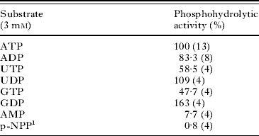
In the presence of 1 mm CaCl2, the phosphohydrolytic activities were detected in samples of different promastigote preparations from standard L. (V.) braziliensis MHOM/BR/1975/M2903 strain. The samples hydrolysed ATP and ADP, with a specific activity of 724±341 (n=13) or 603±451 nmol Pi/mg−1 h−1 (n=8), respectively (Table 1). No endogenous ATPase or ADPase activities were detected in promastigotes preparations if they were tested in the absence of CaCl2, or in the presence of either 5 mm EDTA or 5 mm EGTA, thus confirming the bivalent metal ion dependence of the phosphohydrolytic activity. The hydrolysis of other nucleosides di- and triphosphates was also tested. As shown in Table 1, ATP, ADP, UDP and GDP were the best substrates, while other nucleotides such UTP and GTP produced lower reaction rates.
Substrate specificity
(Activity measurements in the Leishmania (V.) braziliensis promastigote preparation were performed in standard reaction medium. The values represent the percentage of activity when compared to the ATPase activity (724±341 nmol Pi·mg−1·h−1). The number of measurements, in duplicate using different promastigote preparations, is shown in parenthesis.)

1 p-NPP: p-nitrophenylphosphate.
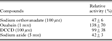
The hydrolysis of AMP, a substrate of 5'-nucleotidase, or p-nitrophenylphosphate (p-NPP), a substrate of phosphatases, was low (<10%) when compared to those found for nucleosides di- and triphosphate (Table 1) and, therefore, the hydrolytic activity of these enzymes did not contribute significantly with the amount of Pi quantified in the assays. To verify the possible relation of the ATPase activity observed in this work with classical well-known ATPases, several inhibitors were tested (Table 2). Addition of 100 μm sodium orthovanadate, an inhibitor of P-type cation transport ATPases, inhibited about 53% of the ATPase activity, suggesting a direct effect on a P-type ATPase in the promastigotes preparation (Table 2). This partial ATPase inhibition suggested that other enzymes that hydrolyse ATP were still present in promastigote preparations. Ouabain (1 mm), an inhibitor of Na+/K+ ATPase, and dicyclohexylcarbodiimide (DCCD), an inhibitor of mitochondrial ATPase, were also tested and no significant interference was observed (Table 2). On the other hand, sodium azide (5 mm), an inhibitor of either mitochondrial ATPase or L. amazonensis ATP diphosphohydrolase (see Coimbra et al. Reference Coimbra, Gonçalves-Da-Costa, Corte-Real, Freitas, Durão, Souza, Silva Santos and Vasconcelos2002), was able to inhibit 58% of the ATPase activity. Since DCCD did not affect significantly ATP hydrolysis, these results suggested a direct effect of sodium azide on ATP diphosphohydrolase inhibiting partially its ATPase activity. The significant hydrolysis of nucleoside di- or triphosphate upon bivalent metal ion activation, associated to the inhibition promoted by sodium azide, strongly suggested that ATP diphosphohydrolase activity exists in L. (V.) braziliensis promastigote forms.
Effects of inhibitors on ATP hydrolysis
(The results are expressed as the percentage of control that was measured in the standard reaction medium, in the absence of the compound indicated. The table shows the mean±standard deviation of 4 different experiments in duplicate for each compound used.)

Localization of the ATP diphosphohydrolase activity in L. (V.) braziliensis promastigote forms was obtained by ultrastructural cytochemical techniques (Fig. 1 A–F). Enzyme activity was found distributed as an electron-dense cerium phosphate deposit at the surface of the plasma membrane, and at the flagellar pocket and flagellum of L. (V.) braziliensis promastigote forms when they were incubated in the cytochemical complete medium containing ATPases, nucleotidase and phosphatase inhibitors plus ATP, ADP or GDP (1A–B, 1C–D and 1E–F) as substrate. The hydrolytic activity appeared to be identically distributed at the outer surface of the mitochondria when ADP (C) or GDP (E–F), was used as substrate, while ATP hydrolysis was insignificant under reaction conditions (A, B). These results suggested that ATP diphosphohydrolase activity also has this subcellular localization. In the absence of nucleotides, controls showed no electron-dense deposits (data not shown).
Ultrastructural localization of enzymatic activity. ATPase (A–B), ADPase (C–D) or GDPase (E–F) activity can be seen as an electron-dense product (arrows) associated at the surface of the plasma membrane of the parasite and at the flagellar pocket (FP) and flagellum (F). Hydrolytic activity appeared to be identically distributed at the external surface of the kinetoplast (K) and mitochondrial membranes (arrowheads), when ADP (C) or GDP (E–F) was used as substrate. ATP hydrolysis was insignificant (A, B). N, nucleus.

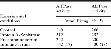
Rabbit polyclonal anti-potato apyrase antibodies recognized bands of approximately 48 and 43 kDa in Western blots of the promastigote preparation (50 μg of total protein; Fig. 2, panel WB, lane Lb). Anti-potato apyrase antibodies were tested for their ability to immunoprecipitate ATP diphosphohydrolase from a detergent-homogenized L. (V.) braziliensis promastigote preparation. After homogenization with non-ionic detergent C12E9 and centrifugation, significant parasite enzyme activity was maintained in the high-speed supernatant (Table 3; control). No significant difference was observed in control assays with either pre-immune serum or Protein A-Sepharose added alone in the absence of serum (Table 3). On the other hand, rabbit polyclonal anti-potato apyrase antibodies immobilized on Protein A-Sepharose immunoprecipitated approximately 83% of the ATPase and 87% of the ADPase activities, corresponding to depletion of an ATPase/ADPase activity ratio of approximately 1 (Table 3).
Identification of an active ATP diphosphohydrolase isoform from Leishmania (V.) braziliensis promastigote forms by immunoprecipitation assays using anti-potato apyrase antibody. Potato apyrase (1 μg; lanes A, WB and WB-I), an aliquot of promastigotes preparation (50 μg of protein; WB, lane Lb) or the immunoprecipitated Protein A-rabbit antibody-antigen complex isolated from the promastigote preparation (WB-I, lane I) was submitted to electrophoresis in 10% SDS-PAGE, electroblotted onto nitrocellulose membrane, and the Western blots were developed with rabbit (WB) or mouse (WB-I) polyclonal serum against potato apyrase. In WB-A, potato apyrase (1 μg) was electrophoresed in 10% SDS-PAGE, electroblotted onto nitrocellulose membrane, and the Western blots were developed with either serum from an American cutaneous leishmaniasis patient (L) or a healthy individual (H). The membranes were revealed by chemiluminescence.

Depletion of ATP diphosphohydrolase activity from the C12E9-homogeneized promastigote preparation by rabbit polyclonal anti-potato apyrase antibodies immobilized on Protein A-Sepharose
(The ATP or ADP hydrolysis was measured in the high-speed supernatant from either the C12E9-homogeneized promastigote preparation (Control) or control assays with Protein A-Sepharose added alone in the absence of serum (Protein A-Sepharose). Pre-immune or immune serum was used at a dilution of 1:500. The results in parenthesis represent the percentage of hydrolytic activity when compared to the pre-immune serum. The experiments were repeated twice with similar results.)

The immunoprecipitated resin-rabbit antibody-antigen complex was washed and subjected to electrophoresis and Western blots. As observed in Fig. 2, the rabbit polyclonal antibodies against potato apyrase immobilized on Protein A-Sepharose immunoprecipitated the same bands of approximately 48 kDa and, in lower amount, the band of 43 kDa, which were recognized by the mouse polyclonal anti-potato apyrase antibodies (Fig. 2, panel WB-I, lane I). These results confirmed the identity of an active ATP diphosphohydrolase isoform in L. (V.) braziliensis promastigote forms, which shares conserved epitopes with potato apyrase.
The antigenicity of these conserved domains was tested in patients with American cutaneous leishmaniasis (ACL) using the potato apyrase as antigen. This vegetable protein was recognized in Western blots by diluted sera (1:100) from ACL patients, and a representative result is shown in Fig. 2 (panel WB-A, lane L). Serum diluted 1:100 from a healthy individual did not react with potato apyrase (Fig. 2, panel WB-A, lane H). These results indicate that the conserved domains shared between the parasite and vegetable proteins are antigenic. In addition, the high purity of the potato apyrase could also be observed in Fig. 2, since no other background was present when sera from rabbit (panels WB, lane A), mouse (panel WB-I, lane A) or ACL patients (panel WB-A, lane L) were tested.
Synthetic peptides belonging to the conserved domain from both L. braziliensis NDPase (LbB1LJ; RERFKRIEPGLSSFATDQEGAK22, r82-103) and its potato apyrase counterpart (potB1LJ; IEYFMATEPGLSSYAEDPKAAA22; r77-98) were assayed. IgG antibody levels were quantified in diluted serum samples (1:50) from patients with ACL using Lb, potato apyrase and synthetic peptides as coating antigens in ELISA. The IgG antibody levels against Lb (C, 0·095±0·027; ACL, 0·193±0·078; P<0·01; cut-off, 0·149; 65% seropositivity), potato apyrase (C, 0·111±0·022; ACL, 0·198±0·049; P<0·01; cut-off, 0·155; 90% seropositivity), LbB1LJ (C, 0·045±0·050; ACL, 0·214±0·111; P<0·001; cut-off,0·145; 80% seropositivity) or potB1LJ (C, 0·108±0·040; ACL, 0·202±0·103; P<0·05; cut-off 0·188; 50% seropositivity) were significantly higher than that found in healthy individuals (C; Fig. 3A). For comparative analysis of positive antibody reactivity, the individual value was then calculated as ELISA units. Median, maximum and minimum values are shown in Fig. 3B. The total IgG antibody reactivity against Lb (median 1·229) or potato apyrase (median 1·263) was similar between them, with 65% and 90% seropositivity respectively (Fig. 3B). Serum samples (diluted 1:50) showed variable total IgG antibody reactivity (median 1·672) against LbB1LJ, up to 3-fold greater than the threshold 1. Sixteen (80%) of the 20 ACL patients had seropositivity for this peptide (Fig. 3B). The IgG seropositivity against potB1LJ was observed in 50% (10/20) of the ACL patients, but its reactivity (median 0·992) was below the threshold of 1, and significantly (P<0·05) lower when compared to the IgG antibody reactivity against LbB1LJ (Fig. 3B).
IgG antibody reactivity of ACL patients against Lb, potato apyrase and synthetic peptides. (A) The total IgG antibody level was quantified in serum diluted 1:50 from healthy individuals (C; n=10) and ACL patients (n=20) using Leishmania (V.) braziliensis promastigotes preparation (Lb), potato apyrase (APY) or synthetic peptide (LbB1LJ and potB1LJ) as the coating antigen in ELISA. The IgG antibody levels are expressed as optical density of serum samples. The horizontal line represents the mean and the dotted line represents the cut-off. (B) The IgG antibody levels are expressed as ELISA units (U), and the horizontal line represents the median. The dotted horizontal line of value 1 represents the mean of the optical density of serum samples from healthy individuals plus 2 standard deviations. Values greater than this cut-off level were considered to be seropositive. The statistical significance of group differences was determined using Mann-Whitney test or Kruskal-Wallis test. P value is *0·05, **0·01 or ***0·001.

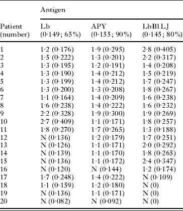
Individual analysis (Table 4) showed that 11 (numbers 1–11) of the 20 ACL patients had IgG antibody seropositivity for Lb, potato apyrase and LbB1LJ, and 7 (numbers 1–7) of these 11 patients showed higher seropositivity for LbB1LJ than those found for both Lb and potato apyrase. Four (numbers 12–15) of the 20 ACL patients showed seropositivity for potato apyrase and LbB1LJ, higher for this last antigen, but not for Lb, and 1 (number 16) patient showed seropositivity only for LbB1LJ. These results suggested that LbB1LJ has high sensitivity for antibody detection when compared to the Lb or potato apyrase. On the other hand, 2 (numbers 17 and 18) of the 20 ACL patients had IgG antibody seropositivity for Lb and potato apyrase, but not for LbB1LJ, and 1 patient (number 19) only for potato apyrase, suggesting the existence of other antigenic domains shared between potato and parasite proteins. One ACL patient (number 20) was seronegative for any antigen. The variable IgG antibody reactivity from ACL patients possibly reflects different parasite load and/or host genetic factors.
Individual IgG antibody seropositivity of ACL patients for Leishmania (V.) braziliensis promastigotes preparation (Lb), potato apyrase (Apy) or synthetic peptide belonging to the L. braziliensis NDPase (LbB1LJ)
(The results are expressed in ELISA units (U), and the number in parenthesis represents the optical density. The cut-off and seropositivity (%) for each antigen are also represented in parentheses.)

N, seronegativity.
DISCUSSION
We identified an ATP diphosphohydrolase activity in the preparation of L. (V.) braziliensis promastigotes, and its hydrolytic activity was localized at the surface of the parasite by cytochemical microscopy when ATP, ADP or GDP was used as substrate. Besides its ecto-localization, this protein was also found at the outer surface of the mitochondrial membrane. Several inhibitors, including sodium azide, were maintained in the cytochemical reaction medium assuring the absence of other phosphohydrolytic activities. It is important to note that sodium azide also reduced ATP diphosphohydrolase activity in the Lb preparation (see Table 2) and, possibly, the nucleotides hydrolysis was affected at different rates in cytochemical assays. Analyses of the NCBI databases revealed a putative nucleoside diphosphatase (NDPase; Gene ID 5413969) and a putative guanosine diphosphatase (GDPase; Gene ID 5413270) in the L. braziliensis genome, homologous to proteins from the ATP diphosphohydrolase family (Peacock et al. Reference Peacock, Seeger, Harris, Murphy, Ruiz, Quail, Peters, Adlem, Tivey, Aslett, Kerhornou, Ivens, Fraser, Rajandream, Carver, Norbertczak, Chillingworth, Hance, Jagels, Moule, Ormond, Rutter, Squares, Whitehead, Rabbinowitsch, Arrowsmith, White, Thurston, Bringaud, Baldauf, Faulconbridge, Jeffares, Depledge, Oyola, Hilley, Brito, Tosi, Barrell, Cruz, Mottram, Smith and Berriman2007). Presumably these two isoforms have distinct metabolic functions and catalytic properties and, under our experimental conditions, they differently hydrolysed nucleosides di- and triphosphate. The ATP diphosphohydrolases have been described by us and by other authors as ecto-enzymes on the parasite surface (Coimbra et al. Reference Coimbra, Gonçalves-Da-Costa, Corte-Real, Freitas, Durão, Souza, Silva Santos and Vasconcelos2002; Faria-Pinto et al. Reference Faria-Pinto, Meirelles, Lenzi, Mota, Penido, Coelho and Vasconcelos2004, Reference Faria-Pinto, Meirelles, Lenzi, Mota, Penido, Coelho and Vasconcelos2006; Pinheiro et al. Reference Pinheiro, Martins-Duarte, Ferraro, Souza, Gomes, Lopes, Vannier-Santos, Santos and Meyer-Fernandes2006; Borges et al. Reference Borges, de Brum Vieira, Wiltuschnig, Tasca, De Carli and Bonan2008; Guevara-Flores et al. Reference Guevara-Flores, Olvera-Sánchez, Gómez-Concha, Juarez, Esparza-Perusquía, Pardo, Mendoza-Hernández, Martinez and Flores-Herrera2008; Sansom et al. Reference Sansom, Riedmaier, Newton, Dunstone, Muller, Stephan, Byres, Beddoe, Rossjohn, Cowan, d'Apice, Robson and Hartland2008; Vasconcelos et al. Reference Vasconcelos, Faria-Pinto, Rezende-Soares, Penido, Gonçalves-da-Costa, Coelho, Tennant and Benkeblia2009). To our knowledge, in parasites this is the first demonstration of hydrolytic activity at the outer surface of the mitochondrial membrane, which is similar to those from ATP diphosphohydrolases. This subcellular location suggests the participation of this enzyme in metabolic pathways still not investigated, possibly essential for parasite survival. Therefore, this hypothesis should be confirmed and more extensively explored. Experiments to investigate this new localization are currently being carried out in our laboratories. In addition, the availability of the L. (V.) braziliensis ATP diphosphohydrolase isoforms, made possible by cloning and heterologous expression, will allow characterization of their catalytic properties towards nucleoside di- and triphosphates and the possible functions in this parasite.
By in silico analysis, closer structural relationships were previously observed between potato apyrase and the putative L. braziliensis NDPase (Faria-Pinto et al. Reference Faria-Pinto, Rezende-Soares, Molica, Montesano, Marques, Rocha, Gomes, Enk, Correa-Oliveira, Coelho, Neto, Franco and Vasconcelos2008; Vasconcelos et al. Reference Vasconcelos, Faria-Pinto, Rezende-Soares, Penido, Gonçalves-da-Costa, Coelho, Tennant and Benkeblia2009) of predicted molecular mass of 47 689 Da. Now, using as a strategy the cross-immunoreactivity with polyclonal anti-potato apyrase antibodies, an active ATP diphosphohydrolase isoform was identified, in L. (V.) braziliensis promastigote forms, as 2 bands of approximately 48 and 43 kDa, and ATPase/ADPase activity ratio of approximately 1. Only 17% and 13% of the ATPase and ADPase activities, respectively, were maintained in the high-speed supernatant resulting from immunoprecipitation assays. In addition, no other reactive band was detected in Western blots. Alignment of amino acid sequences from potato apyrase and putative L. braziliensis GDPase, of predicted molecular mass 74 840 Da, showed low identity between them (33% identity and 48% similarity over 229 amino acids), which was restricted to the conserved domains from the ATP diphosphohydrolase family. A possible cleavage site was observed in the putative GDPase, which could be a signal peptide generating a processed protein with a molecular mass of 71 294 Da. Therefore, if a GDPase isoform is present in the Lb preparation, it is in lower amount or under inadequate experimental conditions for its activation, and/or if cross-immunoreactivity between potato apyrase and this protein occurs, it is lower. The presence of this band of approximately 43 kDa in both the antigenic Lb preparation and the immunoprecipitated Sepharose Protein A-antibody-proteins complex subjected to electrophoresis and Western blots could be the result of proteolysis of the 48 kDa band, as a consequence of experimental conditions or even a natural occurrence in vivo. Further studies of an expressed and active recombinant NDPase could clear this hypothesis.
In predicted three-dimensional structures, particular domains shared between potato apyrase and L. braziliensis NDPase were shown to be exposed and available for antibody binding (Faria-Pinto et al. Reference Faria-Pinto, Rezende-Soares, Molica, Montesano, Marques, Rocha, Gomes, Enk, Correa-Oliveira, Coelho, Neto, Franco and Vasconcelos2008; Vasconcelos et al. Reference Vasconcelos, Faria-Pinto, Rezende-Soares, Penido, Gonçalves-da-Costa, Coelho, Tennant and Benkeblia2009). Supported by both theoretical analysis and the identification of this active isoform of approximately 48 kDa, we synthesized the peptide LbB1LJ, which spans the domain r83-103 within L. braziliensis NDPase, and the peptide potB1LJ, its potato apyrase counterpart. Both of the synthetic peptides were recognized by IgG antibodies from ACL patients, with higher IgG antibody seropositivity for LbB1LJ (16/20; 80%), indicating that for the human immune system the domain r83-103 from L. braziliensis NDPase is rich in B-cell epitopes. The IgG antibody reactivity against domain r83-103 from L. braziliensis NDPase could be contributing to the total IgG antibody reactivity against antigens from the L. (V.) braziliensis promastigote originated from MHOM/BR/1975/M2903 strain, which is commonly used for leishmaniasis diagnosis and epidemiological studies (Junqueira-Pedras et al. Reference Junqueira-Pedras, Orsini, Castro, Passos and Rabello2003; Marques et al. Reference Marques, Volpini, Machado-Coelho, Machado-Pinto, da Costa, Mayrink, Genaro and Romanha2006). Cutaneous clinical forms of leishmaniasis caused by L. braziliensis are generally associated with low circulating antibodies against the Lb preparation (Marques et al. Reference Marques, Volpini, Machado-Coelho, Machado-Pinto, da Costa, Mayrink, Genaro and Romanha2006; Junqueira-Pedras et al. Reference Junqueira-Pedras, Orsini, Castro, Passos and Rabello2003). Because of this, the IgG antibody reactivity from ACL patients against the LbB1LJ – a peptide of only 22 amino acids belonging to the parasite NDPase of 425 amino acids – even close to the background level for some patients, is still significant. Peptide epitopes represent the minimal immunogenic region of a protein antigen and, simple peptides can be poorly immunogenic. Seeking applications such as vaccines, distinct strategies have become available to modify peptides to enhance both their immunogenicity and stability (Purcell et al. Reference Purcell, McCluskey and Rossjohn2007). These observations reinforce the importance of our immunological results. The LbB1LJ peptide could be useful as a composition to improve diagnosis methods for patients with lesions confined to skin, the subject of the present work.
Total IgG antibody seropositivity (50%) for peptide belonging to the potato apyrase (potB1LJ), at the same serum dilution, was lower when compared to the seropositivity (90%) for potato apyrase. The epitopes shared between the vegetable and parasite proteins are linear rather than conformational, as observed by the reactivity of the denatured potato apyrase and L. braziliensis ATP diphosphohydrolase subjected to electrophoresis and Western blots, and because of the similar IgG antibody reactivities from ACL patients against native or denaturated potato apyrase (at 100°C; 5 min) tested by ELISA in preliminary assays (data not shown).
Therefore, it is possible that other antigenic domains within L. braziliensis NDPase exist, also shared with potato apyrase, as previously predicted (Faria-Pinto et al. Reference Faria-Pinto, Rezende-Soares, Molica, Montesano, Marques, Rocha, Gomes, Enk, Correa-Oliveira, Coelho, Neto, Franco and Vasconcelos2008). Cross-immunoreactivity between potato apyrase and parasite ATP diphosphohydrolases has been demonstrated (Faria-Pinto et al. Reference Faria-Pinto, Meirelles, Lenzi, Mota, Penido, Coelho and Vasconcelos2004, Reference Faria-Pinto, Rezende-Soares, Molica, Montesano, Marques, Rocha, Gomes, Enk, Correa-Oliveira, Coelho, Neto, Franco and Vasconcelos2008; Coimbra et al. Reference Coimbra, Gonçalves-Da-Costa, Costa, Giarola, Rezende-Soares, Fessel, Ferreira, Souza, Abreu-Silva and Vasconcelos2008; Vasconcelos et al. Reference Vasconcelos, Faria-Pinto, Rezende-Soares, Penido, Gonçalves-da-Costa, Coelho, Tennant and Benkeblia2009). Comparative studies of the antigenic conserved domains shared between parasite ATP diphosphohydrolase and potato apyrase in different parasitic diseases will facilitate the identification of epitopes in these less well investigated proteins, and will be of interest for determining sensitivity/specificity. These experiments are being carried out in our laboratory.
ACKNOWLEDGEMENTS
This work was supported in part by grants from the Fundação de Amparo a Pesquisa do Estado de Minas Gerais (FAPEMIG; process no. CBB-1859/06), and Conselho Nacional de Desenvolvimento Científico e Tecnológico (CNPq; process no. 474158/2003-9). F.A. Rezende-Soares and C. Carvalho-Campos were recipients of Master Degree fellowships from the UFJF/MG. G.N. Porcino, N.L.L. Giarola, B.L.S. Costa and A. Taunay-Rodrigues were recipient of fellowships from the PIBIC, PROBIC or BIC/UFJF. M.A. Souza, S. Corte-Real, M.A. Juliano and L. Juliano are recipients of fellowships from CNPq and FAPESP.