

INTRODUCTION
The Indian Ocean has been the least well-studied of the major oceans of the world (Ryther, Reference Ryther1963), a fact that remains true to this day. To address this issue, the International Indian Ocean Expedition (IIOE) set out to systematically survey the Indian Ocean, with scientists from over 25 countries and more than 40 research vessels participating in this expedition during the 1960s (Ryther, Reference Ryther1963). Among the material collected during the IIOE and deposited at the National Museum of Natural History, Smithsonian Institution in Washington, DC I discovered thus far unidentified specimens of chirodropid cubozoans (Cnidaria: Cubozoa: Chirodropida) in two lots of medusozoan cnidarians from the island of Madagascar. This discovery represents an important addition to our knowledge of cubozoans.
So far several cubozoans have been reported from mainly West African [Chirodropus gorilla Haeckel, Reference Haeckel1880, Chiropsalmus quadrumanus (Müller, 1859), Tamoya haplonema Müller, 1859 (Haeckel, Reference Haeckel1880; Kramp, Reference Kramp1955, Reference Kramp1959 and references cited therein)] or South African waters [Carybdea branchi Gershwin & Gibbons, Reference Gershwin and Gibbons2009 (Gershwin & Gibbons, Reference Gershwin and Gibbons2009 and references cited therein)]. To my knowledge no chirodropid has been described in detail from eastern Africa, even though Cornelius (Reference Cornelius and Richmond1997) lists Chiropsalmus quadrumanus in a guidebook to the East African seashore without providing a record. Considering that the species name Chiropsalmus quadrumanus had been used incorrectly to refer to other species of Chiropsella (Gershwin, Reference Gershwin2006a), and that Chiropsalmus quadrumanus is generally found in the West Atlantic, including the Caribbean (Gershwin, Reference Gershwin2006a; Bentlage et al., Reference Bentlage, Cartwright, Yanagihara, Lewis, Richards and Collins2010), it seems plausible that the identification as Chiropsalmus quadrumanus is erroneous, and the material described here may be the same as the species mentioned by Cornelius (Reference Cornelius and Richmond1997). The discovery of the species of Chiropsella described herein adds to our knowledge of the cubozoan fauna found in the western Indian Ocean and extends the known distributional range of the genus Chiropsella from the south-west Pacific into the Indian Ocean.
MATERIALS AND METHODS
All specimens investigated were collected by J. Rudloe as part of the IIOE using a minnow seine or shrimp trawl, and subsequently preserved in 10% formaldehyde solution. Observations on the anatomy of the specimens were made using a Nikon SMZ 1500 stereoscope. Nematocysts were sampled from proximal and distal ends of the tentacles (note that the tentacles were truncated), the manubrium, and the gastric phacellae. Pieces of tissue were excised, placed on microscope slides, and then covered with a cover slip. The cover slip was subsequently pressed down firmly to separate the nematocysts from the tissue. Nematocysts were identified using a Nikon Eclipse 80i compound microscope. Images were taken using the SPOT Insight 14.2 Color Mosaic Camera (Diagnostic Instruments Inc., MI); measurements of nematocysts were obtained using the measurement calibration tool implemented in SPOT Imaging Software Basic (v. 4.2). Nematocysts were identified using Mariscal (Reference Mariscal, Muscatine and Lenhoff1974), Östman (Reference Östman2000) and Gershwin (Reference Gershwin2006b). Nematocysts in which the undischarged shaft displayed a v-shaped notch were classified as ‘p-’; according to Östman (Reference Östman2000).
RESULTS
SYSTEMATICS
Phylum CNIDARIA Verrill, 1865
Subphylum MEDUSOZOA Petersen, 1979
Class CUBOZOA Werner, 1973
Order CHIRODROPIDA Haeckel, Reference Haeckel1880
Family CHIROPSALMIDAE Thiel, 1936
Genus Chiropsella Gershwin, 2006
Chiropsella rudloei, sp. nov.
(Figures 1–3)
Images of Chiropsella rudloei specimen USNM 1140278: (A) habitus; (B) lateral gonads running vertically along the interradius; (C) proximal pedalial canal bend; (D) abaxial view of pedalium with pedalial canal dividing into two separate branches as (dashed lines added to highlight separate canals); (E) manubrium (dashed lines added to trace outline of distal manubrium; arrows indicate the position of the four tips of the lips), view from subumbrellar opening up into subumbrellar cavity. Note that bright and grainy particles in the images represent sand grains that have entered cavities and tissues of specimens during collection. go, gonad; ir, interradius; pe, pedalium; pc, pedalial canal (partially filled with sand as an artefact of collection method); sp, split in pedalial canal; vb, volcano-like bend in pedalial canal; *, corresponding pedalial fingers in A and D.

Images of Chiropsella rudloei: (A) apical view with gastric phacellae and saccules being visible (spheres visible represent air bubbles in the gastro-vacular cavities of the specimen); (B) close-up of gastric saccules with dashed line highlighting the shape of the distal portion of the coalesced saccules (view from top of swimming bell into stomach with velarium and velarial canals visible in the background); (C) perradial lappet with outline highlighted by dashed line; (D) velarium with velarial canals; (E & F) rhopaliar niche cover with flap and rhopalium situated within the rhopaliar niche, dashed lines trace the shape of the opening and covering scale; (G) close-up of pedalial canal bend with spike in specimen USNM 1140280. Dashed lines delimit the pedalial canal inside the pedalium and the pedalium on the left side of the image. Note that bright grainy particles in the images represent sand grains that have entered cavities and tissues of specimens during collection. fl, rhopaliar niche flap; go, gonad; gp, gastric phacellae; gs, gastric saccule; le, lens eye; ma, manubrium; pc, pedalial canal; pe, pedalium; pl, perradial lappet; rh, rhopalium; st, statolith; ve, velarium; vc, velarial canals.

Nematocysts found in syntype specimens of Chiropsella sp.: (A–F) tentacle tip; (G–J) tentacle base; (K) gastric phacellae; (L & M) manubrium. For further detail and identifications see Table 2. All scale bars represent 10 µm.

ABBREVIATIONS
National Museum of Natural History, Smithsonian Institution, USNM; bell height, BH—measured from velarial turn-over to the top of the bell; interradial bell width (the distance between the centre of two pedalia), IRW; maximum number of tentacles per pedalium, NT.
MATERIAL EXAMINED
Syntypes: USNM 1140278, 31 mm BH, 25 mm IRW, NT 7; USNM 1140267, 40 mm BH, 32 mm IRW, NT 7; USNM 1140268, 35 mm BH, 28 mm IRW, NT 5; USNM 1140269, 35 mm BH, 28 mm IRW, NT 6; USNM 1140270, 42 mm BH, 30 mm IRW, NT 7; USNM 1140271, 35 mm BH, 26 mm IRW, NT 8; USNM 1140272, 28 mm BH, 24 mm IRW, NT 6; USNM 1140273, 24 mm BH, 19 mm IRW, NT 5; USNM 1140274, 26 mm BH, 21 mm IRW, NT 6; USNM 1140275, 32 mm BH, 24 mm IRW, NT 6; USNM 1140276, 29 mm BH, 22 mm IRW, NT 5; USNM 1140277, 36 mm BH, 29 mm IRW, NT 7; USNM 1140279, 33 mm BH, 27 mm IRW, NT 6. All collected 10 February 1964, Ampora Village (Nosy n' Tanga), Nosy Be, Madagascar, 13°11′55″S 48°11′10″E, by Jack Rudloe using a minnow seine.
Other material: USNM 1140280, 80 mm BH, 65 mm IRW, NT 11, 31 January 1964, Baie d'Amboro, north-west coast of Madagascar, 13°24′30″S 48°39′30″E, collected by Jack Rudloe using a shrimp trawl.
TYPE LOCALITY
Madagascar, Nosy Be, 13°21′55″S 48°11′10″E.
ETYMOLOGY
The specific name was chosen to honour Jack Rudloe for his dedication to the study and protection of the marine environment and its inhabitants.
REGISTRATION OF NOMENCLATURAL ACT
The new species name Chiropsella rudloei was registered with ZooBank (zoobank.org; urn:lsid:zoobank.org:act:5FD25432-47AF-4A96-8F99-D2D11276B710) to comply with the amendments (ICZN, 2012a, b) to the 4th edition of the International Code of Zoological Nomenclature (ICZN, 1999) that govern the availability of nomenclatural acts published using electronic methods.
DIAGNOSIS
Chiropsella (see Table 1) with coalesced, kidney-shaped gastric saccules that are smooth and sessile. Rhopaliar niche with flap on upper covering scale. Ab- and adaxial keels on pedalia absent.
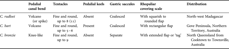
Comparison of genera within the Chiropsalmidae (modified from Gershwin (Reference Gershwin2006b)).

DESCRIPTION
Syntypes
Strongly cuboid chirodropid medusae belonging to the genus Chiropsella (Table 1). Exumbrella smooth, lacking nematocyst warts (Figure 1A; warts may have rubbed off due to abrasion by the collection gear). Most specimens examined had a BH of about 25–40 mm and an IRW of 20–32 mm. Interradial gonads well developed, appearing mature and ripe with eggs or sperm, even in small specimens; gonads with lateral, wing-like extensions (Figure 1B). Pedalia with multiple distal ‘fingers’ bearing tentacles, without adaxial and abaxial keels (Figure 1A, D), branching laterally in opposite fashion; divided along branching portion (Figure 1D). Each pedalium bearing up to eight tentacles (but see description of USNM 1140280 below). Proximal pedalial canal bend rounded, ‘volcano’-shaped sensu Gershwin & Alderslade (Reference Gershwin and Alderslade2006) (Figure 1C). Tentacles fine and round. Manubrium cruciform with four short, broad lips (Figure 1E). Gastric phacellae V-shaped in each corner of stomach (Figure 2A); gastric cirri simple and unbranched. Gastric saccules form a solid kidney-shaped structure (Figure 2A, B) similar to the one observed in Chiropsella bart Gershwin & Alderslade, Reference Gershwin and Alderslade2006 (see figures 1 & 3B in Gershwin & Alderslade, Reference Gershwin and Alderslade2006). The saccules are simple, smooth, and coalesced sensu Gershwin & Alderslade (Reference Gershwin and Alderslade2006) (Figure 2A, B); they are solid and knob-like (sessile sensu Gershwin, Reference Gershwin2006a) rather than pendant and do not extend below the upper third of the umbrella. Perradial lappets smooth, broad, triangular, extending almost to subumbrellar edge of velarium (Figure 2C). The frenulum consists of a single, undivided gelatinous sheet that spans from the rhopaliar window to about two-thirds the distance from the velarial turnover to the velarial margin. Velarial canals highly branched distally (Figure 2D). Rhopaliar niche ostium dome-shaped with a single upper covering scale; upper covering scale exhibits a flap which range in shape from squared to round (Figure 2E, F). Rhopalia not well preserved; all eyes as well as statoliths seem to have completely or partially dissolved in the preservative. No lateral slit or pit eyes in any of the specimens; this is a likely artefact of preservation. Two lens eyes and a statolith are present in the rhopalia, although they are heavily damaged (Figure 2E, F).
Other material
Specimen USNM 1140280 is more than twice as big as the average syntype specimen (80 mm BH by 65 mm IRW versus on average ~33 mm BH by ~26 mm IRW). It also bears more tentacles per pedalium than any other specimen (11 versus 8). Overall, it corresponds well to the smaller specimens although it is much larger. Most importantly it does contain the kidney-shaped, coalesced gastric saccules that were observed in the smaller specimens. However, its proximal pedalial canal forms a prominent spike in contrast to the more rounded volcano-like bend of the other specimens (compare Figure 1C with Figure 2G).
CNIDOME
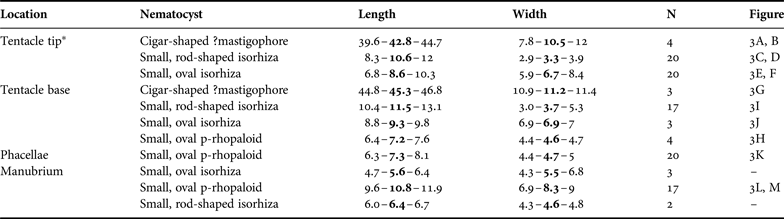
Four different nematocyst types were identified and measured in the syntype specimens with the highest diversity found at the bases of the tentacles (Table 2; Figure 3). Based on comparisons with the shape and size of nematocysts from the other species of Chiropsella it seems probable that the cigar-shaped capsules (Figure 3 A, B & G) represent p-mastigophores (cf. plate 6 in Gershwin, Reference Gershwin2006a and figure 4 in Gershwin & Alderslade, Reference Gershwin and Alderslade2006). Unfortunately all capsules of this type found in Chiropsella rudloei were discharged and missing their shafts making verification of this identification impossible.
Nematocysts found in different body parts of syntype specimens of Chiropsella rudloei from Madagascar. Measurements are provided as follows: minimum–mean–maximum length and width in µm. For images see Figure 3. *, sampled from the most distal part of the truncated tentacles.

COMPARISON
A comparison of key features of the species of Chiropsella is presented in Table 3. Chiropsella rudloei from Madagascar is most similar to its congener C. bart. Both are the only known chirodropids that possess four coalesced gastric saccules—C. bronzie Gershwin, 2006 possesses eight separate solid saccules (Gershwin, Reference Gershwin2006a). Additionally, both C. bart and C. rudloei possess a flap on the upper covering scale of the rhopaliar niche. However, the flap on the rhopaliar niche cover appears more rounded in C. rudloei than in C. bart, which appears to possess a pronounced rectangular extension of the rhopaliar niche cover (cf. figure 2C in Gershwin & Alderslade, Reference Gershwin and Alderslade2006). However, this difference may be an artefact created by compression of the specimens of C. rudloei in the collection gear. Furthermore, specimens of the C. rudloei syntype series possess 7–8 tentacles per pedalium; the large specimen from Baie d'Amboro (USNM 1140280) 11. In comparison, C. bart possesses only up to 5 (in some cases 6) tentacles at a roughly similar size (around 40–50 mm BH). Lastly, C. rudloei lacks the prominent ad- and abaxial pedalial keels that are characteristic of C. bart.
Key to the species of Chiropsella (see also Gershwin, Reference Gershwin2006b; Gershwin & Alderslade, Reference Gershwin and Alderslade2006). Characters in parentheses for C. rudloei represent the character states observed in the large specimen (USNM 1140280), which I interpreted to be part of the same species as the type material at this time.

Herein, I characterized the cnidome of several body parts of specimens from the syntype series of C. rudloei. For its congeners, the tentacular cnidome was described in Gershwin (Reference Gershwin2006a, Reference Gershwinb) and Gershwin & Alderslade (Reference Gershwin and Alderslade2006) and the following comparison focuses on the tentacular cnidomes of all three species. Chiropsella bronzie (as Chiropsalmus n. sp. A in Gershwin, Reference Gershwin2006b) possesses the same complement of nematocysts in its tentacles as C. rudloei (cigar- or club-shaped p-mastigophores, oval isorhizas, rod-shaped isorhizas, and oval p-rhopaloids). Chiropsella bronzie bears two size-classes of oval isorhizas, the size range of the smaller (~9–10 µm by ~7–8 µm) corresponds well to the size range observed in C. rudloei (~7–10 µm by ~6–8 µm), while the large oval isorhizas in C. bronzie are about twice the size (~21–24 µm by ~12–14 µm) of the small isorhizas observed in the two other species of Chiropsella. Chiropsella bart and C. rudloei possess the same complement of tentacular nematocysts (cf. Gershwin & Alderslade, Reference Gershwin and Alderslade2006). However, the size range of p-rhopaloids documented in C. bart (~21–26 µm by ~13–14 µm) is much larger than in C. rudloei (~6–8 µm by ~4–5 µm). P-rhopaloids described from C. bronzie have a similar length (~9–10 µm) to the ones observed in C. rudloei (~6–8 µm), while the width of p-rhopaloids in C. bronzie (~8–9 µm) is about twice that of p-rhopaloids in C. rudloei (~4–5 µm).
HABITAT AND ECOLOGY
At the time of collection the shoreline at the collection locality near Ampora Village was populated by mangrove trees, in some places rich and dense, in others only fringes (J. Rudloe, personal communication). In the minnow seine together with the specimens of Chiropsella rudloei were an undetermined rhizostome scyphozoan, numerous shrimp as well as organic matter such as leaves and seeds (J. Rudloe, personal communication). The subumbrella of USNM 1140280 contained two specimens of fish identified as cf. Nectamia sp. However, the fish seemed to have been caught in the subumbrella as a result of sampling technique rather than having been prey of Chiropsella rudloei.
Generally, chirodropids can be found in the vicinity of mangroves (Fenner et al., Reference Fenner, Lippmann and Gershwin2010) and feed on small shrimp or fish; it seems that C. rudloei follows the same habits. Chiropsella rudloei was collected during the wet season like many, but not all chirodropids, that display seasonality with highest abundances coinciding with the season of highest precipitation (e.g. Fenner et al., Reference Fenner, Lippmann and Gershwin2010). More data and recent collections are needed to conclusively determine a correlation between season and occurrence of C. rudloei. Interestingly C. rudloei is morphologically most similar to C. bart which mainly occurs during the dry season (Currie et al., Reference Currie, McKinnon, Whelan and Alderslade2002).
TOXICITY
The potency of the venom of C. rudloei remains unknown, but stings at the time of collection were mild (J. Rudloe, personal communication). This may have been due to the specimens having lost most of their tentacles in the collection net. The tentacles of the examined material are truncated to short stumps. As with all chirodropids, care should be taken when handling specimens, even though Chiropsella species are not known to inflict stings as severe as other chirodropids.
DISCUSSION
Little is known about the marine invertebrate fauna of the Indian Ocean and species discovery remains a fundamental challenge. Chiropsella rudloei represents a particularly interesting find, as the genus is so far only known from Australia. The current distribution of the genus is patchy with Chiropsella rudloei being described here from Madagascar, C. bronzie being restricted to tropical Queensland ranging from Cooktown to Townsville, and C. bart being restricted to the Gove Peninsula, Arnhem Land in the Northern Territory. One might expect to find additional specimens and species of the genus upon further sampling of the Indian and Pacific Oceans. Alternatively, the current distribution of the three known species may represent a relic of past geological history (cf. Bentlage et al., 2010). Considering the morphological similarities between C. rudloei and C. bart—in particular the gastric saccules—it seems most likely that they are each other's closest known relatives. Unfortunately no genetic data exist to test the relationship among these taxa at this time. Considering the poor preservation of the specimens described here, I established the name for C. rudloei based on the combined characters of the specimens of the syntype series rather than designating a single name-bearing holotype. The species can be distinguished from its congener C. bart based on the lack of pedalial keels in C. rudloei, the distinctly different shape of the rhopaliar niche coverings, tentacle number in equal-sized specimens, and striking size differences in the p-rhopaloids found in the tentacular cnidome. Lastly, C. bart appears to be endemic to Arnhem Land in the Northern Territory of Australia while C. rudloei was discovered in Madagascar, some 5500 km from known distributional range of C. bart.
One of the specimens (USNM 1140280) described above is much larger than the other material examined. Its other characteristics—in particular the shape and structure of the gastric saccules—agree well with the smaller specimens. The larger specimen was collected at a different location, albeit not far from the other sampling station. An important character at odds with the smaller specimens is the thorn-shaped pedalial canal bend. It seems quite possible that the volcano-like pedalial canal bend of the smaller specimens develops a spike as the medusa grows. Barnes (Reference Barnes and Rees1966) noted that the knee-like bend of C. bronzie (which he erroneously identified as Chiropsalmus quadrigatus) never develops a spike whereas a spike or prominent thorn is present in the pedalial canal bend throughout the development of Chironex fleckeri Southcott, 1956 (Chirodropidae). However, Chironex yamaguchii Lewis & Bentlage, 2010 medusae apparently develop a spike in their proximal pedalial canal gradually as they grow (C. Lewis, unpublished data). Considering the overall character agreement between the small and large specimens I think it is justified to conclude that USNM 1140280 is just a large example of the same species as the remainder of the specimens (this is reflected in characters listed in parentheses in Tables 1 & 3). However, additional sampling will be necessary to elucidate the development of C. rudloei and conclusively decide upon the nature of the large specimen. Herein, I have treated all specimens as part of the same species, pointing out how they differ in their characters. Further evidence may show that the specimens described here belong to two separate species that inhabit Madagascan waters and possibly the seashore of the African continent.
ACKNOWLEDGEMENTS
Jack Rudloe generously shared his old field notes with me, giving me an insight into the environment in which the medusae were collected. I am indebted to Tara H. Lynn who prepared and measured nematocysts from the specimens. Cheryl Lewis, Andre Morandini, and Lisa Gershwin provided comments on an earlier draft of the manuscript. In addition, I wish to thank two anonymous referees for their thoughtful comments. Jeff Williams identified the reef fish collected together with the jellyfish described here.
FINANCIAL SUPPORT
This work was funded through US National Science Foundation (NSF) Assembling the Tree of Life grant 0531779 (to P. Cartwright, A.G. Collins and D. Fautin) and a pre-doctoral fellowship of the Smithsonian Institution.