Introduction
Cancer is a general term constituting a group of diseases where it is an uncontrolled, abnormal rate of cell growth in the body. Reference Fitzmaurice, Allen and Barber1 The sixth most common form of cancer worldwide is the head and neck squamous cell carcinoma. The ubiquity of tobacco-related cancers in India is relatively high, making the country the highest prevalence for oral cancers.
Radiation therapy serves as primary therapy in the initial stages of the disease or as a mitigative agent for patients who are inoperable in the final stage of the disease. Reference Santin, Palma-Dibb, Romano, de Oliveira, Nelson Filho and de Queiroz2 During radiotherapy, the oral cavity, salivary glands and the maxillomandibular complex are also exposed to high doses of ionising radiation. Beech et al. Reference Beech, Robinson, Porceddu and Batstone3 reported that radiation therapy also damages the surrounding normal cells, especially the rapidly growing cells leading to the deleterious effects of radiotherapy. Mucosal cells have a high cell turnover rate and diverse microflora, which can also be one of the reasons why the oral cavity is a common site for radiation-induced adverse effects. Reference Sciubba and Goldenberg4 Radiation-related side effects include mucositis, altered taste, caries, xerostomia, trismus, periodontitis, osteoradionecrosis and vascular abnormalities. Reference Naidu, Ramana, Rani, Suman and Roy5 Alterations in the dental components, namely enamel and dentin, include increased solubility, reduction in microhardness, loss of prismatic enamel, increased susceptibility to fracture, increased formation of calcium and reduced oxygen content in the enamel. Reference Gonçalves, Palma-Dibb, Paula-Silva, de Oliveira, Nelson-Filho, da Silva and de Queiroz6 These changes seen in the microstructure level of the enamel are irreversible. Reference Walker, Wichman, Cheng, Coster and Williams7,Reference Kumar, Brooke, Burke, John, O’Donnell and Soldani8 The disruption in the collagen formation significantly affects the mechanical property of enamel.
Minimum tooth damage occurs below 30 Gy. Whereas, 30–60 Gy dosage has 2–3 times higher risk of tooth breakdown due to direct effects on salivary gland function. Dosage above 60 Gy showed 10 times higher risk of damage to the teeth and the salivary glands. Reference Walker, Wichman, Cheng, Coster and Williams7,Reference Naves, Novais, Armstrong, Correr-Sobrinho and Soares9
With the advances in medical science, a cancer diagnosis can be carried out at an early stage. The efficiency in the treatment of the disease has led to an increase in the survival rate of cancer patients in the past decades. This has resulted in an increased probability of orthodontists to come across patients who have undergone radiotherapy seeking orthodontic treatment.
The optimal period of fixed orthodontic intervention following radiotherapy is 1–2 years. Reference Dahllöf and Huggare10
Corrective orthodontics includes the bonding of brackets to the surface of the teeth. Although previous studies have reported that adhesive restorations exhibit decreased tensile strength and are susceptible to fractures in irradiated enamel, studies evaluating the bond strength of the adhesives on orthodontic brackets in irradiated teeth are scanty.
The adequate bond strength for clinical use is 6–8 MPa. Reference Reynolds and Von Fraunhofer11 There is a scarcity of data regarding the adhesion of brackets to the irradiated tooth surface, the effects of ionising radiation on bond strength, the effects on the adhesive properties of the enamel and the effects of thermocycling justifying the need for this study.
The aims of this study are
(1) To compare the shear bond strength (SBS) of the metallic bracket bonded on radiated and non-radiated teeth.
(2) To evaluate and compare the adhesive properties of the dental enamel of bonded radiated and bonded non-radiated teeth.
Methodology
Sixty-six freshly extracted premolars were collected from the Department of Surgery, AB Shetty Memorial Institute of Dental Sciences.
The samples include maxillary and mandibular first and second premolar teeth with complete root formation without a previous restoration. Orthodontic, endodontic, chemically treated teeth and teeth with resorbed roots were excluded.
The collected premolar samples (66) were divided into three groups (n = 22) viz, Group 1—(Control group)—Non-irradiated, non-aged, Group 2—Non-irradiated, aged and Group 3—Irradiated, aged.
Group 3 was irradiated at the Department of Radiation Oncology, KSHEMA, at Nitte University. To maintain a humid atmosphere, samples during irradiation were placed in deionised water. The samples were exposed to X-ray irradiation of 2 Gy dosage per day for 5 days a week over 6 weeks. The samples were stored in artificial saliva until the next irradiation.
Group 2 and Group 3 samples were subjected to ageing with thermocycling in the KSHEMA research lab. Changes in the intraoral temperature occur during food intake and respiration. Thermocycling is a process through which these stresses can be induced in vitro. Reference Gale and Darvell12 Thermocycling was performed by subjecting the samples to 35°C for 20 seconds and 15°C for 2 seconds which constitutes one cycle. Ten thousand such cycles simulate 1 year. Following thermocycling, the specimens were immersed in artificial saliva until the bonding of orthodontic brackets. For bonding, the buccal surface of the teeth samples was etched using phosphoric acid (37%) for 15 seconds, followed by washing and drying for 10 seconds each. A layer of bonding agent (Transbond XT, 3 M Unitek, USA) was spread on the etched surface of the teeth and cured under light for 10 seconds. Composite resin was applied on the surface of the bracket and pressed firmly. The composite was light-cured for 10 seconds.
The SBS testing sample preparation required individual teeth samples to be embedded in acrylic resin. The samples were then submitted to SBS testing with a universal testing machine (Instron testing machine with a crosshead speed of 0·5 mm/minute, load cell of 20 kgf). The specimens were then assessed under 1000× and 2000× magnification. The images obtained were analysed and classified based on the adhesive remnant index (ARI) proposed by Artun and Bergland. Reference Årtun and Bergland13 The samples were scored from 0 to 3 based on the following criteria: 0—No adhesive present on the surface of the enamel, 1—Less than half of the adhesive present on the surface of the enamel, 2—Greater than half of adhesive present on the surface of the enamel and 3—All the adhesive present on the enamel.
The length of the resin tags was further assessed between the non-irradiated and irradiated groups.
Statistical Analysis
The sample size was calculated with a 95% confidence interval and 85% power of the study. A one-way ANOVA test followed by Tukey’s post hoc tests was conducted for comparing SBS between groups. A chi-square test was used to compare ARI scores. Students’ t-test was used for comparing mean length of adhesive tags between the irradiated and non-irradiated groups.
Results
Evaluation of SBS
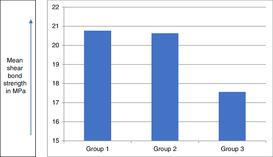
The mean value of SBS in the first group was found to be 20·76 MPa with a standard deviation (SD) of 2·51 (Figure 1). Mean value of Group 2 is 20·62 MPa, with a SD of 2·61. Mean value of Group 3 is 17·55 MPa, with a SD of 2·09. Fischer’s value from the ANOVA test is 12·372 making the difference statistically highly significant, with a p-value <0·001.
Mean shear bond strength (MPa).

The difference in the mean values between Group 2 and Group 3 is 3·06818, which is highly significant (p < 0·001 VHS). The intergroup comparison is proved by the Tukey’s post hoc test.
Intergroup comparison
The difference in mean values between Group 1 and Group 2 is not statistically significant (p = 0·980), whereas the difference in mean values between Group 1 and Group 3 is 3·20, which is statistically significant (p < 0·001 VHS).
SEM evaluation
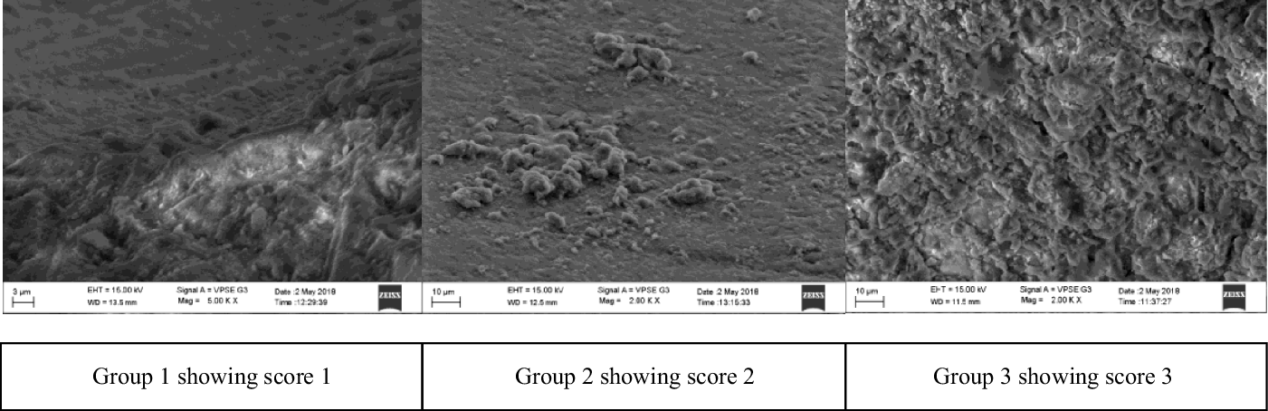
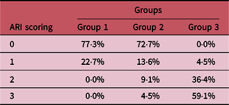
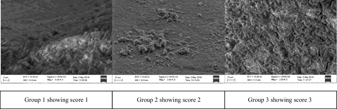
The control group showed no traces of composite on the surface of enamel when visualised under 1000× and 5000× magnification (Figure 2). In Group 1, 77·3% ARI scoring was 0, 22·7% of score 1, and no samples showed scores 2 and 3. Group 2 revealed 72·7% of score 0, 13·6% score 1, 9·1% of score 2 and 4·5% score 3. Whereas in Group 3, the maximum score was seen under the scores 2 and 3 with 36·4 and 59·1%, respectively (Table 1). The adhesive remnant was found on all samples of irradiated enamel.
SEM images of Groups 1, 2 and 3.
ARI scoring

Assessment of the Depth of the Resin Tags
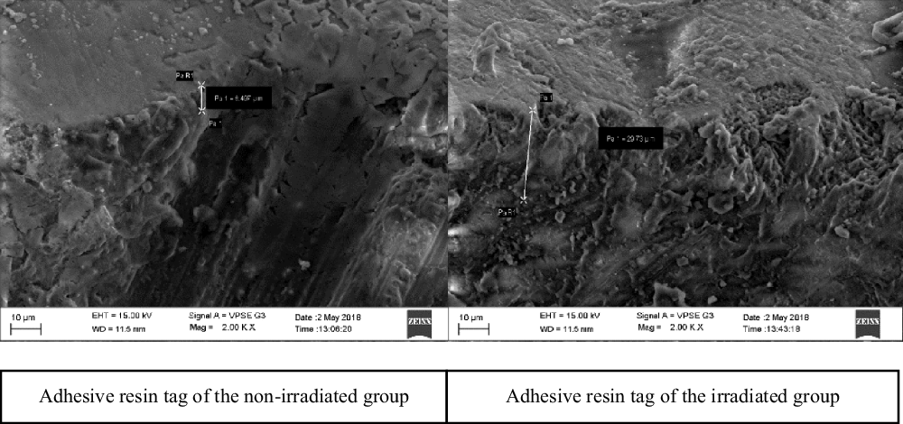
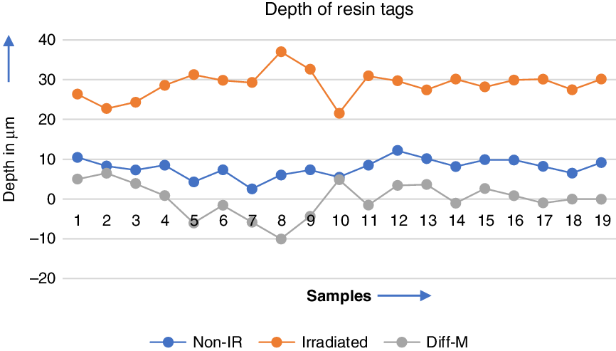
The depth of the resin tags was assessed between the irradiated and the non-irradiated group via scanning electron microscope (SEM) (Figure 3). The longest resin tag measured 37·02 µm in the irradiated group and 10·45 µm in the non-irradiated group. The mean difference between the groups was 20·93 µm (Figure 4).
SEM images of adhesive resin tags.

Illustrating the length of resin tags between non-irradiated and irradiated groups.

The t value obtained by the t-test is −21·784, which suggests that the p-value is highly significant at p < 0·00001.
Discussion
Structural changes in the enamel are seen in patients who undergo cancer therapy involving irradiation of the head and neck area. Bonding bracket on the enamel surface of the tooth is a part of orthodontic treatment.
The null hypothesis stating that SBS decreases with radiotherapy is accepted as the Tukey’s HSD test revealed that there is a significant difference in SBS between the irradiated and the non-irradiated samples. This proves that the irradiation of 2 Gy per day for 5 consecutive days over 6 weeks significantly decreases the bond strength.
In our study, there was a significant decrease in the SBS in the irradiated group by 3·20 MPa when compared to the control group, and a significant 3·06 MPa difference when compared to the non-irradiated, aged group. The samples subjected to 60 Gy of irradiation showed SBS of 17·55 MPa which is nearly three times the ideal bond strength required for the orthodontic purpose. However, the bond strength did not vary significantly between the aged and the non-aged teeth. This result provides an insight into the adverse effect of irradiation which caused a decrease in the SBS.
The low strength may be related to the structural changes induced by ionising radiation on enamel. There may be alterations in the prismatic enamel, causing altered enamel rods leading to weakened areas in the enamel. Loss of organic content is also related to the alteration in the enamel matrix causing the collapse of enamel rods, which in turn decreases the SBS. Reference Dare, Hachisu, Yamaguchi, Yokose, Yoshiki and Okano14
A study by Harhash et al. Reference Harhash and Arnout15 concludes that a high dose of ionising radiation causes a detrimental effect on enamel during the bonding of resin composite. The results of the study also showed that irradiation produced a significant reduction in bond strength.
The analysis of ARI scores revealed that the composite adhered to the bracket in the control group. In control group, samples were only under the ARI scores 0 and 1. The fracture occurred between resin–enamel interface, leaving no trace of adhesive on the enamel (score 0) in 77·3% of samples, whereas cohesive fracture was seen in the 22·7% samples (score 1) with resin present on both, enamel and bracket. The results imply that the adhesion is weak between the tooth and the adhesive in the control group than the adhesive and the bracket.
In Group 2, non-irradiated, aged samples showed 72·7% of adhesive failure at enamel–adhesive interface, whereas 22·7% of samples exhibited cohesive failure falling in scores 1 and 2. The adhesive was present purely on the enamel in 4·5% of the total sample falling under score 3.
In the irradiated group, 59·1% of the total samples had complete adhesive present on the enamel surface. The samples exhibiting failure at the enamel–resin interface in this group were nil. Etching performed alters the surface of the enamel aiding in mechanical retention of the resin. The adhesive penetrates the irregularities creating adhesive tags resulting in a mechanical lock. The resin tags also increase surface energy, providing stability to adhesion. The bond between the tooth and the adhesive seems to be greater in the irradiated group when compared to that of the non-irradiated group. This is mainly due to the surface irregularities created by the ionising radiation in Group 3 and enamel wear induced by thermocycling in Group 2.
The majority of the samples underwent cohesive failure in this group based on results obtained by the ARI index. However, ARI scoring 3 in irradiated samples cannot be gauged as a frank cohesive failure since the depth of the adhesive resin tags in the irradiated group was deeper than the non-irradiated samples. The samples viewed under 1000×, 2000× and 5000× magnification via SEM imaging also reveals that porosities created by etching in irradiated samples are deeper than the etching pattern seen in non-irradiated teeth, with the etching time being constant for all three groups.
The difference between the non-irradiated and irradiated groups was statistically highly significant. The increased depth of the resin tags can be correlated with the increasing porosities caused by the etchant on morphologically altered irradiated enamel. When this frail enamel is subjected to phosphoric acid, the etching pattern exhibits a deeper porosity. There is an increase in surface area of irradiated enamel due to etching. During bonding of irradiated enamel, deeper resin tags are formed increasing surface area of the resin. Whereas, the surface layer of the irradiated enamel is lost due to loss of collagen matrix, demineralisation, loss of proteins, reduced oxygen levels and largely due to the loss of prisms. Thus, the majority of the samples under score 3 do not undergo a frank cohesive failure.
Despite the significant decrease in SBS of irradiated samples when compared to Groups 1 and 2, it was observed that the length of tags was significantly longer when compared to Group 1. The bond strength should have ideally increased in the irradiated samples owing to the increased mechanical retention caused by the adhesive tags. The decreased bond strength in the irradiated group may be due to the morphologic variation of enamel.
60 Gy is the minimum dosage for the radical treatment of HNC, above which the hazardous effects of radiation can be observed on the enamel surface. Further studies could be conducted with varied radiation doses. The standard protocol for etching time must be established for radiotherapy-treated patients owing to the fragility of enamel. Further investigations could be carried out on various methods to combat the low SBS associated with irradiation.
Acknowledgements
No financial support was received from any source for this work.
Author Contributions
Concept: Dr Ashutosh Shetty, Laboratory procedures and manuscript writing: Dr Anushree, Critical review: Dr Kuttappa, Article and formatting: Dr Kaushik Shetty, Study Design: Dr Crystal and Sample collection: Dr Akhil Shetty.
Conflict of Interest
The authors Dr Anushree, Dr Ashutosh Shetty, Dr Crystal, Dr Kuttappa, Dr Krishna Nayak, Dr Kaushik and Dr Akhil declare that they do not have conflict of interest with respect to this manuscript.