Introduction
A variety of treatment options have been proposed for the management of unilateral vocal fold paralysis, including observation, voice therapy and surgery. Surgical treatment can be further categorised into laryngeal framework surgery, injection laryngoplasty and reinnervation procedures.Reference Morgan, Zraick, Griffin, Bowen and Johnson1 Of these options, injection laryngoplasty has recently regained popularity. This can be attributed to the development of various novel injection materials, as well as to the clear advantages of injection laryngoplasty over other management options, including reduced invasiveness, relative simplicity, lower morbidity, minimal anaesthetic requirements, lower procedural costs and convenience of performance in a clinic setting.Reference Kwon and Buckmire2
When performing injection laryngoplasty in patients with unilateral vocal fold paralysis, materials for injection augmentation should be selected according to the purpose of surgery.Reference King and Simpson3
For permanent correction, calcium hydroxylapatite and polydimethylsiloxane gel have recently been introduced; however, due to their permanent, irreversible nature, they are not recommended in cases with a possibility of spontaneous recovery.Reference Rosen and Thekdi4, Reference Sittel, Echternach, Federspil and Plinkert5
Artecoll (Rofil Medical International, Breda, The Netherlands) is an augmentation injection material which consists of homogeneous polymethyl methacrylate microspheres (20 per cent by volume), evenly suspended in a solution of partly denatured bovine collagen (80 per cent by volume) which serves as a vehicle for deep dermal implantation. After injection, the collagen carrier is rapidly degraded within one to three months, and is completely replaced by the body's own collagen at a similar rate, ensuring a consistent augmentation result.Reference Cohen and Holmes6–Reference Bagal, Dahiya, Tsai and Adamson8 The polymethyl methacrylate microspheres have an exceptionally smooth surface, and are large enough not to be phagocytosed by macrophages. In addition, they have no electrostatic charge, and thus become encapsulated by the patient's own collagen fibres.Reference Lemperle, Knapp, Sadick and Lemperle9, Reference Min, Hong, Kim and Son10
According to the authors' previous experience, approximately 50 per cent of patients achieved a satisfactory voice outcome following a single injection laryngoplasty with Artecoll, and subjective and objective voice improvements were maintained over a 12-month period. Provided that the augmentation material was not injected into the superficial layer of the lamina propria, there were no identifiable adverse effects, even in patients with spontaneous recovery from paralysis.Reference Min, Hong, Kim and Son10
Although injection laryngoplasty has become widely accepted as first-line treatment to achieve temporary as well as permanent correction of glottic insufficiency caused by unilateral vocal fold paralysis, it is generally considered useful only in cases with a mild to moderate glottic gap.Reference King and Simpson3 Because volume augmentation by vocal fold injection is limited to the membranous portion of the vocal fold, a vertical level difference or large posterior glottic gap on phonation is theoretically regarded as an unfavourable prognostic factor. However, to the best of our knowledge, there are currently no precise guidelines or objective criteria to help determine those cases in which injection laryngoplasty may be ineffective or insufficiently effective. Such cases may require more complicated procedures, e.g. laryngeal framework surgery or arytenoid adduction.
Therefore, this study was designed to investigate potential predictive factors for injection laryngoplasty outcomes, in patients with unilateral vocal fold paralysis, in the hope of providing better management guidance in individual cases.
Materials and methods
Subjects
This study enrolled 59 adult patients with dysphonia secondary to unilateral vocal fold paralysis, who were treated primarily with Artecoll injection laryngoplasty at Samsung Medical Center, Seoul, South Korea, between July 2005 and June 2009.
Informed consent was obtained from all patients, or their guardians, prior to their participation in this study.
We excluded from the study patients with: a more than moderate degree of pulmonary functional impairment; poor general health (i.e. an Eastern Cooperative Oncology Group performance status grade of three or more); a history of previous radiation treatment to the neck or previous voice surgery; or any coexisting vocal fold mucosal lesions (e.g. vocal polyps, cysts or nodules).Reference Oken, Creech, Tormey, Horton, Davis and McFadden11
Three months after injection laryngoplasty, patients were categorised as improved (n = 44) or unimproved (n = 15), based primarily on their perceptual grade-roughness-breathiness-asthenia-strain scale grading as assessed by two experienced speech language pathologists.Reference Webb, Carding, Deary, MacKenzie, Steen and Wilson12 These two groups were then compared as regards their clinical characteristics (i.e. age, sex, duration of paralysis before injection, year of injection, surgeon's experience and paralysis aetiology) and pre-operative stroboscopic findings (i.e. degree of lateralisation, extent of bowing, arytenoid movement and appearance, relative length of paralysed vocal fold, vertical level difference, and posterior glottic gap on phonation).
Injection laryngoplasty
All injection laryngoplasty procedures were performed under local anaesthesia by one staff otolaryngologist (Y-IS) at a day surgery centre. Pre-injection medication consisted of 0.5 mg atropine sulphate and 50 mg meperidine hydrochloride, administered intramuscularly. Three millilitres of 4 per cent lidocaine hydrochloride was nebulised into the pharyngolarynx, with the patient breathing deeply, for approximately 5 minutes.
With the patient in a supine position and the neck extended, the skin was sterilised with povidone-iodine solution, and 0.5 to 1.0 ml of 2 per cent lidocaine was injected subcutaneously over the cricothyroid membrane. A flexible fibre-optic laryngoscope (ENF P3; Olympus, Tokyo, Japan) was introduced through the nasal cavity. A 23 gauge, 3.8 cm needle was advanced in a posterolateral and superior direction, 5 mm lateral from the midline of the cricothyroid membrane. Once the tip of the needle was confirmed to be correctly positioned lateral to the vocal process tip, or at a midpoint of the membranous vocal fold, the augmentation material was slowly injected. We attempted to inject into a slightly deeper plane of the vocal ligament, and into a free edge or infraglottic portion of the vocal fold. Given the volume of the needle and the small amount of lidocaine mixed with augmentation material, we aimed for slight overcorrection beyond the midline.Reference Min, Hong, Kim and Son10
Voice analysis
Voice evaluation included stroboscopy, acoustic and aerodynamic analysis, and perceptual and self-questionnaire assessments, and was performed before and three months after injection laryngoplasty.
The Multidimensional Voice Program software package (Kay, Lincoln Park, New Jersey, USA) was used for acoustic analysis.
The patient was seated in a quiet room and a head-mounted microphone was placed 3 cm from the patient's mouth. The patient was then asked to produce the vowel sound /a/, continuously, at a comfortable pitch and volume. In the designated range, we calculated fundamental frequency, percentage of jitter and shimmer, and noise:harmonics ratio. Aerodynamic analysis was performed using an Aerophone II voice functional analyser (Kay). The vowel /a/ was produced more than three times, at maximum duration and at constant pitch and volume, and the mean phonation time measured.
To assign perceptual grade-roughness-breathiness-asthenia-strain scale grading, two speech language pathologists listened to the recorded sentence and classified it according to these specified qualities. Overall hoarseness was graded on a scale of zero to three.
Patients' subjective perception of hoarseness was assessed using a validated Korean version of the Voice Handicap Index 30 questionnaire.
Stroboscopy
The pre-operative larynx was also evaluated using rigid laryngeal stroboscopy (Rhino-laryngeal Stroboscope 9100; Kay). Recorded stroboscopic video clips were reviewed twice within a one-month interval by two laryngologists and two speech language pathologists, who discussed each case until they reached a consensus of assessment. The following individual parameters were analysed: (1) degree of vocal fold lateralisation, (2) extent of bowing of the paralysed vocal fold, (3) relative length of the paralysed vocal fold, (4) arytenoid movement or mobility, (5) arytenoid appearance, (6) vertical level difference, and (7) vocal fold posterior glottic gap on phonation.
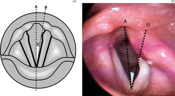
In order to measure the degree of vocal fold lateralisation, we recorded the angle between two imaginary lines, during deep inspiration: a line from the midportion of the interarytenoid mucosa to the anterior commissure, and a line from the anterior commissure to the vocal process of the paralysed vocal fold (Figure 1).
Measurement of degree of vocal fold lateralisation. (a) Diagram illustrating measurement of vocal fold lateralisation. (b) Corresponding still stroboscopic image of patient with left vocal fold paralysis, at full inspiration. Two lines are projected: from the midportion of the interarytenoid mucosa to the anterior commissure (A), and from the anterior commissure to the medial side of the vocal process of the paralysed vocal fold (B). The angle between the two lines is measured in degrees.

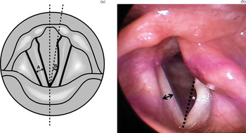
The extent of bowing and length of the paralysed vocal fold were measured using a still photograph taken during production of the Vowel /i/. The extent of vocal fold bowing was assessed by dividing the deviation distance at the midportion of the paralysed vocal fold by the width of the normal vocal fold (Figure 2). The length of the paralysed vocal fold was assessed by dividing the distance from the anterior commissure to the tip of the vocal process of the paralysed vocal fold by the same parameter in the normal vocal fold.Reference Rihkanen, Reijonen, Lehikoinen-Soderlund and Lauri13 To exclude measurement errors resulting from different magnifications (i.e. close-up or remote views), measurements (i.e. widths or lengths) were converted to ratios by comparison with adjacent normal structures.
Measurement of the extent of vocal fold bowing. (a) Diagram showing measurement of vocal fold bowing. The deviated distance of the bowed vocal fold (B) was divided by the width of the normal vocal fold (A). (b) Corresponding still stroboscopic image of a patient with left vocal fold paralysis. To measure the extent of vocal fold bowing, the deviated distance at the midpoint of the paralysed vocal fold (asterisk) was divided by the width of the normal vocal fold (arrow).

Arytenoid movement or mobility for the paralysed vocal fold was defined as ‘yes’ when there was any observable spontaneous movement of the arytenoid hump during respiration or phonation, or ‘no’ if such movement was absent.
Arytenoid appearance was categorised using the classification system of Hong and Jung, into one of four types: body, vocal process, tip and non-visualising.Reference Hong and Jung14
To calculate the vertical level difference, the cephalo-caudal level of the two vocal folds was compared during /i/ phonation, and the difference described as either none, mild or overt.Reference Hong and Jung14
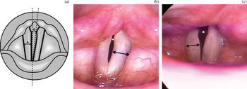
The posterior glottic gap during phonation was compared by measuring the distance between the medial sides of the vocal processes, divided by the width of the midpoint of the membranous vocal fold on the normal side (Figure 3), in order to obtain a more objective, consistent comparison regardless of different magnifications. A small posterior glottic gap was defined as being less than 50 per cent of the normal vocal fold width.
Measurement of posterior glottic gap on phonation. (a) Diagram showing measurement of posterior gap. The distance between the medial edge of the two vocal processes (B) was divided by the width of the normal membranous vocal fold (A). (b) Still stroboscopic image showing a small posterior gap (i.e. the posterior gap distance (asterisk) is less than 50 per cent of the normal vocal fold width (arrow)). (c) Still stroboscopic image showing a large posterior gap (i.e. more than 50 per cent of the normal vocal fold width (arrow)).

Statistical analysis
The results of objective and subjective voice evaluation were compared using the t-test, to confirm the adequacy of placing patients into either the improved or unimproved group. Clinical characteristics and stroboscopic findings were compared using the t-test or chi-square test. Logistic regression analysis was used to calculate odds ratios for potential predictive factors, utilising multivariate analysis with a 95 per cent confidence interval. The Statistical Package for the Social Sciences version 16.0 software (SPSS Inc, Chicago, Illinois, USA) was used for calculations. A p value of less than 0.05 was accepted as significant.
Results
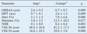
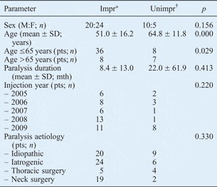
Voice evaluation results clearly differed between the improved and unimproved groups. Table I shows acoustic, aerodynamic and perceptual data, while Table II shows patient clinical characteristics (i.e. age, sex, duration of paralysis, surgeon experience and paralysis aetiology), for the two groups. The mean age of the improved and unimproved group patients was 51.0 and 64.8 years, respectively (p = 0.000). Statistically significant differences were not found for other clinical characteristics.
Voice outcomes

Data represent means ± standard deviations. *n = 44; †n = 15. Impr = improved group; unimpr = unimproved group; GRBAS = grade-roughness-breathiness-asthenia-strain scale; MPT = maximum phonation time; sec = seconds; NHR = noise:harmonics ratio; VHI = Voice Handicap Index
Clinical characteristics

*n = 44; †n = 15. Impr = improved group; unimpr = unimproved group; M = male; F = female; SD = standard deviation; pts = patients
Results for univariate analysis of pre-operative stroboscopic findings are summarised in Table III. Of the 44 patients in the improved group, 35 showed either a small posterior glottic gap or none, while nine (20 per cent) showed a large gap (i.e. more than 50 per cent of the normal vocal fold width). In contrast, eight of the 15 unimproved patients (53 per cent) showed a large posterior gap (p = 0.015). Six patients (13.6 per cent) in the improved group showed an overt vertical level difference between the two vocal folds during /i/ phonation, compared with two patients (13.3 per cent) in the unimproved group; this difference was not statistically significant (Table III).
Stroboscopic findings

*n = 44; †n = 15. Impr = improved group; unimpr = unimproved group; VF = vocal fold; lat = lateralisation; SD = standard deviation; rel = relative
Table IV presents the results of multivariate logistic regression analysis. Patients aged 65 years or younger were approximately 8.0 times more likely to experience voice improvement following injection laryngoplasty (p = 0.038; 95 per cent confidence interval = 1.1–56.7), compared with patients over 65 years. Furthermore, patients with a small or no posterior glottic gap were approximately 9.1 times more likely to show voice improvement following injection laryngoplasty (p = 0.020; 95 per cent confidence interval = 1.4–58.7).
Predictive factors for voice outcome after injection laryngoplasty*

* Multivariate analysis. †≤65 vs >65 years; ‡small vs large. CI = confidence interval; post = posterior
Discussion
When evaluated three months after injection laryngoplasty, 44 of our 59 unilateral vocal fold paralysis patients (74.5 per cent) had satisfactory voice outcomes. However, 15 patients (25.5 per cent) required some form of additional treatment due to insufficient voice improvement. The aim of this study was to investigate the origin of these differences. Of the five clinical characteristics and seven pre-operative stroboscopic findings analysed, only two parameters were closely associated with voice outcome following injection laryngoplasty: the age at the time of surgery, and the size of the posterior glottic gap at maximal phonation.
Although there are many reports of the usefulness of injection laryngoplasty and medialization thyroplasty, it is not easy to find precise guidelines or objective criteria to determine the appropriate procedure in each individual patient. In general, patients with a large glottic gap are thought to be better suited to medialization thyroplasty.Reference King and Simpson3, Reference Hirano, Kishimoto, Suehiro, Kanemaru and Ito15
Volume augmentation by injection is limited to the membranous portion of the vocal fold, and the inability of injection laryngoplasty to completely medialise the affected arytenoid has been recognised since the early writings of Rubin in the 1960s. This is partly due to tight adherence of the perichondrium and epithelium to the underlying vocal process of the arytenoids.Reference Kwon and Buckmire2, Reference Rubin16 Therefore, a patient who presents with a large posterior glottic gap on phonation is theoretically not expected to achieve satisfactory voice improvement with injection laryngoplasty.
Accordingly, we analysed pre-operative laryngeal stroboscopic findings, including the size of the posterior glottic gap on phonation, in the hope of developing evidence-based guidelines to enable optimal treatment selection for patients with unilateral vocal fold paralysis and dysphonia. In our study, patients with a smaller posterior glottic gap were approximately 9.1 times more likely to achieve voice improvement following injection laryngoplasty.
Patients aged 65 years and under were approximately 8.0 times more likely to achieve voice improvement after injection laryngoplasty, compared with older patients. In the ageing vocal fold, atrophy occurs as a result of decreased fibroblast activity, reduced production of hyaluronic acid and elastin, and suppression of collagen synthesis.Reference Hirano, Kurita and Sakaguchi17–Reference Ashcroft, Mills and Ashworth20
• Injection laryngoplasty has regained popularity as treatment for unilateral vocal fold paralysis
• This study aimed to identify predictive factors for voice improvement after injection laryngoplasty
• Patient demographic profiles and pre-operative stroboscopy were compared with post-laryngoplasty voice outcomes
• Younger patients with a smaller posterior glottic gap on phonation had more favourable voice outcomes
When Artecoll is used for injection laryngoplasty, the patient's own collagenesis is essential to maintain the injected volume, as the 80 per cent of the injected Artecoll volume which comprises bovine collagen will be rapidly degraded by the body within one to three months, being replaced by the patient's own collagen. Therefore, due to relatively poor collagenesis, Artecoll volume augmentation may be insufficient in elderly patients.
In summary, younger unilateral vocal fold paralysis patients (i.e. less than 65 years old) with a smaller posterior glottic gap on phonation (i.e. less than 50 per cent of the normal vocal fold width) are expected to have a more favourable outcome following injection laryngoplasty performed for correction of glottic insufficiency following unilateral vocal fold paralysis, compared with older patients with a larger posterior glottic gap.
Although this recommendation should not be regarded as the sole guideline when choosing a management strategy for patients with unilateral vocal fold paralysis, we suggest that clinicians should pay greater concern to the size of the posterior glottic gap and the patient's age when considering injection laryngoplasty as the treatment modality.
Conclusion
Injection laryngoplasty has recently regained popularity for the treatment of unilateral vocal fold paralysis. However, there are no precise guidelines or objective criteria to help determine those cases in which injection laryngoplasty may be ineffective or insufficiently effective. This study analysed the demographic profiles and pre-injection stroboscopic findings of patients with unilateral vocal fold paralysis, in order to identify predictive factors for voice outcome after injection laryngoplasty. Younger unilateral vocal fold paralysis patients (i.e. less than 65 years old) with a smaller posterior glottic gap on phonation (i.e. less than 50 per cent of normal vocal fold width) are expected to have a more favourable outcome following injection laryngoplasty, compared with older patients with a larger posterior glottic gap.
Acknowledgement
This study was supported by a Samsung Medical Center Clinical Research Development Program grant (CRS110-16-1).