In developing countries, rheumatic mitral stenosis continues to be the commonest cause of obstruction to the left ventricular inflow tract. Besides, by virtue of accelerated fibrosis, the disease is capable of producing crippling deformity in the very young.Reference Deshpande, Vaideeswar, Amonkar and Vasandani1 But, there exist rare congenital lesions that can produce a spectacular variety of stenosing lesions. One among them is the so-called supramitral ring, characterized by a ridge of connective tissue that encroaches into the inflow tract at or above the mitral valvar annulus.Reference Ruckman and van Praagh2 The clinical presentation of this rare anomaly is variable owing to the associated cardiac defects, as well as the morphology of the ridge, often leading to inadequate collection of data. We sought to shed light on the morphological aspects by presenting our experience of 24 such supramitral ridges encountered at autopsy.
Materials and methods
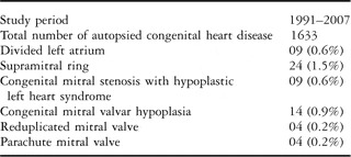
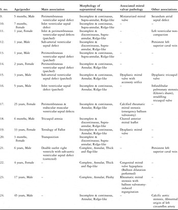
Over a 17-year period, from 1991 through 2007, we came across 64 cases of various congenital obstructions of the left ventricular inflow tract, excluding cases of mitral atresia (Table 1). These cases accounted for 3.9% of the 1633 autopsied examples of congenital cardiac disease, and amongst them were 24 examples of supramitral ring. The age and gender, mode of clinical presentation, findings on cross-sectional echocardiography, interventions if any, and causes of death were noted. All the hearts were carefully examined using sequential segmental analysis. Particular attention was paid to the morphology of the supramitral ridge, taking note of its completeness, location, and nature, as well as the mitral valve. The hearts with the supramitral ridges were then classified into two groups, specifically into those with or without associated Shone’s complex, which is the combination of a supramitral ridge, parachute mitral valve, sub-aortic stenosis, and aortic coarctation.Reference Shone, Sellers, Anderson, Adams, Lillehei and Edwards3 The complex was further categorized into complete and incomplete forms, depending on the presence of all four, or two or more of the lesions, respectively.Reference Ruckman and van Praagh2
Details of 64 cases with congenital obstructions of the left atrial & left ventricular inlet tracts.

Results
Our 24 cases of supramitral ring accounted for 1.5% of the autopsied cases of congenital cardiac disease, and 37.5% of those with obstruction to the left-sided inflow tract. Most of them came from infants and children, with only 3 obtained from adults. There was a male predominance (58%).
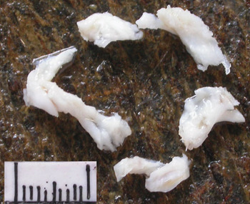
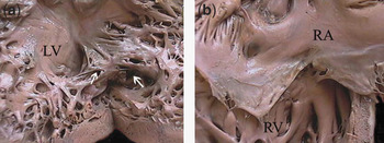
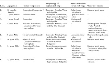
In 8 cases (33.3%), there was an incomplete form of Shone’s complex, with the hearts exhibiting 2 or 3 of the 4 lesions described initially (Table 2). There were 7 children with an age range from 10 months to 14 years. They had a history of failure to thrive, delayed milestones, dyspnoea, easy fatigability, frequent infections of the respiratory tract, and/or repeated episodes of cardiac failure. A circumferential supramitral ridge was identified on echocardiography in 2 of them, which was excised (Fig. 1), combined with patch closure of a ventricular septal defect and excision of a sub-aortic fibrous shelf, respectively. In 3 children, coarctectomy had been undertaken. Among them, one (case 7) sustained a fatal traumatic head injury with bilateral carotico-cavernous fistulas, and had reduplication of the mitral valve with superimposed rheumatic heart disease (Fig. 2). The only adult was a 43 years male (case 8), who was diagnosed as a case of aortic stenosis. He had undergone repeated admissions in the past for cardiac failure, but had refused surgical intervention, leading to his present admission in a gasping state.
Excised pieces of the supramitral ring, which are flap-like and translucent.

Case 7 a) Reduplicated mitral valve showing two orifices (arrows). There are hardly any intercordal spaces due to concomitant rheumatic mitral valvitis. (LV left ventricle), b) There was mild rheumatic affection of the tricuspid valve. (RA right atrium, RV right ventricle).

Supramitral ring with Shone’s complex.

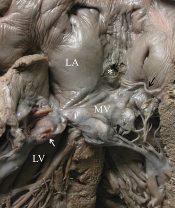
The remaining 16 patients (66.7%) had lesions other than Shone’s complex (Table 3). Ventricular septal defects were present in 9 (56.3%, perimembranous in 3, muscular inlet in 2, doubly committed and sub-arterial in 2, and multiple in 2). Their ages ranged from 5 months to 25 years. Of the children, 5 males and 3 females presented with failure to thrive, breathlessness, and repeated respiratory tract infections for varying durations of time. In 4, the defects had been surgically closed. Cyanotic spells were an additional feature in the child where an inlet defect was associated with pulmonary infundibular stenosis. The defect was closed, and a Glenn shunt was performed. The adult was a female aged 25 years with 8 months amenorrhoea, who was admitted with respiratory distress. An emergency balloon valvotomy was performed for critical mitral stenosis. Autopsy revealed a supramitral ring (Fig. 3), as well as multiple ventricular septal defects.
Supramitral ring without Shone’s complex.

Incomplete ridge (arrow) at the mitral annulus in case 17. Patient had undergone balloon mitral valvoplasty. Note atrial septostomy (*) and calcified postero-medial commissure. (LA left atrium, MV mitral valve, LV left ventricle).

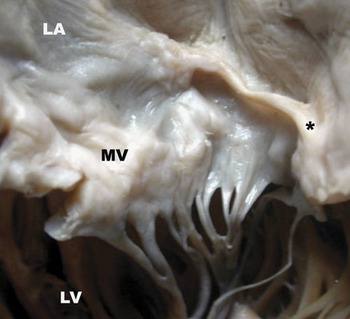
In 4 others, there was associated complex congenital cyanotic heart disease in the form of tricuspid atresia, tetralogy of Fallot (Fig. 4), transposition, and double outlet right ventricle. The anomalies of the outflow tracts were corrected, but all patients died within 24 hours of operation. In 1 child, the supramitral ring was associated with congenital mitral valvar hypoplasia that was subjected to balloon valvotomy. There were no associated anomalies. Rheumatic mitral stenosis and degenerative aortic stenosis were the clinical diagnoses made in two males, aged 17 and 45 years, respectively. The first patient had undergone balloon mitral valvotomy and died subsequently of Penidure-induced anaphylaxis. The aortic valve was replaced in the second.
The patient with Fallot’s tetralogy showing an incomplete cord-like ridge (*) and dysplastic looking mitral valvar (MV) leaflets. (LA left atrium, LV left ventricle).

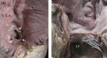
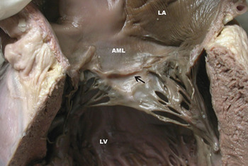
There was no difference in the completeness or width of the supramitral ridge in those with or without Shone’s complex. Circumferential or complete ridges were identified in 7 patients, and these appeared thick, fleshy and fibrous (Figs 1 and 5). Incomplete rings were seen in the rest, as continuous (13) or discontinuous ridges (four). None of these were diagnosed on echocardiography. All of these appeared glistening, grey-white, and cord-like (Fig. 4). The rings were supra-annular in nine, located 0.2 to 0.7 centimetres above the mitral annulus. In 1 patient, the ring extended on to the atrial aspect of the aortic leaflet of the mitral valve (case 5, Fig. 6). In 13 patients, there was concomitant involvement of the mitral valve, either as congenital and/or acquired deformities.
a) View from the left atrium (LA), showing a circumferential annular mitral ring (arrows). Panel b shows the lesion after opening out the atrium and ventricle (LV).

A ridge which is annular at the attachment of the mural leaflet runs on to the mid-portion of the atrial aspect of aortic mitral leaflet. (LA left atrium, AML anterior mitral leaflet, LV left ventricle).

Discussion
The supramitral ring, also known as supravalvar mitral stenosis, supravalvar stenotic mitral ring, supravalvar ring of the left atrium, supravalvar stenosing ring, or mitral supravalvar ring, was first described FisherReference Fisher4 in 1902, when the ridge of connective tissue above the mitral valve was likened to the diaphragm of the microscope. The defect has to be differentiated from the rare divided left atrium, where the partition is fibromuscular, separating the left atrium into the proximal and distal chambers.Reference Jacobstein and Hirschfield5 Up to 2002, less than 100 cases of this rare malformation have been reported.Reference Mychaskiw, Sachdev, Braden and Health6 Consequently no data of the exact incidence or predilection for race or gender are available. The incidence is reported to range from one-eighth to two-fifths among congenital mitral valvar anomalies that were not necessarily stenotic.Reference Ruckman and van Praagh2, Reference Coles, Williams and Watanabe7–Reference Moraes, Lapa and Ventura10 All were children. In our analysis, the ring had an incidence of 1.5% among all congenital cardiac defects at autopsy, and accounted for just over one-third of all congenital lesions producing obstruction of the left ventricular inflow tract. Interestingly, 3 of our specimens were obtained from adults (20.8%). There was also distinctly a male predominance.
Most of the previous series do not particularly comment on the completeness or nature of the ring.Reference Ruckman and van Praagh2, Reference Coles, Williams and Watanabe7–Reference Moraes, Lapa and Ventura10 In a recent report of 15 children,Reference Collison, Kaushal and Dagar11 all had been circumferential, though incomplete rings have been described.Reference Jacobstein and Hirschfield5 Circumferential ridges were seen in only 7 of our 24 hearts (29.1%), and these were entirely located on the valvar leaflets adjacent to the annulus. On the other hand, the incomplete rings (70.9%) were found to be either continuous or discontinuous, and either annular or supraannular.
Even the morphology was different. Complete rings were always fleshy and fibrous, while the incomplete rings appeared akin to a tautened twine thread. The morphology also plays an important role in the clinical presentation as well as preoperative diagnosis. Though complete rings were identified in the study of Collison et al,Reference Collison, Kaushal and Dagar11 pre-operative diagnosis was made in three-quarters of the patients, the symptomatology largely being related to associated anomalies. These anomalies produce altered patterns in the flow of blood that may camouflage the ring. Similar observations were also made by Mychaskiw et al.Reference Mychaskiw, Sachdev, Braden and Health6 In our study, the ring was clinically identified in only 2 patients (8.3%). Hence, it is possible that the incidence of the ring may be underestimated, since all need not produce clinically overt stenosis, and all may not always be identified on routine echocardiography. Transoesophageal echocardiography is advocated for diagnosis.Reference Muhiudeen Russel, Miller Hanee and Silverman12 Since stenosis is an inconstant feature,Reference Collison, Kaushal and Dagar11 the term supramitral ring is appropriate as used in isolation. We believe that all circumferential rings, as seen in three-tenths of our cases, were clinically significant. An abnormal mitral valve and/or an annular location of an incomplete ring, may lead to clinical relevance.
Isolated occurrence of the supramitral ridge was first reported by Chung and associates.Reference Chung, Manning, Lipchik, Gramiak and Mahoney13 In most instances, nonetheless, the lesion occurs with other varied anomalies.Reference Ruckman and van Praagh2, Reference Mychaskiw, Sachdev, Braden and Health6, Reference Banerjee, Kohl and Silverman9, Reference Collison, Kaushal and Dagar11, Reference Coto, Judez and Juffe14 We did not find a single case of isolated ring. We found it useful to categorize the associations into those with or without Shone’s complex. As has been the experience with others, the commonest association was a ventricular septal defect. Others also included malformations of the ventricular outflow tracts. Among those with involvement of the mitral valve, 13 hearts in all, we identified associated rheumatic mitral valve abnormalities in 3. As far as we are aware, this association has not previously been reported. There exists a possibility for the ridge to develop as a post-inflammatory phenomenon, which would mean recognition of such lesions in greater frequency. Apart from these reported cases, we have not seen them in our series of autopsied cases of rheumatic heart disease.Reference Deshpande, Vaideeswar, Amonkar and Vasandani1 More interestingly, one of them also existed in a reduplicated valve. It is important to remember, therefore, that both congenital and rheumatic disease can occur in unison in countries with a high prevalence of rheumatic heart disease.Reference Bokhandi, Tullu, Shatrarao, Bavdekar and Kamat15 Additionally, we found the ring to be associated with even acquired aortic stenosis.
Excision, first performed by Lynch and associates,Reference Lynch, Ryan and Williams16 has been the best therapeutic option,Reference Collison, Kaushal and Dagar11 as balloon valvoplasty may not always be successful.Reference Spevak, Bass and Ben-Shachar17 The outcome also depends on the presence of simple or complex associations. Follow-up is essential since, at times, the lesion can recur, as seen in 4 of the 23 patients reported by Tulloh et al.Reference Tulloh, Bull, Elliot and Sullivan18
Financial support: Nil