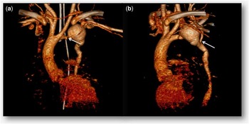
We present the case of an asymptomatic 3-month-old girl in whom a heart murmur and decreased femoral pulses were noted incidentally during a routine medical examination. The two-dimensional echocardiography revealed tricuspid aortic valve, left aortic arch, severe distal aortic arch coarctation with post-coarctation aneurysmReference Ozyuksel, Canturk and Dindar 1 about 2 cm in diameter, and severe left ventricular dysfunction (<30%). The ductus arteriosus was patent, joined to the aneurysm with right-to-left shunt. The three-dimensional CT-scan confirmed these findings and showed the left subclavian artery emerging distal to the aneurysm with reverse flow perfusing the descending aorta (Fig 1a and b) and right subclavian artery normally arising from the innominate artery, although with anomalous initial course – double corkscrew.
Three-dimensional CT scan images. ( a ) The white arrow shows the aortic arch coarctation. ( b ) The white arrow shows the left subclavian artery emerging distal to the aneurysm.

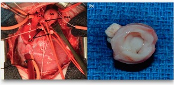
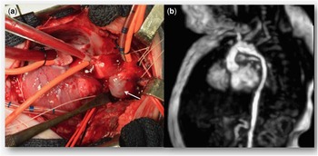
Surgical repair was performed through a median sternotomy with the patient under cardiopulmonary bypass, moderate hypothermia, and selective cerebro-myocardial perfusion (Fig 2a). The ductus arteriosus was ligated. The aneurysm and the coarcted segment were resected (Fig 2b). Extended end-to-end anastomosis with fresh autologous pericardial patch augmentation was performed (Fig 3a). Diffuse thickening of the aortic wall was observed, but any type of vasculitis including PHACE syndromeReference Bayer, Frommelt and Blei 2 was ruled out with analytical, physical, and histopathological examinations.
( a ) Intraoperative view before repair. ( b ) Surgical specimen with diffuse thickening of the aortic wall. AA=aortic aneurysm; DA=ductus arteriosus divided; DAA=distal aortic arch; LCA=left carotid artery.

( a ) Intraoperative view after repair. The white arrow shows the autologous pericardial patch. ( b ) Post-operative MRI showing the reconstructed aortic arch.

Post-operative echocardiography showed a normal reconstructed aortic arch with no residual gradient. Post-operative MRI (Fig 3b) showed the reconstructed aortic arch with no defects in myocardial viability. The left ventricular ejection fraction improved to recovery the 4th post-operative month.
Acknowledgement
None.
Financial Support
This research received no specific grant from any funding agency, commercial, or not-for-profit sectors.
Conflicts of Interest
None.