Introduction
Coeliac disease (CD) is a chronic enteropathy caused by intolerance to gluten in genetically susceptible people. The first description of the coeliac lesion was provided by Paulley and colleagues in the 1950s who examined full thickness biopsy specimens taken at laparotomy from patients with CD and found broad flat villi and a dense chronic inflammatory cell infiltrate in the small intestinal mucosa.Reference Paulley1 Following this, the use of unguided suction biopsy devices, such as the Crosby capsule, allowed the study of patients with malabsorption. The advent of fibre-optic endoscopy as a diagnostic tool has led to extensive study of the duodenal mucosa in less florid forms of CD. More recently, the advent of sensitive and specific serological tests for CD have revolutionized the diagnosis of the condition and led to increased recognition of CD at a relatively early stage. These changes are reflected in the most recent guidelines on investigation and management of CD.Reference Rostom, Murray and Kagnoff2–Reference Ciclitira, Dewar, McLaughlin and Sanders5
There is a broad spectrum of clinical and histological features associated with gluten sensitivity. Epidemiological studies show that there is a substantial number of undiagnosed cases in the general population, possibly seven times as many as have actually been diagnosed, and that clinical CD can be regarded as the ‘tip of the iceberg’ of gluten sensitivity.Reference Holmes, Catassi and Fasano6, Reference Maki and Collin7 This is important because CD can cause significant chronic health problems yet is readily treatable with a gluten-free diet. It can present at any age and the importance of this disease in older patients may be less well appreciated. Several studies have demonstrated that the diagnosis of CD may be delayed or missed because of a failure to appreciate that symptoms are often subtle and not clearly related to the gastrointestinal tract.
Diagnostic criteria
The gold standard for diagnosis of CD remains examination of histological findings in the mucosa of the proximal small intestine. Marsh was the first to describe the progression of histological features from initial increase in intra-epithelial lymphocytes with infiltration of the lamina propria with lymphocytes (stage 1), through crypt hyperplasia (stage 2) to villous atrophy (stage 3).Reference Marsh8 Early morphological changes may be clinically silent.
In the past, CD was usually diagnosed on the basis of characteristic gastrointestinal symptoms, a flat jejunal mucosa, clinical and morphological response to gluten restriction and, ideally, recurrence of clinical symptoms and histological damage with gluten rechallenge. However, patients with a flat mucosa and overt malabsorption only represent a small proportion of the gluten-sensitized population. Clinically silent CD, despite a classical small bowel mucosal morphology, has been identified in relatives of CD patients. Conversely, some patients with gluten sensitivity and a normal intake of gluten may have an initially normal small bowel mucosal pattern.Reference Marsh8, Reference O'Mahony, Vestey and Ferguson9 Recent diagnostic criteria accept a diagnosis of CD if symptoms (whether typical or atypical) and serological tests are positive, a single biopsy is consistent with CD, and there is an obvious clinical and serological response to a gluten-free diet.Reference Husby, Koletzko, Korponay-Szabo, Mearin, Phillips and Shamir3
Subtle early changes remain a diagnostic challenge. In particular, although an increased intra-epithelial lymphocyte (IEL) count is an essential component of CD, the optimal cut-off for normality, given that increased IEL can be seen in other conditions, has been uncertain, with recommendations ranging from 10 to 40 IELs/100 enterocytes. A recent population study from Northern Sweden in which 1000 people underwent small intestinal biopsy as well as serological testing has clarified the issue: ≥25 IELs/100 enterocytes strongly correlates with serological indicators of CD and seems the optimum cut-off.Reference Walker, Murray, Ronkainen, Aro, Storskrubb, D'Amato, Lahr, Talley and Agreus10
Pathogenesis
CD results from immunologically mediated inflammation of the small intestinal mucosa precipitated by ingestion of gluten in wheat and other cereals in genetically susceptible people.
CD is a polygenic disorder. It is strongly associated with class II human leucocyte antigens (HLAs) although it is now clear that non-HLA-linked genes also play an important role.Reference van Heel, Hunt, Greco and Wijmenga11 The strongest association between HLAs and coeliac disease is with the serological marker DQ2, which is found in 95% of patients with CD; the remainder express DQ8. Thus HLA typing showing lack of either DQ2 or DQ8 has a high negative predictive value for CD that may help in difficult cases.Reference Rostom, Murray and Kagnoff2
There is now convincing evidence for a gene-dosage effect of carriage of the HLA-DQB1*02 alleles in CD, with those homozygous for HLA-DQB1*02 having up to a five times greater risk of developing CD than those heterozygous for the allele, as well as having more severe villous atrophy.Reference Murray, Moore, Van Dyke, Lahr, Dierkhising, Zinsmeister, Melton, Kroning, El-Yousseff and Czaja12
Gluten in the cereal proteins wheat, rye and barley, is the major triggering factor for CD. Toxicity lies in the ethanol-soluble prolamin (proline-containing, amine-rich) portions of these proteins: gliadins (wheat), hordeins (barley) and secalins (rye).Reference Srinivasan, Leonard, Jones, Kasarda, Weir, O'Farrelly and Feighery13 In a dose-dependent response, these proteins produce an inflammatory process in the intestine and withdrawal of them results in regression of the process.Reference Marsh8 The prolamin of oats, which has an intermediate proline and glutamine content, is thought to be less toxic,Reference Janatuinen, Pikkarainen, Kemppainen, Kosma, Järvinen, Uusitupa and Julkunen14, Reference Storsrud, Olsson, Arvidsson Lenner, Nilsson, Nilsson and Kilander15 although a subset of coeliac patients taking oats develop mucosal T-cells that can cause mucosal inflammation and lead to villous atrophy.Reference Arentz-Hansen, Fleckenstein, Molberg, Scott, Koning, Jung, Roepstorff, Lundin and Sollid16
The intestinal lesion of CD represents gluten-triggered cell-mediated damage. CD is associated with an increase in the number of lymphocytes in the epithelium and lamina propria.Reference Ferguson, McClure and Townley17 Inflammatory cytokines are released when macrophages in the lamina propria are activated by glutens and present the antigen to HLA-restricted CD4 T-cells. Tissue transglutaminase (tTG) has been identified as the target antigen for specific auto-antibodies found in CD, and patients develop auto-antibodies directed against tTG. This enzyme may facilitate the presentation of gliadin by HLA-restricted cells in CD by cross-linking gluten peptides to increase their antigenicity.
Epidemiology
Differences in diagnostic criteria, variations in case ascertainment and possibly a change in the disease itself complicate interpretation of data regarding CD. Prevalence rates based on clinical diagnosis have varied from 1 in 500 in Ireland, and 1 in 1000 to 2000 in Scandinavia and the United Kingdom, to lower rates of 1 in 3000 to 5000 in Australia and North America.Reference Talley, Valdovinos, Petterson, Carpenter and Melton18 However, serological screening studies from around the world show that approximately 0.5–1% of the population may have undetected CD. The most consistent estimate reported from the largest population-based studies is that approximately 1% of children and adults have CD.Reference Fasano, Berti, Gerarduzzi, Not, Colletti, Drago, Elitsur, Green, Guandalini, Hill, Pietzak, Ventura, Thorpe, Kryszak, Fornaroli, Wasserman, Murray and Horvath19–Reference Mäki, Mustalahti, Kokkonen, Kulmala, Haapalahti, Karttunen, Ilonen, Laurila, Dahlbom, Hansson, Höpfl and Knip21 Most of these cases are unrecognized. For example, a large UK population-based study found that more than 90% of coeliac disease in asymptomatic children was undiagnosed.Reference Ravikumara, Nootigattu and Sandhu22
There is now good evidence that the prevalence of CD has increased across the world in recent decades and that this cannot entirely be attributed to improved detection. For example, large population studies from Finland suggest that the prevalence in that country has almost doubled to 2%.Reference Lohi, Mustalahti, Kaukinen, Laurila, Collin, Rissanen, Lohi, Bravi, Gasparin, Reunanen and Mäki23 Environmental factors, including poor education and hygiene standards, have been associated with lower prevalence of CD.Reference Kondrashova, Mustalahti, Kaukinen, Viskari, Volodicheva, Haapala, Ilonen, Knip, Mäki and Hyöty24 While there are no differences in the sex distribution based on serological abnormalities, clinical diagnosis of CD is approximately twice as common in women than in men in most studies.Reference Loft, Nwokolo and Ciclitira25
Serological tests have confirmed that the risk of CD is substantially higher in first-degree relatives of those with the condition (up to 10%), and also elevated in second-degree relatives. For example, in a large North American study, the prevalence of CD was 1/22 in first-degree relatives, and 1/39 in second-degree relatives compared with 1/133 in the general population.Reference Bingley, Williams, Norcross, Unsworth, Lock, Ness and Jones20
Prevalence of CD in older people
In most adult series, a mean age of presentation in the fifth decade is reported, with 20–30% of adult coeliac patients diagnosed at 60 years or older, and up to 10% at 70 years or older.Reference Gumaa, McNicholl, Egan-Mitchell, Connolly and Loftus26 Examination of data from the Rochester Epidemiology Project has shown that the incidence rates of CD increase with age.Reference Murray, Van Dyke, Plevak, Dierkhising, Zinsmeister and Melton27 However, it had been unclear whether this might reflect delayed recognition in older people or use of better diagnostic tools. A recent Finnish population study, in which over 2000 randomly selected subjects over 55 years were tested (serological screening followed by biopsy if positive) at baseline and 3 years later, found an increase in prevalence from 2.13 to 2.34% over the 3 years.Reference Vilppula, Kaukinen, Luostarinen, Krekelä, Patrikainen, Valve, Mäki and Collin28 Of the five new cases, two had minor abdominal symptoms and three were asymptomatic. This confirms that the frequency of CD is indeed increasing in older people, that the condition can develop at an advanced age, and that late-onset CD cannot be attributed to better screening tools.
Clinical presentation in older people
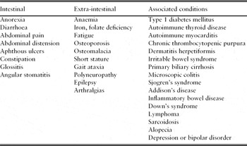
In the older patient, clinical manifestations are often subtle and presentation may be with non-intestinal symptoms (Table 1).Reference Cooke and Holmes29–Reference Tai, Crowe and O'Keeffe34 Only a minority of patients present with classical symptoms of steatorrhoea, osteomalacia or anaemia. Indeed, diarrhoea occurs in less than half of those diagnosed now, compared with almost all of those diagnosed in the 1960s.Reference Murray, Van Dyke, Plevak, Dierkhising, Zinsmeister and Melton27, Reference Duggan35, Reference Sollid and Lie36 The reasons for this are unclear but may relate to relatively limited mucosal involvement in many older people combined with substantial functional reserve in the small intestine. Although unexplained weight loss should trigger consideration of CD, about a third of patients are overweight at time of diagnosis.Reference Dickey and Kearney37 Non-specific symptoms such as tiredness, low mood and poor appetite are common and may be the only presenting feature. The current NICE guideline on CD recommends that screening by serological testing should be offered to those with any of:
• chronic or intermittent diarrhoea;
• persistent or unexplained gastrointestinal symptoms including nausea and vomiting;
• prolonged fatigue (‘tired all the time’);
• recurrent abdominal pain, cramping or distension;
• sudden or unexpected weight loss;
• unexplained iron-deficiency anaemia, or other unspecified anaemia.
Clinical manifestations of coeliac disease

Gastrointestinal symptoms
The onset of diarrhoea in adults can be gradual or dramatic. Traveller's diarrhoea, gastroenteritis or gastrointestinal surgery can sometimes act as a trigger for the development of CD. Although diarrhoea is the most common intestinal symptom in older patients, it is often mild and intermittent.Reference Cooke and Holmes29, Reference Midhagen, Jarnerot and Kraaz30 Indeed, some patients experience constipation as their only gastrointestinal symptom. Glossitis or angular stomatitis may be present in up to 90% of untreated CD patients.Reference Midhagen, Jarnerot and Kraaz30 Recurrent aphthous ulceration may be the presenting feature of the disorder and usually responds well to a gluten-free diet.Reference Ferguson, Wray, Carmichael, Russell and Lee38 The NICE guideline recommends that screening should be considered in the presence of aphthous stomatitis (mouth ulcers) or persistent or unexplained constipation.4
Bone disease
Disorders of bone and fractures secondary to malabsorption of calcium and of vitamin D can occur in the absence of signs of malabsorption and are common in CD patients regardless of age. Reduced bone mass is the most common bone disease, and almost three-quarters of adult CD patients have osteopenia, with a bone mineral density at least one standard deviation below normal controls.Reference Corazza, Di Sario, Cecchetti, Tarozzi, Corrao, Bernardi and Gasbarrini39, Reference Meyer, Stavropolous, Diamond, Shane and Green40 In a recent population-based study in North America, coeliac disease was associated with at least a 2-fold increase in fracture risk, both before and after diagnosis; the increased fracture risk was present in the spine and the limbs.Reference Jafri, Nordstrom, Murray, Van Dyke, Dierkhising, Zinsmeister and Melton41 Conversely, the prevalence of CD among osteoporotic individuals (3.4%) is much higher than that of non-osteoporotic patients (0.2%).Reference Stenson, Newberry, Lorenz, Baldus and Civitelli42
Anaemia and abnormal laboratory tests
Almost 80% of elderly patients diagnosed with CD have anaemia. Folate deficiency occurs in over two-thirds of patients.Reference Cooke and Holmes29, Reference Cellier, Patey, Mauvieux, Jabri, Delabesse, Cervoni, Burtin, Guy-Grand, Bouhnik, Modigliani, Barbier, Macintyre, Brousse and Cerf-Bensussan43 Hallert and colleagues found CD in 30% of adult patients investigated prospectively because of unexplained folate deficiency.Reference Hallert, Tobiasson and Walan44 Iron deficiency is also common, and may not improve with iron supplements. This is mainly due to poor absorption. However, a study showing an association between patients with CD and positive faecal occult blood sampling suggests that intestinal bleeding may also be a causative factor.Reference Fine45 Macrocytosis is the most common abnormality in the blood film and often occurs in the absence of anaemia.Reference Kirby and Fielding31, Reference Paulley, Fairweather and Leeming46 A dimorphic blood film showing features of hyposplenism may be found. Hypoalbuminaemia, hypocalcaemia (sometimes with tetany), hypomagnesaemia and unexplained abnormalities of liver enzymes may also be found. Vitamin B12 deficiency is much less common.
Neurological and muscular disease
Neurological syndromes may occasionally precede the development of other features of CD. Some result from vitamin malabsorption while others may have an immunological basis or are unexplained. Interestingly, antigliadin antibodies in the absence of CD have been reported in Huntington's disease and spinocerebellar ataxia.Reference Bushara, Goebel, Shill, Goldfarb and Hallett47, Reference Bushara, Nance and Gomez48
Ataxia is the most common neurological symptom associated with CD.Reference Hadjivassiliou, Gibson, Davies-Jones, Lobo, Stephenson and Milford-Ward49–Reference Cooke and Smith51 A number of potential causes have been identified. Cerebellar ataxia associated with low vitamin E levels has been reported in one study,Reference Mauro, Orsi, Mortara, Costa and Schiffer52 but others have not replicated this finding and it has been suggested that gluten itself might cause cerebellar damage.Reference Hadjivassiliou, Boscolo, Davies-Jones, Grünewald, Not, Sanders, Simpson, Tongiorgi, Williamson and Woodroofe53 Deficiencies in vitamins B1, B12 and niacin might also contribute to cerebellar impairment, and lymphomatous metastases to the cerebellum have also been found in CD.Reference Shams, Waldman, Dogan, MacKenzie and Plant54
Peripheral neuropathy occurs in up to 50% of adult CD.Reference Cicarelli, Della Rocca, Amboni, Ciacci, Mazzacca, Filla and Barrone55, Reference Chin, Sander, Brannagan, Green, Hays, Alaedini and Latov56 Neuropathy can also result from vitamin deficiencies and as a paraneoplastic effect of lymphomas. A symmetrical distal sensory neuropathy is the most common finding, and may lead to gait ataxia or to dysaesthesias. Motor and autonomic neuropathies have also been reported.Reference Chin, Sander, Brannagan, Green, Hays, Alaedini and Latov56–Reference Usai, Usai Satta, Lai, Corda, Piras, Calcara, Boy, Morelli, Balestrieri and Bassotti59 The effects of a gluten-free diet on neuropathy have been inconsistent.Reference Kaplan, Pack, Horoupian, DeSouza, Brin and Schaumburg58, Reference Luostarinen, Himanen, Luostarinen, Collin and Pirttila60
An association between CD and cognitive impairment has also been reported. Collin and colleagues reported five patients who developed dementia before the age of 60 and who were subsequently found to have CD.Reference Collin, Pirttila, Nurmikko, Somer, Erila and Keyrilainen61 Only one patient showed any improvement in cognitive function with a gluten-free diet. A more recent case series of 13 patients showed three patients with improved or stabilized cognitive function with gluten withdrawal.Reference Hu, Murray, Greenaway, Parisi and Josephs62 Interestingly, in a study of seven elderly coeliac patients diagnosed after 60 years of age, cognitive decline attributed to Alzheimer's dementia was evident in three cases and, in one case, neurological changes were ameliorated after initiation of a gluten-free diet.Reference Hadjivassiliou, Gibson, Davies-Jones, Lobo, Stephenson and Milford-Ward49, Reference Lurie, Landau, Pfeffer and Oren63
Proximal muscle weakness is common in CD. However, this can often be attributed to vitamin D deficiency, neuropathy or electrolyte disturbance, and it is uncertain whether a true primary myopathy can occur.Reference Hardoff, Sharf and Berger64 Similarly, it is unclear whether CD is associated with epilepsy in adults. Epilepsy has been found in 3–5% of patients with CD. In particular, an association has been reported in children between epilepsy and cerebral calcification. In one study, 24 of 31 children with idiopathic cerebral calcification and epilepsy were found to have CD.Reference Gobbi, Ambrosetto, Zaniboni, Lambertini, Ambrosioni and Tassinari65
The NICE guideline recommends that screening for CD should be considered for those with epilepsy or polyneuropathy.4 The American Gastroenterology Association suggests considering screening in those with cerebellar ataxia or recurrent migraine.Reference Rostom, Murray and Kagnoff2
Associated diseases
NICE guidelines recommend screening for CD in those with any of:
• autoimmune thyroid disease;
• dermatitis herpetiformis;
• type 1 diabetes;
• irritable bowel syndrome.
Autoimmune thyroid disease is relatively common in CD, with up to 13.9% of patients in one study with CD demonstrating positive thyroid antibodies. Dermatitis herpetiformis (DH) has a strong association with HLA DQ2, and a flat jejunal biopsy is found in 40% of cases.Reference Christensen, Hindsen and Svensson66 The onset of DH occurs after 60 years in up to 10% of cases. CD occurs in up to 6% of people with type 1 diabetes mellitus, while 5% of coeliac patients are diabetic.Reference Hakanen, Luotola, Salmi, Laippala, Kaukinen and Collin67 Diabetic control improves with a gluten-free diet. The symptoms of irritable bowel disease closely mimic those of CD and it has been suggested that, even in the absence of fully developed CD, gluten can induce irritable bowel symptoms.Reference Verdu, Armstrong and Murray68
A large number of other conditions have also been reported to be associated with CD, although the evidence is less compelling. Currently, the NICE guideline4 suggests that screening for CD should be considered for those with:
• Addison's disease;
• autoimmune liver conditions;
• autoimmune myocarditis;
• chronic thrombocytopenic purpura;
• Down's syndrome;
• lymphoma
• persistently raised liver enzymes with unknown cause;
• recurrent miscarriage.
Investigations
Serological tests
Auto-antibody tests have increasingly become the first-line test in screening for CD, particularly for high-risk groups such as first-degree relatives, those with associated conditions such as autoimmune thyroid disease, and for patients with atypical presentations. The sensitivity, specificity and positive predictive value of these tests depends on the populations studied. Serial serological testing may also be useful for monitoring the course of CD in those originally serologically positive.
Antigliadin antibodies (AGA) have good sensitivity but inadequate specificity and have now been superseded by newer serological tests.Reference Sjoberg, Alm, Ivarsson, Lindstrom and Eriksson69, Reference Grodzinsky, Franzen, Hed and Strom70 Initial testing now relies on endomysial (EMA) and tissue transglutaminase antibodies (tTG). The antigen of these two serological tests is tissue transglutaminase. EMA is identified by histological immunofluorescence techniques, whereas tTG employs quantitative ELISA methodology. IgA EMA is the most specific test but is likely to underestimate prevalence by 20%; in particular, negative serum EMA is associated with advanced CD.Reference Salmi, Collin, Korponay-Szabó, Laurila, Partanen, Huhtala, Király, Lorand, Reunala, Mäki and Kaukinen71 IgA tTG has both high sensitivity (approximately 98%) and specificity (95%). It is also simpler and cheaper.Reference Holbrook72
The current NICE guideline recommends IgA tissue transglutaminase (tTGA) as the first choice test with IgA endomysial antibodies (EMA) testing if the result of the tTGA test is equivocal.4 IgA deficiency is more common in CD, occurring in about 2.5% of cases compared with 0.25% of the general population. Investigation for IgA deficiency should be done if serology is negative in a high-risk patient or the laboratory detects a low or very low optical density on IgA tTGA test or low background on IgA EMA test.4 In such cases, IgG tTGA and/or IgG EMA serological tests will usually be positive. HLA DQ2/DQ8 testing is primarily of value for its negative predictive value in difficult cases. However, there are some patients who do not have IgA deficiency and have negative IgG tTG and EMA serology who are found to have histological lesions consistent with CD and who respond to a gluten-free diet. These patients invariably have the HLA DQ2 haplotype.Reference Husby, Koletzko, Korponay-Szabo, Mearin, Phillips and Shamir3
Small intestinal biopsy
All patients in whom CD is suspected should have a small intestinal biopsy at endoscopy to confirm the diagnosis. This includes those with negative serology and suggestive symptoms. It is essential to confirm, first, that the patient is taking adequate gluten in the diet, since some may institute a gluten-free diet on learning that CD is suspected. The often patchy distribution of mucosal lesions means that multiple biopsies should be taken, with a recent study suggesting that four specimens should be taken.Reference Pais, Duerksen, Pettigrew and Bernstein73 Visible abnormalities have been described at endoscopy such as mucosal pallor, visible blood vessels, scalloping, and a decrease in duodenal folds. These changes have been shown to correlate with degrees of villous atrophy, but appearances are often normal and thus cannot be relied on for diagnosis.Reference Bardella, Minoli, Radaelli, Quatrini, Bianchi and Conte74
People with mucosal changes that fall short of villous atrophy sometimes pose a diagnostic dilemma, especially if they have positive serology and non-specific symptoms. Often, both histology and symptoms improve with a gluten-free diet and it is reasonable to accept a diagnosis of CD. Sometimes, even supposedly asymptomatic individuals with minimal histological changes report feeling better on a gluten-free diet.Reference Tursi and Brandimarte75
Repeat biopsy should be performed if there is diagnostic doubt or if the clinical response to a gluten-free diet is inadequate after 2 months, and following a review from an expert dietician. Complete resolution of mucosal changes may take up to 2 years in the adult, and repeat biopsy should be delayed for at least 6 months.
Treatment
Once the diagnosis of CD is confirmed the patient should be commenced on a gluten-free diet. Gluten is the major storage protein of wheat, but the prolamines in rye and barley are also toxic and should be avoided. Although it seems that oat prolamines are considerably less toxic, commercial oats are often contaminated with wheat due to crop rotation. Compliance with the gluten-free diet is often encouraged by the frequent rapid symptomatic response to gluten withdrawal.
Dietary supplements are often necessary to correct deficiencies of folate, iron, calcium and vitamin D. Bone densitometry should always be performed. Even with a gluten-free diet, bone mineral density often remains below normal limits and treatment with an anti-resorptive agent should be considered. Patients with haematological evidence of hyposplenism should be assessed for the need for immunization with polyvalent pneumococcal vaccine.
Older subjects might theoretically find it more difficult to change lifetime dietary habits and adopt a strict gluten-free diet, especially if they were relatively asymptomatic and diagnosis was triggered by serological testing. However, a recent study of 35 patients with a median age of 61 years (range 52–76 years), diagnosed as a result of a screening programme, reported that dietary compliance was good and the diet had a beneficial effect on bone mineral density and nutritional indices.Reference Vilppula, Kaukinen, Luostarinen, Krekelä, Patrikainen, Valve, Luostarinen, Laurila, Mäki and Collin76 Furthermore, gastrointestinal symptoms improved, even though the patients reported no or only subtle symptoms at diagnosis. These findings support maintenance of a high index of suspicion for CD in older patients.
Non-responsive CD
The most common cause for failure to respond to a gluten-free diet is non-compliance. Adherence to the diet is often incomplete, and lapses are common. In addition, wheat flour containing gluten is present in a wide variety of processed foods and even in some medication formulations. Input from a dietician to suggest palatable alternatives and continued monitoring and education is essential. Decreased concentrations of antibodies such as IgA tTG are significantly associated with the degree of compliance with a gluten-free diet; serial measurement of antibody levels is more reliable in monitoring compliance than simply expressing results as positive or negative.Reference Nachman, Sugai, Vázquez, González, Andrenacci, Niveloni, Mazure, Smecuol, Moreno, Hwang, Sánchez, Mauriño and Bai77 If compliance is meticulous and symptoms persist, then small intestinal biopsy should be repeated and alternative or co-existing diagnoses such as other causes of malabsorption should be considered. The possibility of small bowel bacterial overgrowth is particularly important in the older patient. In one recent study, 10 of 15 coeliac patients with persistent diarrhoea had a positive breath test and benefited from antibiotic therapy.Reference Tursi, Brandimarte and Giorgetti78
Refractory CD
Refractory coeliac disease represents a small subset of those who are non-responsive, and is defined by continued significant clinical features in patients with persistent histological features despite a strict gluten-free diet for 1 year. Such patients have an increased mortality rate.Reference Rubio-Tapia, Kelly, Lahr, Dogan, Wu and Murray79 Treatment options are limited. A gluten-free diet should be maintained and nutritional supplements are usually required. Immunosuppressive therapy is sometimes helpful.Reference Goerres, Meijer, Wahab, Kerckhaert, Groenen, Van Krieken and Mulder80, Reference Verbeek, Schreurs, Visser, von Blomberg, Al-Toma and Mulder81
Enteropathy-associated T-cell lymphoma (EATL) is a particular concern in this group. Some patients who are refractory to the diet have an aberrant intra-epithelial T-cell population and are considered to harbour a cryptic T-cell lymphoma. They have a high mortality and a high rate of progression to EATL.Reference Malamut, Afchain, Verkarre, Lecomte, Amiot, Damotte, Bouhnik, Colombel, Delchier, Allez, Cosnes, Lavergne-Slove, Meresse, Trinquart, Macintyre, Radford-Weiss, Hermine, Brousse, Cerf-Bensussan and Cellier82
Complications
Mortality
CD is known to be associated with increased mortality.Reference Corrao, Corazza, Bagnardi, Brusco, Ciacci, Cottone, Sategna Guidetti, Usai, Cesari, Pelli, Loperfido, Volta, Calabró and Certo83, Reference West, Logan, Smith, Hubbard and Card84 However, there has been uncertainty as to whether this is confined to those with clinical symptoms or whether even those with minimal symptoms detected using serology are similarly affected. A study of patients with a median age of 21 found that undetected CD was associated with a 4-fold increased risk of death during 45-years follow-up.Reference Rubio-Tapia, Kyle, Kaplan, Johnson, Page, Erdtmann, Brantner, Kim, Phelps, Lahr, Zinsmeister, Melton and Murray85, Reference Metzger, Heier, Mäki, Bravi, Schneider, Löwel, Illig, Schuppan and Wichmann86 In contrast, a Finnish study of older patients (mean age 50 years) suggested a good prognosis for those with unrecognized CD except for increased risk for lymphoma and oesophageal carcinoma.Reference Lohi, Maki, Rissanen, Knekt, Reunanen and Kaukinen87 This conclusion is supported by a North American population study that showed that, with the exception of reduced bone health, older adults with undiagnosed CD had limited co-morbidity and no increase in mortality compared with controls.Reference Godfrey, Brantner, Brinjikji, Christensen, Brogan, Van Dyke, Lahr, Larson, Rubio-Tapia, Melton, Zinsmeister, Kyle and Murray88
Malignancy
An increased rate of malignancy has been found in patients with CD, especially lymphomas, adenocarcinoma of the small intestine, and squamous cell carcinomas of the oesophagus and the head and neck.Reference Holmes, Prior, Lane, Pope and Allan89–Reference Green, Fleischauer, Bhagat, Goyal, Jabri and Neugut91 Increased risk of malignancy has also been noted in patients with dermatitis herpetiformis and in relatives of coeliac patients. The association between CD and non-Hodgkin's lymphoma of both T- and B-cell type is well established, and such malignancies occur at both intestinal and extra-intestinal sites.Reference Smedby, Akerman, Hildebrand, Glimelius, Ekbom and Askling92 The peak incidence of lymphoma in patients with CD is the seventh decade.Reference Ryan and Kelleher93
The risk of malignancy currently estimated for CD is 1.3-fold greater than that of the general population,Reference Card, West and Holmes94, Reference Lohi, Mäki, Montonen, Knekt, Pukkala, Reunanen and Kaukinen95 which is considerably less than earlier estimates for increased mortality.Reference Logan96 Also, the increased incidence of malignancy seen in patients with CD returns to that of the general population after several years strict adherence to a gluten-free diet,Reference Holmes, Prior, Lane, Pope and Allan89 although some increased risk of lymphoma persists.Reference Green, Fleischauer, Bhagat, Goyal, Jabri and Neugut91
Other complications
Chronic ulcerative enteritis is a rare complication of CD.Reference Baer, Bayless and Yardley97 The ulcers, which can present in the ileum or colon, may bleed, perforate or cause strictures in the bowel. This condition has a poor prognosis and may proceed to the development of EATL. Mesenteric lymph node cavitation has also been described in those with long-standing untreated CD and may be a cause of dietary unresponsiveness.
Conflict of interest statement
The authors have no conflicts of interest to declare.