Introduction
The FMRFamide tetrapeptide (Phe-Met-Arg-Phe-NH2) was originally isolated from the bivalve mollusc Macrocallista nimbosa (Lightfoot) and is characterized as a cardioexcitatory agent (Price and Greenberg Reference Price and Greenberg1977). Since then, bioactive peptides with the signature “-RFamide” in the C-terminus have been described in a variety of animals (Greenberg and Price Reference Greenberg and Price1992; Mercier et al. Reference Mercier, Friedrich and Boldt2003), including coelenterates (Grimmelikhuijzen and Graff Reference Grimmelikhuijzen and Graff1985), worms (Maule et al. Reference Maule, Geary, Bowman, Shaw, Falton and Thompson1996), insects and other arthropods (Veenstra and Schooneveld Reference Veenstra and Schooneveld1984; Schümann and Erber Reference Schurmann and Erber1990; Lange et al. Reference Lange, Peeff and Orchard1994; Taghert Reference Taghert1999; Na et al. Reference Na, Sung, Kim, Kim, Kim and Park2004; Yasuda-Kamatani and Yasuda Reference Yasuda-Kamatani and Yasuda2006), and vertebrates (Dockray et al. Reference Dockray, Reeve, Shively, Gayton and Barnard1983; D'Aniello et al. Reference D'Aniello, Fiorentino, Pinelli, di Meglio, Vallarino and Rastogi1996; Perry et al. Reference Perry, Yi-Kung Huang, Crink, Bagust, Sharma and Walker1997). Studies using antisera developed against the molluscan FMRFamide have revealed that animals have many types of cells that express such FMRFamide-related peptides (Schoofs et al. Reference Schoofs, Holman, Paemen, Veelaert, Amelinckx and De Loof1993; D'Aniello et al. Reference D'Aniello, Fiorentino, Pinelli, di Meglio, Vallarino and Rastogi1996).
Price and Greennberg (Reference Price and Greennberg1989) suggested that peptides with the common sequence “F(M/L/I)- RFamide” were homologous to FMRFamide but later (Price and Greenberg Reference Price and Greenberg1994) modified the definition of FMRFamide and established a stricter criterion, “F(M/L)RFamide”. FMRFamide-related peptides act as pleiotropic modulators in various target tissues of animals (Schneider and Taghert Reference Schneider and Taghert1990). Specifically, they exert potent myotropic function on the heart, skeletal and visceral muscles, oviduct, and hindgut (Painter and Greenberg Reference Painter and Greenberg1982; Li and Calavrese Reference Li and Calabrese1987; Cuthbert and Evans Reference Cuthbert and Evans1989; O'Brien et al. Reference O'Brien, Schneider and Taghert1991; Krajniak and Greenberg Reference Krajniak and Greenberg1992; Peeff et al. Reference Peeff, Orchard and Lange1993; Lange and Cheung Reference Lange and Cheung1999).
The functions of five FMRFamides are known. Bommo-myosuppressin, or FMRF-related peptide, suppresses ecdysteroidogenesis in the prothoracic gland of silkworms, Bombyx mori (L.) (Lepidoptera: Bombycidae) (Yamanaka et al. Reference Yamanaka, Zitnan, Kim, Adams, Hua and Suzuki2006). Sulfakinin modulates gut and heart muscle contraction (Palmer et al. Reference Palmer, Tran, Duttlinger and Nichols2007). Neuropeptide F (NPF) is the homolog of the vertebrate neuropeptide Y (NPY) related to the feeding behaviors of Drosophila Fallen (Diptera: Drosophilidae). Short neuropeptide F (sNPF) regulates food intake and body size (Lee et al. Reference Lee, You, Choo, Han and Yu2004) and allatostatin inhibits the generation of juvenile hormones and reduces the food intake of insects and crustaceans (Yamanaka et al. Reference Yamanaka, Sachie, Zitňan, Ken, Tsuyoshi and Honoo2008). The function of the FMRF-related peptides is not fully defined, and the specific peptide immunolabeled with the FMRFamide antiserum is still unknown in silkworms.
Specimens of B. mori express an allatostatin perprohormone that gives rise to eight mature peptides. A putative allatostatin receptor was identified in the gut of B. mori and experimental evidence demonstrates that these eight peptides are cognate ligands for the allatostatin receptor (Secher et al. Reference Secher, Lenz, Cazzamali, Sørensen, Williamson and Hansen2001). Bommo-FMRFamide (BRFa) peptides are prothoracicostatic factors, and BRFa has been shown not only to suppress ecdysteroidogenesis in the prothoracic gland, but also to be expressed in the thoracic ganglia (Yamanaka et al. Reference Yamanaka, Hua, Mizoguchi, Watanabe, Niwa and Tanaka2005). One of the two pairs of BRFa-expressing neurosecretory cells in the prothoracic ganglion co-expresses orcokinin, and these neurons project axons through the transverse nerve and then terminate in the surface of the prothoracic gland. In addition, two pairs of medial neurosecretory neurons in Bombyx show contralateral projection into the corpus cardiacum (CC) (Yamanaka et al. Reference Yamanaka, Zitnan, Kim, Adams, Hua and Suzuki2006, Reference Yamanaka, Roller, Zitňan, Satake, Mizoguchi and Kataoka2011). In silkworms, three pairs of FMRFamide-labeled efferent neurons in the eighth abdominal ganglion project their axons into the hindgut, controlling the contraction of hindgut smooth muscle (Na et al. Reference Na, Sung, Kim, Kim, Kim and Park2004).
Although some information is available on the localization of medial neurosecretory cells (MNCs), lateral neurosecretory cells (LNCs), and other neurons that produce FMRFamide-like neuropeptides in the brain of several insect species, the transport patterns of these neurons, including axonal projection and termination in the target organs are poorly understood. In particular, information regarding projection of FMRFamide-like neuropeptide-producing MNCs from the brain into the ventral nerve cord (VNC) is lacking, despite its importance to insect neurobiologists. Therefore, this study was conducted to describe localization of FMRFamide-like neuropeptide-producing neurons in the brain of larval B. mori, and their axonal projection from the brain to the VNC and the retrocerebral complex (the latter consisting of the CC and corpus allatum [CA]).
Materials and methods
Animals
Cold-treated eggs of B. mori were supplied by the National Institute of Agricultural Science and Technology (NIAST; Suwon, Korea). The larvae hatched about 9 days after incubation at 29°C at 60%–70% relative humidity. The hatched larvae were reared on fresh mulberry leaves with a long-day photoperiod regimen (17 h light/7 h dark) at 27°C and ∼60% relative humidity, and eventually developed into fifth (final) instar larvae.
Whole-mount immunocytochemistry
Fifth instar larvae were collected from stock colonies in a rearing chamber and used for whole-mount immunocytochemistry of the brain, retrocerebral complex (which consists of both the CC and CA), and the connective of the VNC between the brain and subesophageal ganglion as described in earlier studies (Kim et al. Reference Kim, Lee, Kwon, Kang and Nässel1998; Park et al. Reference Park, Kim, Kim, Hwang, Kang and Kang2004). Larvae were held at 4°C for 1 h after which the brains, retrocerebral complexes, and connective of the VNC were isolated in 0.1 M sodium phosphate buffer (pH 7.4) under a stereo microscope and then fixed overnight at 4°C in 4% paraformaldehyde (PFA) dissolved in a 0.1 M phosphate buffer. Fixed tissues were subsequently immersed overnight in 0.01 M phosphate buffer saline (PBS) solution with 1% Triton X-100 (PBST) at 4°C. Peroxidase activity was blocked in 10% methanol in 3% H2O2 for 20 min. The tissues were then washed in 0.1 M Tris–HCl buffer (pH 7.6) containing 1% Triton X-100 and 4% NaCl, after which they were incubated for 5 days with gentle shaking with the primary antiserum, anti-FMRFamide of California sea hare, Aplysia californica J.G. Cooper (1:300 in 0.01 M PBST; Bachem, Torrance, California), which displayed 80% amino acid sequence homology with the allatostatin C precursor of B. mori upon analysis by a BLAST (basic local alignment search tool) search, at 4°C (Nässel Reference Nässel1996; Kim et al. Reference Kim, Lee, Kwon, Kang and Nässel1998; Palmer et al. Reference Palmer, Tran, Duttlinger and Nichols2007). Tissues were washed in 0.01 M PBST and subsequently incubated in peroxidase-conjugated mouse immunoglobulin G (IgG) (1:100; DAKO) for 2 days at 4°C. After preincubation with 0.03% diaminobenzidine (DAB) in 0.05 M Tris–HCl buffer for 1 h at 4°C, the tissues were then treated with 0.03% DAB in 0.05 M Tris–HCl buffer containing 0.01% H2O2 for 5–10 min and then rinsed in a 0.05 M Tris–HCl buffer, mounted in glycerin, examined using an interference microscope (Zeiss Axioskop 50), and photographed with an attached digital camera (Olympus c-4000 zoom) (Kim et al. Reference Kim, Song, Kim, Kang, Kwon and Lee2006).
Indirect immunofluorescence staining
The brains, retrocerebral complexes, and connective between the brain and subesophageal ganglion in VNC were isolated from fifth instar larvae and subsequently fixed in PFA as described by Song et al. (Reference Song, Choi, Kim, Kim, Yoon and Song2009). Fixed tissues were washed in PBST and immersed in fresh PBST at 4°C overnight. Tissues were washed in 0.1 M Tris–HCl buffer (pH 7.6) containing Triton X-100 and 4% NaCl, after which they were treated for 5 days with primary antiserum (anti-FMRFamide; 1:300 in PBST) at 4°C. Tissues were then washed in PBST, followed by incubation with fluorescein isothiocyanate-conjugated anti-rabbit IgG (DAKO; 1:100) for 2 days at 4°C. Finally, the samples were washed in 0.25% PBST, rinsed with 0.05 M Tris–HCl buffer, mounted in glycerin, and photographed using a Zeiss LSM510 confocal microscope (Carl Zeiss Inc., Germany).
As controls, immunocytochemistry and immunofluorescent staining were conducted on whole brain–CC–CA and connective of the VNC tissues with anti-FMRFamide (1:300) preincubated for 24 h at 4°C with 50 nmol synthetic FMRFamide (Bachem, California) per mL.
Results
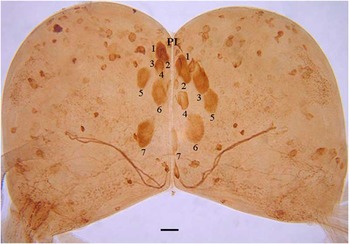
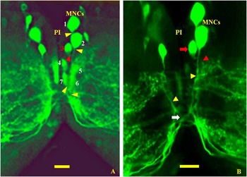
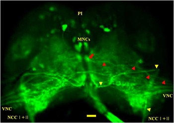
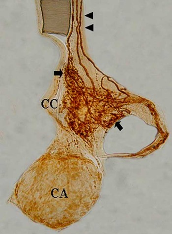
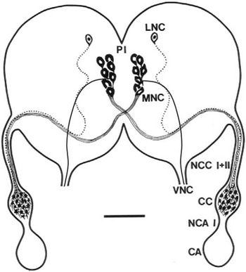
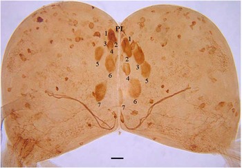
Three types of neurons (MNCs, LNC, and other neurons which were small in diameter and sparsely located throughout the brain) were found to be immunoreactive to FMRFamide-like neuropeptide (Fig. 1). Seven pairs of large or medium-sized MNCs with cell bodies of ∼20–25 μm in long diameter (numbered in Fig. 1) were detected in the pars intercerebralis (PI) between the two cerebral hemispheres. These cells showed moderate or strong immunoreactivity to FMRFamide-like peptide. Axon (arrowheads in Fig. 2A, 2B) from cell body (indicated as red “3” or red arrow in Fig. 2A, 2B) of a MNC ran to the lateral regions of the two cerebral hemispheres and then extended to the connective of the VNC (arrowheads in Fig. 3). In addition, the six pairs of MNCs with cell bodies that were clearly immunoreactive in the PI projected their axons into the lower median region of the ipsilateral side (Figs. 2A, 2B, 6). Labeled axons on both sides then decussated in the median region (arrow in Fig. 2B) and subsequently ran to the contralateral hemisphere through the given median bundle. Upon leaving the brain, the labeled axons extended to the contralateral CC of the retrocerebral complex and terminated in the nervi corporis cardiaci (NCC) I, which was fused with NCC II (Figs. 3, 4, 6). In the CC, labeled axons formed abundant terminals (arrows) that were full of FMRFamide-like neuropeptide (Fig. 6). However, the CA did not contain axons labeled with FMRFamide-like peptide.
Localization of FMRFamide-like immunoreactive neuronal cell bodies from the brain of fifth instar Bombyx mori. Seven pairs of large or medium-sized cell bodies of the medial neurosecretory cells (MNCs) (as numbered, 20–25 μm in diameter) are clustered in the pars intercerebralis (PI), showing strong or moderate levels of immunolabeling. The labeled axon fibers in the lower parts are thought to be those projecting from cell bodies of the MNCs. About 100 neuronal cell bodies (mostly 15 μm in diameter) with various levels of labeling are sparsely scattered throughout the brain. It should be noted that one pair of lateral neurosecretory cells is not shown in this figure. Scale bar indicates 25 μm.
Projection patterns of medial neurosecretory cell (MNC) of the brain of Bombyx mori. The MNCs are classified as two populations on the basis of the axonal projection pattern. One population is neurons with axonal fibers (yellow arrowheads) extended from the cell bodies of the FMRFamide-like immunoreactive (LI) MNCs (numbered excluding a red “3” in “A”) of both sides near the pars intercerebralis (PI) that decussate in the lower median region (white arrow in “B”). Axons of these cells eventually project into the contralateral corpus cardiacum. These FMRFamide-LI MNCs project their axons to the lateral region of the brain. The other population is neurons for which one pair of FMRFamide-LI MNCs (numbered as red “3” and red arrow in “B”) run their axons (red arrowhead) to the lateral region of the brain for projection into the connective of the ventral nerve cord. Scale bars indicate 25 μm.
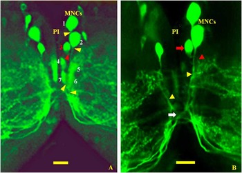
Confocal image of the FMRFamide-like immunoreactive cell body and axon of medial neurosecretory cell (MNC) in both cerebral hemispheres of Bombyx mori. One pair of the MNCs project theirs axons (red arrowheads) to the connective of ventral nerve cord (VNC). The labeled axons extending from six pairs of somata of MNCs near the pars intercerebralis (PI) form a given median bundle and can be also detected in the contralateral hemispheres after decussation in the median region, immediately before extending to the nervi corporis cardiaci (NCC) I outside the brain (yellow arrowheads). NCC I + II, fusion of NCC I with NCC II. Scale bar indicates 25 μm.
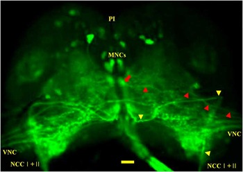
In addition to an axon running into the VNC, other axons were extended through the connective connected to the VNC (Fig. 3). Those were presumed to be axons projected from the FMRFamide-like immunoreactive (LI) cell bodies of brain into the VNC and/or vice versa.
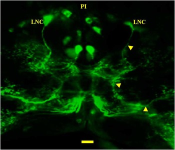
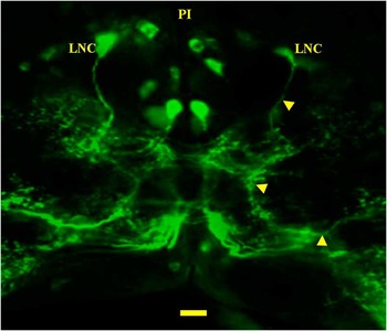
LNC was the second type of labeled neuron. One pair of small LNCs was identified near the pars lateralis (PL) (arrows in Fig. 4). Their cell bodies were small (15 μm diameter) and exhibited moderate immunoreactivity to FMRFamide-like neuropeptide. One pair of labeled LNCs extended their axons through the given ipsilateral bundle in both hemispheres without decussation in the median region, then to the ipsilateral NCC II, which was fused with the NCC I outside the brain, and finally to the CC where they terminated (Figs. 3, 6). Therefore, the labeled axons of LNCs formed abundant terminals that were full of FMRFamide-like neuropeptides in the CC (Figs. 5, 6).
Immunoreactivity of brain lateral neurosecretory cells (LNCs) and their axons to FMRFamide-like neuropeptides in Bombyx mori. One pair of the LNCs project axons (yellow arrowheads) into the ipsilateral hemisphere, without decussation, and terminate in the ipsilateral corpus cardiacum through the nervi corporis cardiaci (NCC) II fused with the NCC I. A few perikarya of the MNCs in the median region are intensively stained, but the remaining cell bodies are stained to a lesser degree. Most FMRFamide-like immunoreactive axons in the middle and lower neuropils of the brain are likely those that extend from cell bodies scattered throughout the brain. PI, pars intercerebralis. Scale bar indicates 30 μm.
Photograph of corpus cardiacum (CC), corpus allatum (CA), and nervi corporis cardiaci (NCC) I + II showing axonal termination of FMRFamide-like immunoreactive medial neurosecretory cells (MNCs) and lateral neurosecretory cells (LNCs) in the CC of Bombyx mori. The NCC I and II (arrowheads) have labeled axon fibers that extend from the somata of the MNCs and LNCs in brain, and the CC contains abundant, labeled axon terminals (arrows). Scale bar indicates 30 μm.

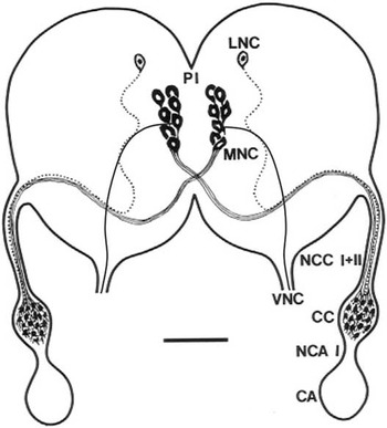
Schematic drawing of the projection of seven pairs of FMRFamide-like immunoreactive medial neurosecretory cells (MNCs) and one pair of labeled lateral neurosecretory cells (LNCs) from the brain into the retrocerebral complex and connective of ventral nerve cord (VNC) of fifth instar Bombyx mori. One pair of the MNCs in the brain extend their axons to the connective of the VNC, and six pairs of the MNCs, whose large or medium-sized cell bodies are located near the pars intercerebralis. These cells project their axons into the median brain region, in which their axons then decussate. Following decussation, the axons of these MNCs, together with LNC axons, extend through a given median bundle in each cerebral hemisphere and then through the contralateral nervi corporis cardiaci (NCC) I and nervi corporis allati (NCA) I to terminate in the corpus cardiacum (CC). The axons of the MNCs eventually innervate the contralateral CC. Axons of the LNCs, whose small cell bodies are located near the PL of the brain, run to the median region and then extend out of the brain without decussation. These axons project into the CC via the ipsilateral NCC II fused with the NCC I. Scale bar indicates 100 μm.
The third type of neurons exhibited a range of immunoreactivities to FMRFamide-like neuropeptide, weak, moderate, and strong. About 53 cell bodies, mostly small (∼15 μm diameter), were dispersed throughout each hemisphere, with more cell bodies being found in the upper portion (Fig. 1). Neurons of this type had abundant separated branches of axons or their terminals in the middle and lower neuropils of the brain (Figs. 2, 3, 4).
No immunolabeled structures were revealed in the brain, retrocerebral complex, or connective of the VNC by the FMRFamide antiserum preabsorbed with 50 nmol synthetic FMRFamide.
Discussion
The brain of silkworms, B. mori, has a variety of neurosecretory cells, including MNCs and LNCs (Ichikawa Reference Ichikawa1991; Park et al. Reference Park, Kim, Kim, Hwang, Kang and Kang2004), which produce a number of neuropeptides from their somata, then transport them along the given nerve tracks to several regions as specific ganglia of the VNC and the retrocerebral complex. Neuropeptides such as brain derived neurotrophic factor (BDNF), allatotropin, allatostatin, and tachykinin-related neuropeptide are eventually accumulated in either the CC or CA for subsequent secretion (Park et al. Reference Park, Jeon, Kim, Han, Yu and Lee2001, Reference Park, Hwang, Kang and Lee2002, Reference Park, Kim, Kim, Hwang, Kang and Kang2004). Whole-mount immunocytochemistry revealed ∼100 FMRFamide-LI somata in the brains of fifth instar silkworms (Fig. 1).
Production of neuropeptide by the brain has been suggested in Apis mellifera L. (Hymenoptera: Apidae) (Schümann and Erber Reference Schurmann and Erber1990) and Drosophila melanogaster (Merte and Nichols Reference Merte and Nichols2002). However, in our study new data regarding the number, localization, and axonal pathway of silkworm MNCs and LNCs were obtained from the brain and connective of the VNC in addition to the retrocerebral complex. Specifically, our results revealed one pair of MNCs projecting to the connective of the VNC, and six pairs of MNCs and one pair of LNCs projecting to the CC.
From about 100 FMRFamide-LI neuronal perikarya present in the fifth instar silkworm brain, seven pairs of MNCs and one pair of LNCs produced FMRFamide-like neuropeptides (Figs. 1, 2, 4). Cell bodies of the MNCs were clustered in groups in the median region and had diameters of ∼20–25 μm. In contrast, there was only one pair of LNC somata with a diameter of 15 μm. The cell bodies of the MNCs showed moderate or strong immunoreactivity. Differential intensities of immunoreactivity within the MNCs may reflect different physiological states but may also be a result of other conditions. In insects, especially Lepidoptera, one pair of MNCs project their axons to the connective of the VNC (Figs. 2A, 2B, 3) and the six pairs of MNCs that produce FMRFamide-like neuropeptides in the brain project their axons to the median region, in which axons from both sides then decussate (Fig. 2B). The decussation of the axons was previously described for tachykinin-related neuropeptide in Spodoptera litura (F.) (Lepidoptera: Noctuidae) (Kim et al. Reference Kim, Lee, Kwon, Kang and Nässel1998) and for BDNF in B. mori (Park et al. Reference Park, Kim, Kim, Hwang, Kang and Kang2004).
These results suggest that the MNCs of the brain can be classified as two populations. One population consists of one pair of the MNCs projecting their axons into the connective of VNC. The other population is composed of six pairs of MNCs, which project their axons to the CC (Figs. 2A, 2B, 3). After decussation, the axons extend to the contralateral NCC I and then terminate in the contralateral CC, which contains abundant FMRFamide-LI axon terminals (Fig. 5). Axonal termination of the FMRFamide-LI MNC in the tissues (perhaps some of ventral ganglia and [or] target organ) and function of the FMRFamide-like neuropeptide in the innervated tissues remain to be further studied.
In contrast, LNCs project their axons into the ipsilateral CC without decussation in the median region of the brain through the NCC II that is joined with NCC I (Fig. 4), as previously described for BDNF in B. mori (Park et al. Reference Park, Kim, Kim, Hwang, Kang and Kang2004).
These results suggest that FMRFamide-like neuropeptides, which have amino acid sequences that are ∼80% homologous to those of allatostatins, have a twofold function: inhibition of juvenile hormone generation and reduction of larval food intake (Gäde and Goldsworthy Reference Gäde and Goldsworthy2003). FMRFamide-like neuropeptides are produced from MNCs and LNCs in the brain, and then are transported to the VNC in addition to the CC in relation to controlling of the prothoracic gland (Palmer et al. Reference Palmer, Tran, Duttlinger and Nichols2007).
Five FMRFamides have known functions. Bommo-myosuppressin, or FMRF-related peptide, suppresses ecdysteroidogenesis from the prothoracic gland in Bombyx (Yamanaka et al. Reference Yamanaka, Zitnan, Kim, Adams, Hua and Suzuki2006), whereas sulfakinin function as modulators of gut and heart muscle contractions (Krajniak and Greenberg Reference Krajniak and Greenberg1992; Palmer et al. Reference Palmer, Tran, Duttlinger and Nichols2007). NPF is the homolog of the vertebrate NPY related to the feeding behaviors of Drosophila and sNPF regulates food intake and body size (Lee et al. Reference Lee, You, Choo, Han and Yu2004) and allatostatin inhibits the generation of juvenile hormones and reduces the food intake of insects and crustaceans (Yamanaka et al. Reference Yamanaka, Sachie, Zitňan, Ken, Tsuyoshi and Honoo2008). It has been suggested that six pairs of MNCs and one pair of LNC in the brain are related to modulation of hormones, such as juvenile hormone, while one pair of MNCs is associated with modulation of the gut contraction as allatostatin receptor is primarily expressed in the gut (Secher et al. Reference Secher, Lenz, Cazzamali, Sørensen, Williamson and Hansen2001).
However, the specific function of these MNCs, which project their axons into the VNC, remains to be further studied.
Acknowledgement
This research was supported by a Grant from Korea University.