INTRODUCTION
Microsporidia are obligate intracellular spore-forming parasites that infect protozoa, invertebrates and vertebrates. Fish are among the groups most affected by microsporidia infection. The phylum Microsporidia (Balbiani, 1882) is represented by at least 144 genera (Larsson, Reference Larsson1999). Of these, 15 genera are known to infect fish (Lom and Nilsen, Reference Lom and Nilsen2003). Microsporidia that infect the musculature and mesenteric tissues of commercially important fish species have a negative impact on fish commerce. Such microsporidia species belong to the genera Dasyatispora, Heterosporis, Pleistophora, Kabatana and the collective group Microsporidium (Dyková, Reference Dyková and Woo1995; Lom and Nilsen, Reference Lom and Nilsen2003; Diamant et al. Reference Diamant, Goren, Yokes, Galil, Klopman, Huchon, Szitenberg and Karhan2010). Infections with species of the genus Heterosporis, in particular, may have significant pathogenic effects on the host. These species do not produce a xenoma but instead infect the host's tissues in a diffuse fashion and may eventually be surrounded by host connective tissue (Lom et al. Reference Lom, Dyková, Wang, Lo and Kou2000). The species of this genus form elongated white nodules in the host musculature, thereby causing the liquefaction of muscle fibres and resulting in a characteristic concave body surface and, in extreme circumstances, resulting in death of the host fish. According to Lom and Nilsen (Reference Lom and Nilsen2003), the key feature of the genus Heterosporis, as observed in electron microscopy images, is the presence of a sporophorocyst (SPC), which is a dense, solid wall enclosing all the developmental stages of the parasite, i.e., meronts, sporonts and sporophorous vesicles with sporoblasts and spores. The genus Heterosporis includes 4 recognized species that infect freshwater fish from Africa and South America and 1 euryhaline fish from Japan (Hoshina, Reference Hoshina1951; Schubert, Reference Schubert1969; Lom et al. Reference Lom, Dyková, Körting and Klinger1989, Reference Lom, Dyková, Wang, Lo and Kou2000; Coste and Bouix, Reference Coste and Bouix1998). There have been no reports of microsporidiosis in the ichthyological fauna of the Arabian Gulf. In this report, we describe a new microsporidia species of the genus Heterosporis that infects the skeletal muscle, the body cavity and mesenteric tissues of the lizardfish Saurida undosquamis (Richardson, 1848) that are caught in the Arabian Gulf in Saudi Arabia. The description is based on the morphology, ultrastructural characteristics and phylogenetic analysis of the discovered species.
MATERIALS AND METHODS
Fish and sites of infection
A total of 420 freshly caught specimens of the marine fish Saurida undosquamis (Richardson, 1848) (Synodontidae, Harpadontinae) were obtained from the fish market in the city of Riyadh. Infection was determined by the presence of several cysts located in the skeletal muscles, the abdominal cavity and the mesenteric tissues.
Light and electron microscopy
To study the infecting microsporidia, the lengths and widths of the cysts were measured, and the cysts were crushed to study the spore morphology. The fresh spores were measured using a calibrated ocular micrometer on an Olympus microscope.
For transmission electron microscopy, the cysts were excised and fixed in 3% glutaraldehyde in 0·2 m sodium cacodylate buffer (pH 7·2) at 4°C for 24 h. After being washed overnight in the same buffer at 4 °C and post-fixing in 2% osmium tetroxide in the same buffer and temperature for 3 h, the fragments were dehydrated in a graded ascending series of ethanol, followed by propylene oxide (3 incubations of 2 h each) and embedding in Epon (12 h for each incubation). Semi-thin sections were stained with methylene blue. The ultra-thin sections were double-stained with uranyl acetate and lead citrate and were examined with a Philips (208) electron microscope at 80 to 100 kV.
DNA isolation and PCR amplification
Several cysts dissected from the fish were homogenized to isolate the spores and subsequently stored in 80% ethanol at 4°C. Genomic DNA was extracted from approximately 5×10⁶ spores using a GenElute™ Mammalian Genomic DNA Miniprep Kit (Sigma) following the manufacturer's instructions for animal tissues except for the incubation time (12 h). The DNA was stored in 50 μl of TE buffer at −20°C until further use. To amplify the 3′ end of the SSU and internal transcribed spacer (ITS) and the 5′ end of the large subunit (LSU) rRNA gene, the HG4F (5′GCGGCTTAATTTGACTCAAC3′) and HG4R (5′TCTCCTTGGTCCGTGTTTCAA3′) primers were used (Gatehouse and Malone, Reference Gatehouse and Malone1998). PCR was performed in 50 μl reactions using 10 pmol of each primer, 10 nmol of each dNTP, 2 mm MgCl2, 5 μl of 10X Taq polymerase buffer, 1·50 units of Taq DNA polymerase (Finnzymes Products), and 5 μl of genomic DNA. The reactions were performed in a Hybaid PxE Thermocycler (Thermo Electron Corporation, Milford, MA, USA). The amplification programme consisted of a 94°C denaturation for 5 min followed by 35 cycles of 94°C for 1 min, 50°C for 1 min and 72°C for 2 min. A final elongation step was performed at 72°C for 10 min. Aliquots (5 μl) of the PCR products were separated by electrophoresis through a 1% agarose 1X Tris-acetate-EDTA (TAE) buffer gel and stained with ethidium bromide.
DNA sequencing
The ∼1100-bp PCR product was sequenced directly after cleaning with NucleoSpin Extract II (Macherey-Nagel). The sequencing reactions were performed using the BigDye Terminator v1.1 kit (Applied Biosystems) and an ABI3700 DNA Analyzer (Perkin-Elmer, Applied Biosystems, Stabvida Co., Oeiras, Portugal).
Distance and phylogenetic analyses
Previously, the various forward and reverse sequence segments were manually aligned using ClustalW (Thompson et al. Reference Thompson, Higgins and Gilson1994) in MEGA 4 software, and ambiguous bases were clarified using the corresponding ABI chromatograms. To evaluate the relationship of Heterosporis saurida sp. n. with other microsporidia, a homology search was performed using BLAST (NCBI). After a preliminary comparison with almost all of the rDNA sequences from microsporidia of the class Marinosporidia, we selected 20 rDNA sequences belonging to the Dasyatispora, Glugea, Heterosporis, Pleistophora, and Ovipleistophora genera for molecular and phylogenetic inferences. The sequences and NCBI Accession numbers obtained from GenBank are as follows: Dasyatispora levantinae (GU183263); Glugea anomala (AF044391); Glugea atherinae (U15987); Glugea hertwigi (GQ203287); Glugea plecoglossi (AJ295326); Glugea stephani (AF056015); Glugea sp. GS1 (AJ295325); Heterosporis anguillarum (AF387331); Heterosporis sp. PF (AF356225); Ovipleistophora mirandellae (AF356223); Ovipleistophora ovariae (AJ252955); Pleistophora ehrenbaumi (AF044392); Pleistophora finisterrensis (AF044393); Pleistophora hippoglossoideos (AJ252953); Pleistophora hyphessobryconis (GU126672); Pleistophora typicalis (AF044387); Pleistophora sp. 1 (AF044394); Pleistophora sp. 2 (AF044389); Pleistophora sp. 3 (AF044390) and Pleistophora sp. (HQ703580); Loma embiotocia (AF320310) was used as an outgroup. The alignment was performed with ClustalW in MEGA 4 software (Tamura et al. Reference Tamura, Dudley, Nei and Kumar2007) with an opening gap penalty of 10 and a gap extension penalty of 4 for both pairwise and multiple alignments. Subsequent molecular evolutionary analyses were conducted using MEGA 4 with rDNA sequences from the same 20 microsporidian species and the outgroup species mentioned above. Distance estimation was performed using the Kimura 2-parameter model distance matrix for transitions and transversions. For the phylogenetic tree reconstructions in MEGA 4, maximum parsimony (MP) analysis was performed using the close neighbour interchange heuristic option with a search factor of 1 and random initial trees addition of 2000 replicates. The maximum likelihood (ML) method was implemented in the new version of the MEGA software (version 5.01) (Tamura et al. Reference Tamura, Peterson, Peterson, Stecher, Nei and Kumar2011). The GTR substitution model was selected, assuming an estimated proportion of invariant sites (0·3917) and 4 gamma-distributed rate categories to account for rate heterogeneity across sites. The gamma shape parameter was estimated directly from the data (gamma=0·4164). The reliability for the internal branch was assessed using the bootstrapping method (100 bootstrap replicates). Clade support was assessed with bootstrapping of 100 replicates. Bayesian inference (BI) was performed with the MrBayes program (v3.1.2) (Ronquist and Huelsenbeck, Reference Ronquist and Huelsenbeck2003) with the following parameters: the standard (4 by 4) model of nucleotide substitution, the number of substitution types=6 and the rate variation across sites fixed to ‘invgamma’. Probability distributions were generated using Markov Chain Monte Carlo methods. Four chains were run for 1×10⁶ generations with sampling every 100 generations and the first 10 000 sampled trees discarded as ‘burn-in’. Finally, a 50% majority rule consensus tree was constructed.
RESULTS
Light microscope observations
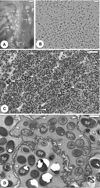
The prevalence of the infection was 32·1% (135/420) in both sexes of fish. Infections were visible as numerous macroscopic white, spindle-shaped cysts throughout the abdominal cavity, skeletal muscle and mesenteric tissues (Fig. 1A). The cysts numbered up to 20 per fish and measured as large as to 3–8 mm in size. In wet mounts, the fresh spores were mostly ovoid to pyriform in shape and had a large vacuole at the posterior end (Fig. 1B). There were 2 types of spores; most were microspores, a few were macrospores and all were oval to pyriform in shape. The microspores measured ∼3·3 (3·0–3·8)×2·0 (1·5–2·5) μm, and the macrospores measured ∼5·6 (5·0–6·0)× 3·3 (3·0–3·8) μm. Semi-thin sections revealed that the cysts contained multiple mature microspores and macrospores (Fig. 1C).
Light and ultrastructural characteristics of the Heterosporis saurida sp. n. parasite in the muscle of the lizardfish Saurida undosquamis. (A) Cysts in the muscles (arrows). Scale bar=10 mm. (B) Fresh free spores. Scale bar=10 μm. (C) Semi-thin section through a cyst containing aggregates of late developmental stages; microspores (*) and macrospores (arrowheads). Scale bar=10 μm. (D) Ultra-thin section through a cyst showing clusters of spores (S) within parasitophorous vacuoles, which are delimited by a thick, irregular membrane (arrowheads). Scale bar=2 μm.

Electron microscope observations
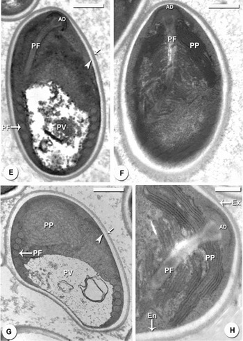
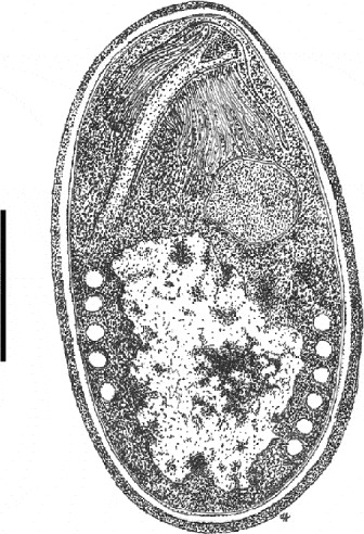
This parasite did not form a xenoma structure. Unfortunately, we could not detect all of the stages of development, only the spores. The spores were located within parasitophorous vacuoles (Fig. 1D) and were lined by a thick, amorphous wall. The spore wall was thick except for the anterior end where the central zone of the anchoring disc contacted the wall, which was thick and consisted of an electron-lucent endospore and a thin electron-dense exospore (Figs 2E, G and 3). The anchoring disc was located in the apical region of the spore in an eccentric position in relation to the spore axis, producing a bilateral asymmetry (Fig. 2E, F, 3). The posterior vacuole, situated at the basal part of the spore between the polar filament coils, was irregular and contained light material (Figs 2E, G and 3). The polar filament was isofilar, and it was coiled 5 times within the posterior vacuole and organized into a single row (Figs 2E, G and 3).
Details of the ultrastructural morphology of Heterosporis saurida sp. n. spores from the muscles of the lizard fish Saurida undosquamis. (E, F and G) The spore ultrastructure showing the anchoring disc (AD), polar filament (PF), polaroplast (PP), nucleus (N), posterior vacuole (PV) and spore wall, which is composed of the exospore (arrows) and endospore (arrowheads). Scale bar=1 μm. (H) Ultrastructural details of the spore anterior showing the spore wall formed by the exospore (Ex), endospore (En), anchoring disk (AD), polaroplast (PP) and polar filament (PF). Scale bar=0·5 μm.

Schematic drawing of mature spore. Scale bar=1 μm.

Description of H. saurida n. sp.
Systematic position
Phylum: Microsporidia (Balbiani, 1882)
Class: Marinosporidia (Vossbrinck and Debrunner-Vossbrinck, 2005)
Order: (Glugeida Issi, 1986)
Family: Glugeidae (Thélohan, 1892)
Genus: Heterosporis (Schubert, Reference Schubert1969)
Species: Heterosporis saurida n. sp.
Description of the species
Host type:Saurida undosquamis (Richardson, 1848)
Locality: Arabian Gulf
Site of infection: Skeletal muscles of the abdominal cavity and mesenteric tissues.
Prevalence of infection: 32·1% (135/420)
Specimen locality: One slide containing mature free spores and another with semi-thin sections of tissues containing spores at different developmental stages of the hapantotype were deposited in the International Protozoan Type Slide Collection at the Smithsonian Institution, Washington, D.C., 20560, USA.
Etymology: The specific epithet ‘saurida’ derives from the species epithet of the host species Saurida undosquamis.
Molecular analysis:
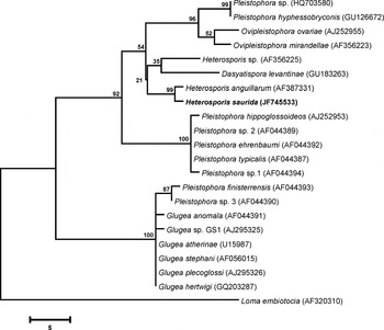
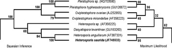
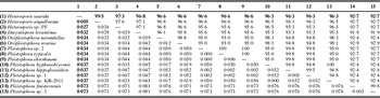
A 950-bp sequence, including a partial sequence from the SSU rRNA, ITS and LSU rRNA from Heterosporis saurida n. sp., was deposited in the GenBank database (Accession no. JF745533). In total, 20 rDNA sequences, including those with the highest BLAST scores, were aligned with Heterosporis saurida n. sp. The resulting alignment consisted of 1030 positions after trimming the 5′ and 3′ ends. Based on pairwise comparisons among the rRNA genes, the minimum genetic distances (Kimura 2-parameter) observed were 0·5%, 2·7% and 3·2% to Heterosporis anguillarum (AF387331), Heterosporis sp. (AF356225) and Dasyatispora levantinae (GU183263), respectively (Tables 1 & 2). An MP analysis (Fig. 4) strongly supported a clade for the species belonging to the Heterosporis, Ovipleistophora, Pleistophora and Dasyatispora genera (bootstrap 92%). Phylogenetic trees reconstructed using BI and ML methods showed similar topologies (Fig. 5).
The maximum parsimony tree of the partial SSU rDNA+ITS+partial LSU sequence of Heterosporis saurida n. sp. and the microsporidian species with the highest BLAST matches. The numbers on the branches are bootstrap confidence levels from 100 replicates. The GenBank Accession numbers are in parentheses after the species names, and a scale bar is provided under the tree. Heterosporis saurida n. sp. is placed within group III (Lom and Nilsen, Reference Lom and Nilsen2003), which includes the sequences of species from the genera Dasyatispora, Heterosporis, Ovipleistophora, and Pleistophora.

Phylogenetic trees based on Bayesian inference and a maximum likelihood analysis of a partial SSU rDNA+ITS+partial LSU sequence of Heterosporis saurida n. sp. and microsporidia positioned in the same clade, which revealed identical topologies.

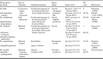
Comparative measurements (in μm) of the spores from Heterosporis spp.

* mi, microspores; ma, macrospores.
DISCUSSION
The light and ultrastructural examinations of the cysts, developmental stages and spore morphologies of the parasite described in the present study all revealed structures typical of parasites belonging to the phylum Microsporidia and the genus Heterosporis. The infection reported in this study was in the skeletal muscle, body cavity and mesenteric tissues of the lizardfish Saurida undosquamis. There has not been a prior report of Microsporidia infection in this fish species or other fishes of Saudi Arabia and the Arabian Gulf region. Infection by the microsporidian Pleistophora oolyticus (Negm-Eldin, Reference Negm-Eldin1992) has been reported in S. tumbil on the Egyptian coast of the Red Sea. Additionally, Peyghan et al. (Reference Peyghan, Nabavi, Jamshidi and Akbari2009) described a microsporidian species obtained from the lizardfish S. undosquamis that was caught in the Persian Gulf; however, light and electron microscopy showed that the spores were similar to those of the genus Glugea. The size range and the number of polar tube coils in the lizardfish microsporidia described here are different from those of all previously reported species.
A comparison of the previously described species of the genus Heterosporis with the parasite reported here reveals some differences in spore morphology (Table 1). Except for the H. anguillarum parasite of a euryhaline fish, Anguilla japonica from Japan (Hoshina, Reference Hoshina1951; Lom et al. Reference Lom, Dyková, Wang, Lo and Kou2000) and from a fish farm in Korea (Joh et al. Reference Joh, Kwon, Kim, Kim, Kwon, Park, Kwon and Kim2007), most of the other known Heterosporis spp. parasites have been found in tropical freshwater fish that were obtained from European pet shops. H. schuberti parasitize fish that inhabit African and South American rivers (Lom et al. Reference Lom, Dyková, Körting and Klinger1989), while the H. finki host is a fish from South American rivers (Schubert, Reference Schubert1969) and H. cichlidarum infect the gill filaments of a fish from west and central Africa (Coste and Bouix, Reference Coste and Bouix1998). All of these species parasitize the skeletal muscles, except for H. cichlidarum. In addition, Sutherland et al. (Reference Sutherland, Marcquenski, Marcino, Lom, Dyková, Hsu, Jahns and Nilsen2000) found Heterosporis sp. in the skeletal muscles of the yellow perch Perca flavescens in Wisconsin and Minnesota, USA.
Molecular and phylogenetic analyses
Similar to data recently published by Diamant et al. (Reference Diamant, Goren, Yokes, Galil, Klopman, Huchon, Szitenberg and Karhan2010), in our phylogenetic trees of fish-infecting microsporidia, group III corresponded to a clade that included all of the Heterosporis, Ovipleistophora and Dasyatispora sequences and almost all of the Pleistophora sequences and was supported by bootstrap values of 92% (MP), 88% (ML), and 100% (BI). In all the trees, H. saurida was a sister species of H. anguillarum (AF387331) with bootstraps of 99% (MP), 95% (ML) and 100% (BI). With regard to Heterosporis species, only H. anguillarum (AF387331) and another parasite found in the yellow perch P. flavescens (AF356225), classified as a Heterosporis sp. but not yet ultrastructurally characterized, had been sequenced. The phylogenetic trees from MP showed that the sequences of H. anguillarum, Heterosporis spp. (yellow perch) and Dasyatispora levantinae (GU183263) formed a cluster distant from the Ovipleisphora species and another two Pleistophora species. The genetic distances (calculated using the Kimura 2-parameter method) also indicated that there were similarities in the rDNA sequences among the species belonging to the Heterosporis and Dasyatispora genera, having percentage identities with H. anguillarum, Heterosporis spp. (PF) and Dasyatispora levantinae of 99·5%, 97·3% and 96·8%, respectively.
Although the molecular and phylogenetic similarities between H. saurida and D. levantinae indicate that these two species are more closely related, they are different ultrastructurally because the Dasyatispora sporophorocyst is absent, and the spore is monomorphic.
Microsporidian infections have not been previously reported in lizardfish in the Arabian Gulf. Because the microsporidia of most fish are host-specific, the parasite described here is thought to represent a new species. In recognition of the fish host, Heterosporis saurida is proposed as a name for this new species.
Comparison of rDNA sequences: percent identities (top diagonal) and pairwise distances (bottom diagonal) obtained by Kimura 2-parameter analysis

ACKNOWLEDGEMENT
The authors are thankful for the funding support of the Applied National Research Project (NPAR3-(15)), Deanship of Scientific Research, King Saud University, Riyadh, Saudi Arabia.