Introduction
Mothers with gestational diabetes mellitus face a higher risk of adverse pregnancy outcomes, including gestational hypertension, amniotic fluid overload, and postpartum haemorrhage. Similarly, newborns are at greater risk for adverse outcomes such as preterm birth, hypoglycaemia, macrosomia, and asphyxia.Reference Muche, Olayemi and Gete1–Reference Guillén-Sacoto, Barquiel, Hillman, Burgos and Herranz3 Since December 2011, China has adopted the International Study Group on Diabetes and Pregnancy diagnostic criteria for gestational diabetes mellitus, which has improved the early identification and management of gestational diabetes mellitus and reduced the risk of adverse maternal and neonatal outcomes.Reference Weinert4,Reference Berezowsky, Ardestani and Hiersch5 Previous studies have confirmed that gestational diabetes mellitus is associated with neonatal interventricular septal hypertrophy, but the specific impact of gestational diabetes mellitus on different cardiac structures and the correlation with maternal glycaemic control indicators remain unclear, especially the effect on neonates with normal birth weight. This article focuses on the impact of maternal gestational diabetes mellitus on neonatal cardiac development according to the International Study Group on Diabetes and Pregnancy diagnostic criteria and explores the risk factors for neonatal cardiac structural abnormalities, aiming to provide evidence for clinical monitoring and intervention.
Method
Study population
The study included full-term newborns admitted to the neonatology ward of our hospital between January and December 2024. The mothers of these newborns had been diagnosed with gestational diabetes mellitus, the control group consisted of full-term newborns born during the same period, whose mothers had not been diagnosed with gestational diabetes mellitus.The criteria for inclusion in the gestational diabetes mellitus group were as follows: (1) gestational diabetes mellitus in the mother (diagnosed by International Study Group on Diabetes and Pregnancy criteria); (2) a gestational age of 37 to 42 weeks; and (3) transient hypoglycaemic manifestations within 24 hr of birth or with temporary dyspnoea. The control group was comprised of subjects who met the following inclusion criteria: (1) Absence of gestational diabetes mellitus in the mother; (2) gestational age of 37–42 weeks; and (3) absence of postnatal hypoglycaemia. The exclusion criteria for both groups were as follows: (1) Newborns with a history of asphyxia, infectious diseases, severe respiratory diseases requiring respiratory support, and diseases affecting circulatory function, etc.; (2) mother’s combination of other gestational diseases; and (3) incomplete observation indexes.
Diagnostic criteria for gestational diabetes mellitus
Glucose tolerance test was performed at 28 weeks of gestation for gestational diabetes mellitus diagnosis, and the diagnosis was made when any one of the following criteria was met: fasting blood glucose level ≥ 5.1 mmol/L, 1-h postprandial blood glucose level ≥ 10.0 mmol/L, 2-h, and postprandial blood glucose level ≥ 8.5 mmol/L.
Data collection
This retrospective study recorded children’s health indicators by reviewing their medical records. The study considered various observation indicators: General indicators: gender, gestational age, birth weight, length, placental weight, and the ratio of birth weight to placental weight; echocardiographic indicators: cardiac parameters measured in the first week after birth using a Philips HD7 ultrasound device, including diameter of the proximal ascending aorta, diameter of the main pulmonary artery trunk, the size of the left ventricle, left auricle and right ventricle, the thickness of the interventricular septum (measured at the parasternal long-axis section, end-diastolic phase), the ejection fraction, and the rate of shortening of the left ventricular short axis; and biochemical indexes: The glycated albumin, glycated haemoglobin, and insulin levels in mothers of the gestational diabetes mellitus group at 24–28 weeks of gestation.
Statistical analysis
The following methods of analysis were employed: (1) A comparison between the gestational diabetes mellitus group and the control group in terms of neonatal general indicators and cardiometrical data; (2) a comparison of cardiac measurements of neonates with a birth weight of ≥ 3500 g and < 3500 g in the gestational diabetes mellitus and control groups, respectively. Infants weighing ≥ 4500 g (very large) and ≤ 2500 g (low birth weight) were defined as significant weight deviation and excluded from the subgroup analysis to account for the impact of extreme birth weight on cardiac measurements; (3) to ascertain the prevalence of CHD in the gestational diabetes mellitus and control groups (diagnosed by neonatal echocardiography, excluding physiological foramen ovale); and (4) to analyse the factors affecting septal hypertrophy in neonates in the gestational diabetes mellitus group, including gestational age, birth weight, maternal insulin, glycosylated haemoglobin, and glycosylated albumin.
Continuous variables were expressed as mean ± standard deviation and categorical variables as frequencies (%). Group comparisons were performed using independent samples t-tests for normally distributed data and χ 2 tests for proportions. All echocardiographic measurements were converted to Z-scores using Body Surface Area (BSA)-adjusted reference,Reference Lopez, Colan and Frommelt6 and abnormal cardiac enlargement was defined as z > 2.0. Multivariable linear regression analysed predictors of septal hypertrophy, reporting unstandardised coefficients (β) with 95% confidence intervals. Statistical significance was set at two-tailed P < 0.05. Analyses were performed using SPSS 26.0
Result
General information
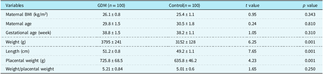
During the study period, 130 full-term newborns born to mothers with gestational diabetes mellitus were admitted to our hospital’s neonatal department. Thirty cases were excluded because the mothers had other gestational diseases, the observation indices were incomplete, or the newborns suffered from asphyxia or severe infectious diseases. The remaining 100 cases were included in the gestational diabetes mellitus group. Among these, there were 53 males and 47 females. In 28 cases, the mothers controlled their blood glucose by applying insulin treatment. In 72 cases, the mothers controlled their blood glucose through dietary means. A total of 234 newborns qualified for the control group. A total of 134 cases were excluded due to a variety of factors, including the mother’s combination of other gestational diseases, the child’s combination of severe cardiopulmonary system diseases, asphyxia, severe infectious diseases, incomplete observation indexes, and so forth. A total of 100 cases were included in the control group, comprising 42 males and 58 females. The mean weight, length, and placental weight of births in the gestational diabetes mellitus group were significantly higher than those of the control group (P < 0.05). However, no statistically significant differences were observed in the maternal BMI, maternal age, gender, gestational age, or birth weight/placental weight ratio between the two groups (P > 0.05) (Table 1).
General information of two groups

Table 1 Long description
The table presents a comparison of various variables between a gestational diabetes mellitus group and a control group. It includes data for 100 participants in each group. The variables compared are maternal body mass index, maternal age, gestational age, weight, length, placental weight, and the weight/placental weight ratio. The table shows that the mean weight, length, and placental weight of births in the gestational diabetes mellitus group are significantly higher than those in the control group, with p-values indicating statistical significance. However, no significant differences are observed in maternal BMI, maternal age, gender, gestational age, or the weight/placental weight ratio between the two groups. The table consists of 8 rows and 5 columns, with column headers including Variables, GDM (n = 100), Control (n = 100), t value, and p value. Each row provides specific data points for the variables listed.
BMI = body mass index.
Comparison of cardiac measurement data between two groups
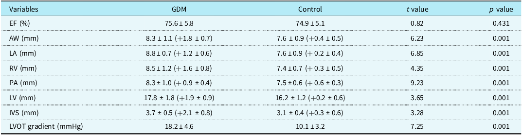
Comparison between gestational diabetes mellitus group and control group: Aortic width, left atrium, right ventricle, main pulmonary artery width, and left ventricle of the gestational diabetes mellitus group were greater than those of the control group, and the difference was statistically significant (P < 0.05); there was no statistically significant difference in the ejection fraction between the two groups (P > 0.05) (Table 2).
Comparison of cardiac measurement data between two groups

Table 2 Long description
The table presents a comparison of cardiac measurement data between two groups: GDM and Control. It includes variables such as EF percentage, AW in millimeters, LA in millimeters, RV in millimeters, PA in millimeters, LV in millimeters, IVS in millimeters, and LVOT gradient in millimeters of mercury. The table has eight rows and five columns, with columns labeled Variables, GDM, Control, t value, and p value. Each row provides specific measurements and statistical values for the GDM and Control groups. Notable trends include significantly higher measurements in the GDM group for AW, LA, RV, PA, LV, and IVS compared to the Control group, with corresponding high t values and low p values indicating statistical significance. The EF percentage shows no significant difference between the two groups.
Note: All echocardiographic measurement data are expressed as “mean ± standard deviation (SD)”, and the values in square brackets correspond to body surface area (BSA)-adjusted z-scores. Abbreviations: EF = ejection fraction; AW = aortic width (proximal ascending aorta); LA = left atrium; RV = right ventricle; PA = pulmonary artery (main pulmonary artery trunk); LV = left ventricle; IVS = interventricular septum; LVOT = left ventricular outflow tract.
Comparison of cardiac parameters in neonates with birth weight ≥ 3500 g between the gestational diabetes mellitus group and the control group
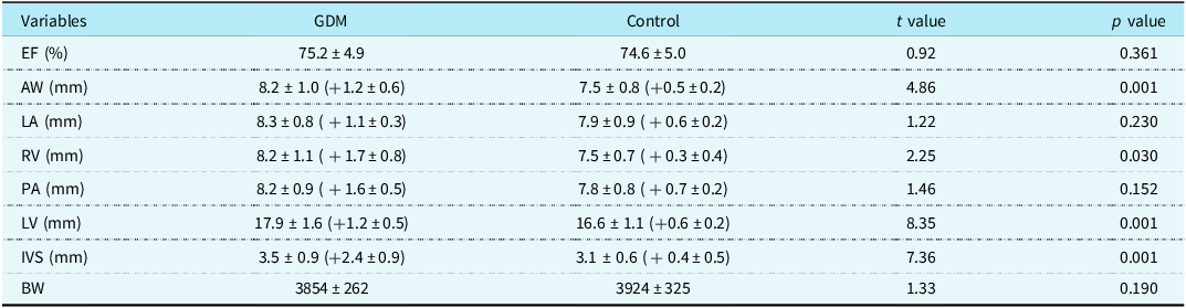
In the gestational diabetes mellitus group, there were 48 neonates with birth weight ≥ 3500 g, and 12 cases with significant weight deviation (≥ 4500 g) were excluded, leaving 36 cases for analysis. In the control group, no cases had significant weight deviation, so all 30 cases were included. No statistically significant difference was found in birth weight between the two groups (P > 0.05), and the slightly higher birth weight in the control group was due to the exclusion of extreme macrosomia in the gestational diabetes mellitus group. However, the gestational diabetes mellitus group exhibited greater ascending aorta, right ventricle diameter, left ventricle diameter, and interventricular septum thickness compared to the control group, with statistically significant differences (P < 0.05). No significant differences were observed in other indicators (P > 0.05) (Table 3).
Comparison of cardiac measurement data with a birth weight of ≥3500 g

Table 3 Long description
The table presents a comparison of cardiac measurement data between gestational diabetes mellitus (GDM) and control groups, focusing on neonates with a birth weight of 3500 grams. The table includes six rows and six columns, with column headers labeled as Variables, GDM, Control, t value, and p value. The variables measured include ejection fraction (EF) in percentage, aortic wall (AW) in millimeters, left atrium (LA) in millimeters, right ventricle (RV) in millimeters, pulmonary artery (PA) in millimeters, left ventricle (LV) in millimeters, interventricular septum (IVS) in millimeters, and birth weight (BW) in grams. The GDM group shows higher values in AW, RV, LV, and IVS compared to the control group, with statistically significant differences indicated by p values less than 0.05. The EF, LA, PA, and BW measurements do not show significant differences between the two groups.
Note: All echocardiographic measurement data are expressed as “mean ± standard deviation (SD)”, and the values in square brackets correspond to body surface area (BSA)-adjusted z-scores. Abbreviations: EF = ejection fraction; AW = aortic width; LA = left atrium; RV = right ventricle; PA = pulmonary artery; LV = left ventricle; IVS = interventricular septum; LVOT = left ventricular outflow tract.
Comparison of cardiac parameters in neonates with birth weight < 3500 g between the gestational diabetes mellitus group and the control group
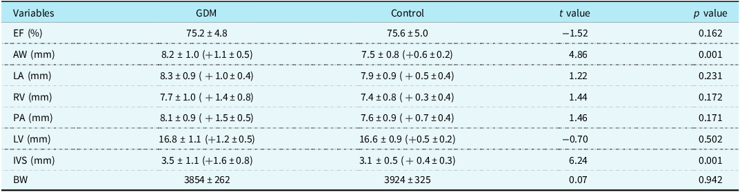
In the gestational diabetes mellitus group, there were 52 neonates with birth weight <3500 g, and 7 cases with significant weight deviation (≤ 2500 g) were excluded, leaving 45 cases for analysis. In the control group, no cases had significant weight deviation, so all 70 cases were included in the analysis. There was no statistically significant difference in birth weight between the two groups (P > 0.05). Although the interventricular septum thickness of both groups was within the normal reference range (2.23–4.24 mm), the interventricular septum thickness in the gestational diabetes mellitus group was greater than that in the control group, with a statistically significant difference (P < 0.05), suggesting a pathological thickening trend within the physiological range. No statistically significant differences were observed in other indicators between the two groups (P > 0.05) (Table 4).
Comparison of cardiac measurement data with a birth weight of <3500 g

Table 4 Long description
The table compares cardiac measurement data between gestational diabetes mellitus (GDM) and control groups with birth weight less than 3500 grams. It includes variables such as ejection fraction, aortic wall thickness, left atrium, right ventricle, pulmonary artery, left ventricle, interventricular septum, and birth weight. The table has 8 rows and 6 columns, with headers for Variables, GDM, Control, t value, and p value. Notable trends include a significant difference in aortic wall thickness and interventricular septum thickness between the two groups, with p values of 0.001 and 0.001 respectively. Other variables do not show statistically significant differences. The data is presented with mean values and standard deviations, along with t values and p values for each comparison.
Note: All echocardiographic measurement data are expressed as “mean ± standard deviation (SD)”, and the values in square brackets correspond to body surface area (BSA)-adjusted z-scores. Abbreviations: EF = ejection fraction; AW = aortic width; LA = left atrium; RV = right ventricle; PA = pulmonary artery; LV = left ventricle; IVS = interventricular septum; LVOT = left ventricular outflow tract.
The incidence of CHD
The incidence of CHD in the gestational diabetes mellitus group was 2% (2/100), both of which were small perimembranous ventricular septal defect (diameter 2–3 mm). No cases of CHD were observed in the control group (physiological foramen ovale was not considered as Atrial Septal Defect (ASD)). The observed difference between the two groups was statistically significant (χ2 = 4.02, P = 0.047).
Factors affecting septal hypertrophy in neonates from the gestational diabetes mellitus group
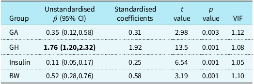
Interventricular septum thickness of 3.7 ± 0.5 mm was observed in 18 cases (18%) of the gestational diabetes mellitus group, defined as septal hypertrophy in this study. Maternal gestational glycosylated albumin (13.2 ± 0.5)%, glycosylated haemoglobin (6.3 ± 0.5)%, and insulin (61.2 ± 6.8)pmol/L were identified as contributing factors. Multiple linear regression analysis showed that maternal glycosylated albumin (β = 0.35, 95% confidence interval: 0.12–0.58, P = 0.003), glycosylated haemoglobin (β =1.76, 95% confidence interval: 1.20–2.32, P = 0.001), insulin (β = 0.11, 95% confidence interval: 0.05–0.17, P = 0.001), and neonatal birth weight (β = 0.52, 95% confidence interval: 0.28–0.76, P = 0.001) were all positively correlated with interventricular septum thickness in the gestational diabetes mellitus group (all P < 0.05, VIF < 1.2, no multicollinearity) (Table 5).
Multiple linear regression analysis of GDM septal thickness

Table 5 Long description
The table presents a multiple linear regression analysis of gestational diabetes mellitus (GDM) septal thickness. It includes four rows and five columns. The columns are labeled as Group, Unstandardised β (95% CI), Standardised coefficients, t value, p value, and VIF. The rows are labeled as GA, GH, Insulin, and BW. The GA group has an unstandardised β of 0.35 with a 95% confidence interval of 0.12 to 0.58, a standardised coefficient of 0.31, a t value of 2.98, a p value of 0.003, and a VIF of 1.12. The GH group has an unstandardised β of 1.76 with a 95% confidence interval of 1.20 to 2.32, a standardised coefficient of 1.92, a t value of 13.5, a p value of 0.001, and a VIF of 1.08. The Insulin group has an unstandardised β of 0.11 with a 95% confidence interval of 0.05 to 0.17, a standardised coefficient of 0.25, a t value of 6.54, a p value of 0.001, and a VIF of 1.05. The BW group has an unstandardised β of 0.52 with a 95% confidence interval of 0.28 to 0.76, a standardised coefficient of 0.58, a t value of 3.19, a p value of 0.001, and a VIF of 1.10. The table indicates that all variables are positively correlated with interventricular septum thickness in the GDM group.
GA = glycosylated albumin; GH = glycosylated haemoglobin; BW = birth weight.
Discussion
Abnormal maternal glucose metabolism during pregnancy is a key factor affecting fetal cardiac development, and the core manifestations in this study are the enlargement of neonatal cardiac great vessels and chambers and the thickening of interventricular septum, which is consistent with the results of previous studies but further clarifies the correlation with maternal glycaemic control indicators and the independent effect of gestational diabetes mellitus regardless of birth weight.Reference Araújo, Keating and Martel7,Reference Sundarapperuma, Wasalathanthri, Hettiarachchi and Wijesinghe8 Fetal growth and development depend on maternal nutrient transport through the placenta, and maternal chronic hyperglycaemia and insulin resistance in gestational diabetes mellitus can induce adaptive changes in the placenta, leading to increased placental weight and fetal macrosomia.Reference Christians, Grynspan, Greenwood and Dilworth9 This study found that the placental weight and birth weight of the gestational diabetes mellitus group were significantly higher than those of the control group, and the abnormal placental function may be the initial link between gestational diabetes mellitus and fetal cardiac development abnormalities.
Cardiac hypertrophy, especially Interventricular Septal Hypertrophy (IVSH), is the most common cardiac manifestation in gestational diabetes mellitus-exposed neonates, with reported incidence rates ranging from 30 to 40% in previous literature. The incidence of IVSH in this study was 18%, which was lower than the previous reports, possibly due to the strict inclusion/exclusion criteria (excluding neonates with asphyxia and maternal comorbidities) and the early intervention of gestational diabetes mellitus based on International Study Group on Diabetes and Pregnancy criteria. Notably, even in the subgroup of neonates with birth weight < 3500 g, the interventricular septum thickness of the gestational diabetes mellitus group was significantly higher than that of the control group, and both were within the normal reference range. This finding indicates that gestational diabetes mellitus can cause pathological thickening within the physiological range of the neonatal interventricular septum, which is an early manifestation of myocardial development damage, and this effect is independent of fetal macrosomia. The possible mechanism is that maternal hyperglycaemia leads to increased fetal insulin secretion, which promotes the proliferation and hypertrophy of cardiomyocytes, especially the interventricular septum cardiomyocytes.Reference Ghandi, Habibi and Nasri10,Reference Cade, Levy and Tinius11
In the subgroup of neonates with birth weight ≥ 3500 g, the gestational diabetes mellitus group had larger ascending aorta, right ventricle diameter, left ventricle diameter, and interventricular septum thickness than the control group, although the birth weight of the two groups was not statistically different. The slightly higher birth weight in the control group was due to the exclusion of ≥ 4500 g neonates in the gestational diabetes mellitus group, which avoided the interference of extreme macrosomia on cardiac measurements. The enlargement of great vessels and cardiac chambers in the gestational diabetes mellitus group may be related to the increased fetal cardiac preload and afterload caused by maternal hyperglycaemia: on the one hand, fetal hyperinsulinaemia leads to increased blood volume and cardiac preload; on the other hand, the abnormal vascular development caused by hyperglycaemia leads to increased peripheral resistance and cardiac afterload, which ultimately induces the enlargement of cardiac structures.Reference Ackerman-Banks, Palmsten, Lipkind and Ahrens12,Reference Zheng, Huang and Li13 This finding suggests that the adverse effect of gestational diabetes mellitus on neonatal cardiac development is exacerbated with the increase of fetal weight, which is consistent with the conclusions of prior studies.Reference Szlapinski, King, Retta, Yeo, Strutt and Hill14
Maternal glycaemic control indicators are important predictors of neonatal interventricular septum thickness in the gestational diabetes mellitus group, which is a new finding of this study. Multiple linear regression analysis showed that maternal glycosylated albumin, glycosylated haemoglobin, insulin levels, and neonatal birth weight were positively correlated with interventricular septum thickness. Glycosylated haemoglobin reflects long-term glycaemic control (8–12 weeks), glycosylated albumin reflects short-term glycaemic control (2–3 weeks) before delivery, and insulin reflects maternal insulin resistance. The positive correlation of these indicators with interventricular septum thickness suggests that both long-term and short-term abnormal glycaemic control in gestational diabetes mellitus mothers can affect fetal myocardial development, and the cumulative effect of hyperglycaemia is the key factor leading to neonatal IVSH.Reference Zhu, Chen, Li, Tao and Teng15–Reference Deprest, Jani and Van Schoubroeck17 Although the mean glycosylated albumin and glycosylated haemoglobin levels of gestational diabetes mellitus mothers in this study were within the normal reference range, the interventricular septum thickness of neonates was still significantly increased, indicating that the current clinical glycaemic control standards may need to be further optimised for the protection of fetal cardiac development.
This study found that the incidence of CHD in the gestational diabetes mellitus group was 2% (2 cases of perimembranous ventricular septal defect), and no CHD was found in the control group, which confirmed that gestational diabetes mellitus is a risk factor for neonatal ventricular septal defect. Previous studies have shown that maternal hyperglycaemia during embryonic cardiac development can induce oxidative stress and endoplasmic reticulum stress in fetal cardiac cells, disrupting cardiomyocyte differentiation and chamber septation, leading to structural defects.Reference Nassr, El-Nashar, Shazly, White and Brost18,Reference Wu, Reece and Zhong19 The small perimembranous ventricular septal defect in this study may be related to the early intervention of gestational diabetes mellitus, which reduces the severity of fetal cardiac structural damage. It should be noted that physiological foramen ovale in neonates cannot be diagnosed as ASD, which is a common misdiagnosis in clinical practice, and this study strictly distinguished the two to ensure the accuracy of CHD diagnosis.
The innovation of this study is as follows: (1) based on the International Study Group on Diabetes and Pregnancy diagnostic criteria, it systematically analysed the impact of gestational diabetes mellitus on different neonatal cardiac structures and found that gestational diabetes mellitus can cause the enlargement of great vessels and cardiac chambers in addition to IVSH; (2) confirmed that gestational diabetes mellitus has an adverse effect on neonatal cardiac development independent of birth weight, and even normal birth weight neonates have interventricular septum thickening; and (3) quantitatively analysed the correlation between maternal glycaemic control indicators (glycosylated albumin, glycosylated haemoglobin, and insulin) and neonatal interventricular septum thickness, providing a new basis for clinical monitoring of gestational diabetes mellitus pregnancies.
Limitation
This study has some limitations. First, the retrospective design limited our ability to control for other potential confounders such as gestational weight gain, dietary patterns, and physical activity levels during pregnancy. Second, as the study focused on neonates admitted for clinical observation (e.g., transient hypoglycaemia), our findings may not generalise to all gestational diabetes mellitus-exposed infants without such conditions. Third, echocardiographic measurements were performed within one week postpartum but lacked longitudinal follow-up to assess whether the observed cardiac changes persist or resolve during infancy. Fourth, the sample size of CHD in the gestational diabetes mellitus group is small, and further large-sample studies are needed to confirm the correlation between gestational diabetes mellitus and different types of CHD.
Conclusion
Maternal gestational diabetes mellitus significantly adversely affects neonatal myocardial development, which is characterised by the enlargement of the ascending aorta, main pulmonary artery and cardiac chambers, and the pathological thickening of interventricular septum even within the normal reference range. This adverse effect is independent of birth weight, and even neonates with normal birth weight born to gestational diabetes mellitus mothers have an increased risk of interventricular septum thickening. Maternal glycosylated albumin, glycosylated haemoglobin, and insulin levels are important predictors of neonatal interventricular septum thickness, which suggests that strict control of maternal blood glucose in both long term and short term is crucial for protecting fetal cardiac development.
These findings highlight two critical clinical implications: (1) implementation of routine fetal echocardiographic screening for pregnancies complicated by gestational diabetes mellitus, particularly in cases with suboptimal glycaemic control (glycated haemoglobin > 6.0%), and close monitoring of cardiac structures such as interventricular septum, ascending aorta and main pulmonary artery and (2) the necessity for early postnatal cardiac evaluation in all neonates of gestational diabetes mellitus mothers, including those with normal birth weight, and regular follow-up to observe the dynamic changes of cardiac structures. Furthermore, our results underscore the need for mechanistic studies to unravel the pathophysiological pathways linking maternal hyperglycaemia to altered cardiac development and to establish more targeted glycaemic control standards and cardiac monitoring strategies for gestational diabetes mellitus pregnancies.
Acknowledgements
We would like to thank Lifen Yuan for his efforts in data collection during the early stage of this study.
Authors’ contribution
Lina Qi wrote the main manuscript text; Junsheng Jiang prepared tables, revised the manuscript and designed the study; and Lifen Yuan collected data during the early stage of this study. All authors reviewed and approved the final manuscript.
Financial support
The authors declared that this study received no financial support.
Competing interests
No conflict of interest exists in the submission of the manuscript.