Significant outcomes
• Being male and LA/LA carrier both correlated with larger whole brain volumes.
• We noted the importance of correcting volumes from sub-brain regions for the total brain size.
• Previous findings of association between 5-HTTLPR and hippocampal size in patients with major depressive disorder (MDD) could not be replicated.
Limitations
• The aim of the present study was to replicate a previous finding. However, our population differed from the previous study, as the LG/LG genotype and LG allele frequencies were markedly lower in our patients group. Potentially, this decreased the power to find an effect of the LG/LG genotype and LG allele. Moreover, our sample size was smaller. Nevertheless in terms of statistical power, our sample size was large enough to detect the expected differences in brain morphometry. However, the small sample size was probably the most important limitation for the genetic analyses.
Introduction
The influence of genetic factors on MDD has been established from multiple sources although genetic variation alone is unlikely to explain this complex disorder (Reference Sullivan, Neale and Kendler1). The serotonergic system is an important biological substrate in the pathophysiology of MDD (Reference Owens and Nemeroff2), and the involvement of specifically the serotonin transporter in mood control has lead to the development of multiple antidepressant medications targeting these elements. Antidepressants based on selective serotonin reuptake inhibitors are known to have their site of action at the serotonin transporter, and the solute carrier family 6 gene (SLC6A4 – also known as 5-HTT) on chromosome 17q11.2 coding for the serotonin transporter protein has been identified as a candidate gene. In specific, a common insertion/deletion polymorphism 5-HTTLPR in the 5′ regulatory region of SLC6A4 has been associated with high or low activity depending on the number of copies (Reference Cohen-Woods, Schosser and McGuffin3). The terms ‘short’ (S) and ‘long’ (L) allele are commonly used to refer to two different lengths of the sequences in the gene's regulatory region, where the L allele has been found to produce more protein than the S allele. The single nucleotide polymorphism (SNP) rs25531 sub-classifies the L allele as an LA and LG allele with the LG allele thought to be similar to the S allele in terms of lowered transcriptional efficiency (Reference Hu, Lipsky and Zhu4). Thus, both a diallelic and a triallelic genetic marker may be investigated but using the triallelic marker is now state of the art. A meta-analysis of genetic studies on MDD has indicated a possible influence of the long variants (L and LA) in MDD (Reference Frodl, Moller and Meisenzahl5), and several studies have demonstrated the influence of the serotonin transporter polymorphism on the hippocampus volume in MDD (Reference Frodl, Meisenzahl and Zill6–Reference Taylor, Steffens and Payne9). In these reports, a reduction in the hippocampal volume was found for LA/LA homozygous depressed patients compared with healthy controls, whereas no significant differences were found between patients and controls carrying LA/(LG + S) or (LG + S)/(LG + S) genotypes (Reference Frodl, Meisenzahl and Zill6–Reference Frodl, Zill and Baghai8). Here (LG + S) denote that the person carries either LG or S. However, Taylor et al. (Reference Taylor, Steffens and Payne9) also showed that the effect of serotonin transporter genotypes may depend on the age of onset. The possible variation with age of onset and other demographic or treatment characteristics was also noted by Hickie et al. (Reference Hickie, Naismith and Ward10) who found no prediction of reduced hippocampal volume by 5-HTT status. A recent study by Selvaraj et al. (Reference Selvaraj, Godlewska and Norbury11) showed decreased grey matter volume for (LG + S) carriers in a larger sample of healthy controls. We previously conducted a longitudinal study to evaluate the effect of treated MDD on brain structure using deformation-based morphometry and measures of hippocampal volume (Reference Ahdidan, Hviid and Chakravarty12). The main finding was that brain structure changes seemed state dependent, only occurring in acute episode of MDD and normalising after remission. These results and the potential importance for studying the neurobiology of MDD induced us to attempt replication of the 5-HTTLPR findings in the well-characterised samples obtained from this longitudinal study.
Aims of the study
In the present study, we investigated the influence of 5-HTTLPR variants on the hippocampal and brain volume. The main aim was to replicate the findings of Frodl et al. (Reference Frodl, Koutsouleris and Bottlender7), but additionally we also hypothesised that 5-HTTLPR may be a risk factor for MDD. Aware of the small sample size of our groups we used of well known atrophy sensitive method, the tensor-based morphometry (TBM), to the replication of Frodl et al.'s (Reference Frodl, Koutsouleris and Bottlender7) findings.
Material and methods
Samples
The study included 23 patients admitted to Aarhus University Hospital, Risskov with MDD (cases) between 1996 and 1999 and 33 healthy subjects (controls). All subjects gave written informed consent and were obtained from the Danish PET/Depression project (Reference Videbech, Ravnkilde and Pedersen13). The present neuroimaging follow-up was conducted between 2004 and 2007 (Reference Ahdidan, Hviid and Chakravarty12) while later adding genotypic data. The patients were 18–70 years old at inclusion. Patients were screened for MDD using the Structured Clinical Interview for DSM-III-R (14) and included if they scored higher than 17 on the 17-item Hamilton Depressed Rating Scale (Reference Bech, Kastrup and Rafaelsen15). To avoid selection bias towards less depressed patients, those treated with psychotropic medication were not excluded. Individuals showing any medical conditions involving the central nervous system were excluded, as were individuals with drug or alcohol dependence or ongoing abuse. Blood and urine tests were used to confirm this information.
Healthy subjects were recruited by advertisement and screened by the same means as patients. Controls were excluded from the study when having any current or past psychiatric or neurological illness, a first degree family history of psychiatric illness, and any medical condition. None of the healthy controls had a history of substance abuse.
All subjects were followed for 8 years, and their case/control status was verified by the subjects’ personal accounts, the GPs’ medical records, and the Danish Psychiatric Central Register (Reference Mors, Perto and Mortensen16). The Danish PET/Depression project was approved by the Regional Scientific Ethics Committee for Aarhus County and by the Danish Data Protection Agency.
Image processing and analysis
All subjects were scanned in random order to avoid any systematic bias due to changes in magnetic resonance imaging (MRI) systems. Magnetic resonance images were acquired on a GE 1.5 T Signa Echospeed (GE Medical Systems, Milwaukee, WI, USA). A T1-weighted sequence of the whole brain was acquired with the following parameters: 3D-spoiled GRASS; repetition time 31 ms; echo time 12 ms; slice thickness 1.5 mm and zero gap, field of view 280 cm; yielding a voxel size of 1.5 mm × 1.09 mm × 1.09 mm.
We excluded images of poor quality, i.e. images with the brain cut, aliasing artefacts, or excessively noisy data resulting in low structural resolution.
The images were corrected for non-uniformity of the signal intensity using the non-parametric non-uniform intensity normalisation method (Reference Sled, Zijdenbos and Evans17). This step was used to improve the accuracy of anatomical analysis techniques such as tissue classification and segmentation. Subsequently, we removed the skull of all images to improve the spatial normalisation step (Reference Smith18).
Spatial normalisation was used to transform all subjects’ data, at each time point, to match the international consortium for brain mapping (ICBM) model space (Reference Mazziotta, Toga, Evans, Fox and Lancaster19). All T1-weighted magnetic resonance images were matched to the model by a 12-parameter affine transformation (three translations, three rotations, three scalings, and three shears), using a hierarchical strategy through the optimisation of the cross-correlation objective function (Reference Collins, Neelin, Peters and Evans20). The transformation was used as the input to the nonlinear transformation estimation process.
Nonlinear MRI-to-ICBM model transformations were estimated using the automatic nonlinear image matching and anatomical labelling (ANIMAL) algorithm (Reference Collins and Evans21). ANIMAL matches two images through the estimation of local translations defined on a grid of equally spaced nodes. Image matching was achieved through local optimisation of the correlation coefficient objective function (Reference Robbins, Evans, Collins and Whitesides22). The final transformation was estimated using a hierarchical matching strategy and contained local translations estimated on a grid of nodes with 2 mm spacing.
Comparisons between groups were performed using the general linear model MATLAB (http://www.mathworks.com) commands ‘multistat’ and ‘stat_summary’ from the fmristat MATLAB package (Reference Taylor and Worsley23). ‘Multistat’ was used to calculate the statistical parametric map of each group comparison, correcting for the effect of either age or gender, or both, if this effect was significant. ‘Stat_summary’ was then used to identify the statistically significant cluster of voxels after correcting for multiple testing using the lowest threshold implicated by non-isotropic random field theory, Bonferroni, or discrete local maxima.
Brain and hippocampal volumetry
The hippocampus was segmented using the ICBM model. We started by the coronal slice at the connection of the thalamus to the superior colliculus. The end point was the appearance of the mammillary bodies. We used the white matter as the inferior limit and the corona radiata as the superior limit. The inferior horn of the lateral ventricle was the lateral limit.
The brain tissue and the hippocampus were segmented based on the nonlinear transformation file of the spatial normalisation step and the hippocampal segmentation of the ICBM standard brain (Reference Collins and Evans21). The hippocampus of the ICBM standard brain was first manually segmented, thereby creating a mask in the ICBM standard space. The hippocampal mask was then matched to each subject's brain using the inverse of the MRI-to-ICBM model transformation. Finally, we multiplied the number of voxels of the hippocampal mask in the image scanning space by the size of a voxel of the image in the scanning space to measure the hippocampal volume.
Genotyping
Genotypes were determined for two polymorphisms in the promoter region of SLC6A4: the 44 bp insertion/deletion (long/short or L and S allele) diallelic microsatellite 5-HTTLPR and the SNP rs25531. By combining these two markers, the L allele of 5-HTTLPR may be sub-classified as an LA and LG allele, and the LG allele is then thought to be similar to the S allele in terms of association with lowered transcriptional efficiency (Reference Hu, Lipsky and Zhu4). We will use the term triallelic 5-HTTLPR also used in Frodl et al. (Reference Frodl, Koutsouleris and Bottlender7). The derived diallelic marker obtained by combining LG and S will be denoted ‘the high-low expression marker’. For genotyping 5-HTTLPR, polymerase chain reaction (PCR) was run on a Perkin Elmer 9700 with silver block using fluorescence-labelled primers (DNA Technology, Risskov, Denmark): 95°C for 2 min, 1 cycle; 95°C 1 min, 51°C 30 s, 72°C 1 min for 35 cycle; 72°C 7 min for 1 cycle. The primers used were:
F primer 6-Fam GGCGTTGCCGCTCTGAATGC
R primer CCGGGATGCGGGGGAATACTGGTAGGGTGC.
The rs25531 SNP was revealed by cutting the STPR PCR product with the restriction enzyme, MspI (New England Biolabs, Ipswich, MA, USA). Genotyping was performed on an ABI 3100 Prism Genetic Analyser (Applied Biosystems, Foster City, CA, USA) and peaks were analysed using Genemapper version 3.7 (Applied Biosystems, Foster City, CA, USA). To reduce human error, all genotypes were double-checked by another experienced technician in the laboratory.
Statistical analysis
Mean age was compared between patients and controls using the Student t-test under assumption of equal variances, which had been checked by an F-test. Difference in gender distribution was tested by Fisher's exact test for 2 × 2 tables. Hardy–Weinberg equilibrium was assessed using an exact test (diallelic markers) or Pearson's χ2-test with simulation-based p-values (10 000 replicates). Allelic and genotypic association tests were carried out using Fisher's exact test with permutation-based p-values. A deviance-based χ2-test was used to test the reductions in model complexity from the triallelic genotype model (potentially six categories) to the diallelic high-low expression genotype model (three categories) and further down to the high-low expression dominant genetic model [same risk for heterozygous and homozygous (LG + S) carriers].
Quantitative traits (brain and hippocampal volumes) were analysed using multiple linear regression (analysis of covariance) when one measure per subject was considered (whole brain and hippocampus), and with linear mixed models when two or more measures per subject were analysed (e.g. right and left hippocampus). The subject identifier was the only random effect included as no sign of variance heterogeneity between groups (case/control, genotypes, or gender) was observed. This model implies equal correlation (exchangeable correlation structure) of within-subject observations.
Gender and age were generally included as nuisance parameters (covariates). Exploratory analyses were carried out on females to exclude the large variation from the very limited group of male patients and in these analyses we adjusted for age. Analyses of the hippocampus were furthermore adjusted for whole brain volume (included as a covariate in the models) to ensure that possible findings were specific to this region and not a result of the subject's brain size per se.
Comparisons between nested models were performed with the usual F-test for single observation data or a likelihood ratio χ2-test in the case of multiple observations per subject (e.g. left/right hippocampus).
The statistical software R (http://www.R-project.org) was used for the statistical analyses, and a significance level of 5% was applied.
Results
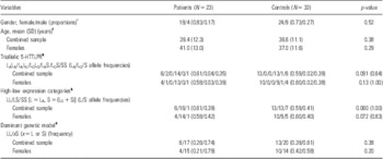
Table 1 summarises the distribution of age, gender, genotypes, and allele frequencies in patients and controls. Age and gender distributions were not significantly different. The absence in the present study of the LG/LG genotype observed in 14% of patients in Frodl et al. (Reference Frodl, Koutsouleris and Bottlender7) was striking. Furthermore, LG allele frequencies were much lower in the present study (4% in patients and 2% in controls) as compared with Frodl et al. (Reference Frodl, Koutsouleris and Bottlender7) who found an LG allele frequency of 27% in patients and 13% in controls.
Distribution on age, gender, genotypes, and allele frequencies for MDD patients and healthy controls in the combined sample of both genders and for females separately

MDD, major depressive disorder.
*Fisher's exact test for 2 × 2 tables.
†Two sample t-test under equal variances assumption.
‡Fisher's exact test for 2 × m table (genotypic or allelic association test) with simulation-based p-values (10 000 permutations).
Association analyses
The power to detect significant associations of small to medium effect was unfortunately limited with the present sample. Homozygous carriers of the LG or S allele tended to be more abundant among controls than patients (Table 1). This was not observed in the study by Frodl et al. (Reference Frodl, Koutsouleris and Bottlender7). p-values from Fisher's exact test for association of the high-low expression marker with disease were just above the border of significance in the combined sample of both genders as well as in the sample of females only (p = 0.060 and 0.072, respectively, see Table 1). As a consequence of the low LG allele frequency and correspondingly very low genotype frequencies of the triallelic 5-HTTLPR marker, we decided to focus on the functional categories (same effect of LG and S alleles). Reducing from LA, LG, and S to LA and (LG + S) resulted in a statistically non-significant 1.43 increase in residual deviance (p = 0.49) evaluated on a 2 df χ2 distribution. Further reduction to the dominant genetic model [LA/LA vs. x/(LG + S) with x denoting any of LA and (LG + S)] increased the deviance to 3.97, which was borderline significant (p = 0.046) in the 1 df χ2 distribution. The proportion of LA/LA carriers was larger in controls than in patients (0.39 vs. 0.26) and even more pronounced in females (0.42 vs. 0.21), though not significantly different (Table 1). None of the allelic association tests had p-values even close to the border of significance.
Quantitative traits
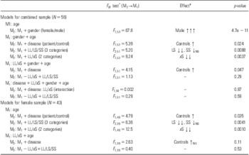
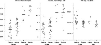
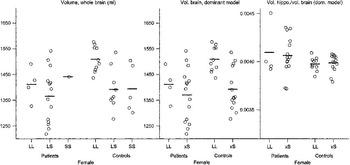
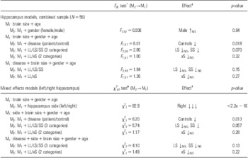
Results from analysing total brain and hippocampal volumes are shown in Tables 2 and 3, respectively, and are visualised in Figs 1 and 2. For brevity, L denotes the LA allele and S denotes LG or S, i.e. the (LG + S) allele. In the dominant model, x denotes any of L and S corresponding to a dominant effect of the (LG + S) allele. We found that being male and L/L (i.e. LA/LA) carrier both correlated with larger whole brain volumes. Furthermore, we noted the importance of correcting volumes from sub-brain regions for the total brain size: The correlation with larger whole brain volume pertained to the hippocampus but when corrected for total brain size, this correlation disappeared or possibly even changed sign.
Results for whole brain volumes in the combined sample of both genders and in the subsample of females

Notation: L = LA, S = (LG + S), x = L or S.
*df = degrees of freedom: d 2 − d 1, N − d 2 (all models include intercept).
†Number of arrows denotes significance (subscript NS if not significantly different from zero) from a t-test of the coefficient (null hypothesis: coefficient = 0).
Brain volumes (ml): whole brain (left panel); unadjusted total hippocampus (middle panel); total hippocampus adjusted for total brain size (right panel). Observations are shown separately for patients, controls, and each gender. Horizontal bars indicate mean value within subgroup.

Female brain volumes (ml): whole brain (left panel) for each 5-HTTLPR high-low expression genotype; 5-HTTLPR-dominant brain volume model (middle panel); the dominant model for total hippocampus adjusted for total brain size (right panel). Observations are shown separately for patients, controls, and genotypic group. Horizontal bars indicate mean value within subgroup. For brevity the LA allele is simply denoted L whereas S may be any of LG and S, i.e. the (LG + S) allele. In the dominant model, x denotes any of L and S corresponding to a dominant effect of the (LG + S) allele.

During the forward inclusion model building procedure (Table 2), we confirmed the larger brain volume of males compared with females (F 1, 53 = 67.8, p = 4.7e − 11). Subsequently, we confirmed the larger brain volume of L/L carriers (F 1, 52 = 9.24, p = 0.0037), and we furthermore found that the dominant genetic model fitted the data just as well as the three categorical genotypic model. This conclusion was valid whether case/control (disease) status was included in the model or not. Healthy controls tended to have larger brains than severely depressed inpatients (e.g. F 1, 51 = 4.15, p = 0.047). The results pertained to the analysis of females only, though the tendency towards larger brain volumes in controls was even weaker.
Males and females displayed no hippocampal volume difference when correcting for brain size (see Table 3). Healthy controls had a smaller relative hippocampal volume (relative to brain size) compared with depressed inpatients (F 1, 51 = 6.01, p = 0.018). In the analysis of left/right hippocampal volumes, a very clear and highly significant effect of side was observed (p < 2.2e − 16) with the right side being the smaller. The smaller volumes observed in healthy controls pertained after this adjustment for side effect (p = 0.013). Genotype had no significant effect on the hippocampal size though it may be argued that there was a weak tendency towards reduced hippocampal volume in subjects carrying one or two S alleles with a post hoc significant difference between S/S and L/L homozygotes. However, this difference was no longer statistically significant after inclusion of the significant case/control (disease) effect on the volume.
Results for the hippocampus in the combined sample of both genders

The first part considers the total hippocampus volumes whereas left and right hippocampus volumes are considered in the lower part of the table. The models are adjusted by brain size, gender, and age. Notation: L = LA, S = (LG + S), x = L or S.
*df = degrees of freedom: d 2 − d 1, N − d 2 (note: all models also include intercept).
†Number of arrows denotes significance (subscript NS if not significantly different from zero) from a t-test of the coefficient (null hypothesis: coefficient = 0).
‡df = degrees of freedom = d 2 − d 1.
Tensor-based morphometry
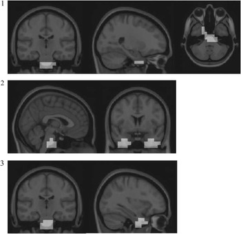
The results from TBM analyses are shown in Fig. 3. Atrophy was found for patients compared with healthy controls in: (1) the right temporal lobe and pons medulla for LA/LA genotype carriers, (2) the right and left temporal lobe and pons medulla for carriers of LA/(LG + S) or (LG + S)/(LG + S) genotype, and (3) the right temporal lobe and pons medulla for carriers of the LA/(LG + S) genotype of the serotonin transporter.
For patients compared with healthy controls, atrophy was found in: (1) the right temporal lobe and pons medulla for carriers of the LA/LA genotype of the serotonin transporter polymorphism 5-HTTLPR, (2) the right and left temporal lobe and pons medulla for LA/(LG + S) or (LG + S)/(LG + S) genotype carriers, and (3) the right temporal lobe and pons medulla for LA/(LG + S) genotype carriers.

Discussion
The main aim of the present study was to replicate the findings of previous studies of the influence of the long variant of the tri- and diallelic serotonin transporter polymorphism on the brain structure and hippocampal volume in a sample of well-characterised patients with MDD. We found no evidence of reduced hippocampal volumes in association with MDD and different genotypic combinations of the serotonin transporter polymorphism. In particular, the hippocampal volume, when corrected for the whole brain volume, was not significantly influenced by any of the genotypes investigated in patients and controls. Patients with the LA/LA genotype were found to have atrophy in the right temporal lobe and pons medulla compared with healthy controls. Patients carrying the LA/(LG + S) genotype and patients with LA/(LG + S) or (LG + S)/(LG + S) genotype had a larger degree of atrophy in the right and left temporal lobe and pons medulla compared with healthy controls. In conclusion, difference in atrophy between patients and controls was almost independent of the serotonin transporter genotype, and this polymorphism thus seems unlikely to be causing the difference. This observation was supported by the finding of a similar difference in atrophy when investigating patients and controls regardless of their genotype (results not shown). In summary, nothing in the present study indicates that variants of the 5-HTTLPR serotonin transporter polymorphism affect the hippocampus volume in MDD.
The lack of correlation between genotype and hippocampal volume does not agree with the findings of Frodl et al. (Reference Frodl, Zill and Baghai8). Lack of power due to the limited sample size of the present study may well be the most important reason and limitation. However the TBM method show group differences and indicates that our study has enough power to show brain atrophy due to the serotonin transporter genotype. Large differences in genotype and allele frequencies compared with the studies of Frodl et al. (Reference Frodl, Meisenzahl and Zill6–Reference Frodl, Zill and Baghai8) may also explain the disagreement. It is not uncommon to see variation in genotype distributions between studies, see e.g. table 1 in Risch et al. (Reference Risch, Herrell and Lehner24). One can speculate if these differences are due to differences in the patients’ severity, age of onset, or other characteristics but they may also be caused by ethnical difference (Reference Kunugi, Hattori and Kato25). Furthermore, while Frodl and colleagues had an almost even proportion of females and males the present study included a larger proportion of females. The final explanation may be that the association between depression and 5-HTTLPR does not exist. Indeed, there has been a lot of debate about this. A meta-analysis investigated this association together with stressful life events (Reference Risch, Herrell and Lehner24) using both published data and individual-level original data from 14 studies, found no evidence of an elevated risk of depression associated with the 5-HTTLPR genotype alone or associated with stressful life events, in men or women alone, or in both sexes combined. But this meta-analysis has been criticised for being biased towards the negative studies and a more recent and more comprehensive review actually found evidence of a moderating effect of the polymorphism with the S allele being associated with increased stress sensitivity (Reference Karg, Burmeister, Shedden and Sen26). As a maybe more speculative explanation of the results we note that epigenetic regulation of gene expression, e.g. via stress-induced transcription factors at the embryonic stage, or at later time points in life, potentially also relates to MDD. That is, heredity per se may not solely determine genetic effects on disease traits or changes in e.g. hippocampal volume, but how and when the genetic information is read may have impact.
Acknowledgements
We acknowledge Mette M. Hansen and Tracey J. Flint for doing the genotyping. Jamila Ahdidan and Leslie Foldager contributed equally to this work.
Conflicts of interest: All authors declare to have no conflicts of interest.