The prevalence of vancomycin-resistant enterococci (VRE) has been increasing in Canadian hospitals since the organism was first reported in 1993,Reference Lior, Litt and Hockin 1 and nationally, VRE accounts for approximately 6% of all Enterococcus faecium/faecalis isolates.Reference Simner, Adam and Baxter 2 In 2014, in 6 of 16 countries reporting data to the European Center for Disease Control and Prevention, the proportion of vancomycin-resistant E. faecium isolates was >5%, and it was >25% in 3 of these 16 countries. 3 VRE colonization upon admission to an intensive care unit is estimated to be 12.3% in the United States.Reference Ziakas, Thapa, Rice and Mylonakis 4 Following early reports of higher rates of morbidity and mortality from VRE infection when compared to vancomycin-susceptible enterococci (VSE), and due to concerns about the transfer of vancomycin-resistant genetic elements to S. aureus, aggressive VRE control measures were believed to represent the best practice. These measures, as endorsed by the Society for Healthcare Epidemiology of America (SHEA), include screening for asymptomatic carriage, contact tracing when transmission is suspected, outbreak control measures, and use of single-room isolation and contact precautions.Reference Muto, Jernigan and Ostrowsky 5
Despite initial fears, vancomycin-resistant S. aureus (VRSA) has not been detected in significant case numbers, and significant transmission of the vanA or vanB gene to S. aureus has not been reported.Reference Limbago, Kallen and Zhu 6 Many published studies address the issue of VRE versus VSE morbidity and mortality, and these results have been mixed.Reference Bhavnani, Drake and Forrest 7 – Reference Prematunge, MacDougall and Johnstone 18 A better understanding of VRE-specific outcomes is required.
Recent evidence has called into question the effectiveness of costly, large-scale control programs for antimicrobial-resistant organisms,Reference Huskins, Huckabee and O’Grady 19 , Reference Derde, Cooper and Goossens 20 not to mention their potentially detrimental impact on patients.Reference Karki, Leder and Cheng 21 – Reference Day, Perencevich and Harris 25 In Canada, rates of VRE colonization have continued to increase exponentially despite widespread use of hospital-based control measures; however, rates of clinical infection have not followed the same trajectory and have been decreasing in recent years. 26
In this study, we sought to quantify the impact of complete cessation of VRE control measures at 4 academic healthcare organizations in Ontario, Canada, representing 37.8% of all academic beds and 13.9% of all inpatient acute-care beds in that province. In this longitudinal analysis, we evaluated the impact on total VRE infection incidence as a primary outcome. We further evaluated a number of secondary outcomes, including the impact on VRE positive blood culture incidence (a positive blood culture irrespective of its clinical significance), VRE bacteremia incidence (clinical infection), VRE clinical isolate incidence, all-cause mortality, and VRE-attributable mortality.
METHODS
Study Setting and Population
Four academic health sciences centers in Ontario, Canada, were included in this study: University Health Network (Toronto), Ottawa Hospital, Kingston General Hospital, and London Health Sciences Centre/St. Joseph’s Health Care. These 4 sites comprise approximately 2,200 inpatient beds. Two study sites have large solid-organ transplant programs; 2 study sites have large malignant hematology programs performing both autologous and allogeneic stem cell transplants; and all sites have medical and radiation oncology programs. All sites include medical-surgical and specialty intensive care units, as well as outpatient dialysis units, and 3 sites have trauma units. During the study period, there were approximately 96,000 patient days per month across all of the study sites (range, 88,248–102,920 patient days). No significant changes to other existing infection prevention and control practices, infrastructure, or patient demographics were identified at any of the sites during the study period. The baseline VRE colonization rate across all study sites prior to cessation of control measures was 9 per 100,000 patient days.
Infection Prevention and Control Measures for VRE
Between July 1, 2010, and June 30, 2012, all study sites followed published guidance and screened patients for VRE carriage upon admission to hospital using either universal or targeted (risk-based) rectal surveillance cultures. Screening cultures were also performed on contacts of identified nosocomial VRE cases, during VRE outbreaks, and upon transfer or discharge during periods of high nosocomial VRE activity. All patients found to be colonized or infected with VRE were isolated using contact precautions and placed in a single room or were cohorted with other VRE patients. Enhanced environmental cleaning was put into place. When VRE outbreaks were declared, temporary closure of medical-surgical units occurred, and unit-wide prevalence screens were carried out and repeated until they were negative. In this study, 2 sites conducted periodic prevalence screens regardless of outbreak status.
On July 1, 2012, all elements of VRE control programs at each of the 4 study sites were discontinued. Routine practices were used in the care of all VRE patients.
Data Collection
Microbiologic data were retrospectively extracted from hospital laboratory information systems. A VRE clinical specimen was defined as one obtained from a sterile or nonsterile site due to suspicion of infection and showing the presence of E. faecalis or E. faecium harboring either the vanA or vanB gene, with a vancomycin minimum inhibitory concentration (MIC)>8 µg/mL. Only nosocomial VRE specimens (ie, for cases acquired ≥48 hours after admission) were included in this analysis.
All patient charts linked to VRE-positive clinical specimens throughout the study period were abstracted using a standardized anonymized form. A VRE clinical infection was defined as a positive VRE clinical specimen coupled with signs and symptoms of infection, validated through independent chart review by at least 2 infection control or infectious diseases physicians (1 site had 1 physician reviewer). Colonization was defined as the presence of VRE in a clinical specimen without causing disease. Colonization and contamination were analyzed as 1 group. A 30-day VRE-attributable mortality case was defined as a VRE clinical infection that directly or indirectly contributed to death of a patient within 30 days of the last VRE infection event. Mortality attribution was conducted via chart review by at least 2 infection control or infectious diseases physicians (1 site had 1 physician reviewer). Multiple infections in the same patient that occurred during the study period were identified and included. Discordant reviewer opinions were adjudicated by a third physician reviewer where possible.
Data for additional endpoints over the study period were collected as follows: use of linezolid and daptomycin by defined daily doses (DDD) per 100,000 patient days; rate of nosocomial methicillin-resistant S. aureus (MRSA) colonization per 10,000 patient days; rate of nosocomial C. difficile infection per 10,000 patient days; and the proportion of vancomycin resistant as compared to vancomycin-sensitive enterococcal isolates.
Data Review for Quality Assurance
A random sample of 5% of abstracted anonymized VRE patient charts were independently reviewed by 5 infectious diseases physicians not affiliated with any of the study sites to evaluate agreement of determination of colonization/contamination or infection as well as mortality related to VRE infection. Independent reviewers were provided with data definitions and used clinical judgment to make all determinations.
Laboratory Methods
Prior to discontinuing VRE control measures, all sites performed VRE surveillance cultures of stool or rectal swabs using 1 of 2 chromogenic media: Brilliance VRE Agar media (Oxoid, Canada) or Colorex VRE Agar (Alere, Canada). In this study, 3 centers used polymerase chain reaction (PCR) testing to identify genotype (vanA or vanB genes) on positive screen cultures.
During the entire study period, all enterococci clinical isolates were identified using standard laboratory methods and were tested for susceptibility to vancomycin. Isolates that appeared resistant to vancomycin (MIC>4 µg/mL) were further characterized using one of the following methods: growth on selective media, E Test (AB Biodisk), or PCR for vanA and vanB. At 2 of the study sites, all enterococci isolates were tested from sterile sites by PCR for the vanA or vanB genotype.
Statistical Analysis
Descriptive statistics were calculated using counts, percentages, proportions, means, and standard deviations. Counts of categorical outcomes were converted to rates using patient days as the denominator. For descriptive purposes, rates of categorical outcomes for all study sites were calculated in 3-month time periods to ensure statistical stability, given the paucity of events. For analytical purposes, annual outcome rates for each site were calculated. Generalized estimating equation (GEE) methods were used to cluster outcome rates within sites and to account for the fact that site-specific rates are not expected to be independent of each other. 27 , Reference Ballinger 28 GEE was used instead of a formal longitudinal analysis because the small number of outcome events observed within each cluster prohibited the latter. Negative binomial models were constructed for each outcome to account for the annual number of days of observation within each site; the negative binomial distribution was used to account for the overdispersion seen when the rates were modeled as Poisson counts. Each model had 2 fixed covariates: a binomial variable indicating VRE screening status and a linear term for year to account for any trends that might exist. The association of screening, independent of any trend, with the rate of each outcome was expressed as the incidence rate ratio (IRR). The IRR is interpreted like a relative risk, with numbers <1 indicating a decreased risk of the given outcome and numbers >1 indicating an increased outcome risk. Given that only 4 clusters (sites) were included in the study, small sample variance correction was invoked using PROC GLIMMIX in SAS 9.3.Reference Cook 29 Statistical significance was determined as P<.05.
A Cohen’s (pairwise) κ statistic was used to evaluate agreement between site-specific independent chart reviews and the independent chart reviews conducted for quality assurance.
This study was approved by the institutional review boards at each of the study sites.
RESULTS
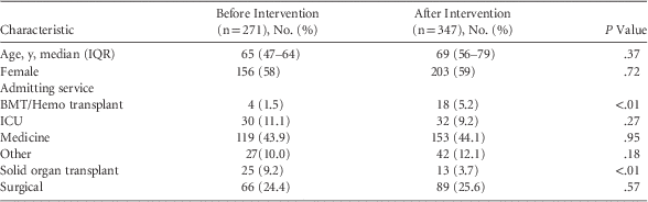
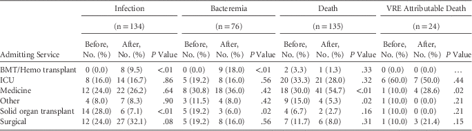
Patient demographics remained similar before and after cessation of control measures, with the exception of a higher percentage of malignant hematology patients (1.5% before and 5.2% after, P<.0001) and a lower percentage of solid organ transplant patients (9.2% before and 3.7% after, P=.0004) identified after cessation (Table 1). Significantly more VRE infections and bacteremias (0 before and 8 after, P<.01, 0 before and 9 after, P<.01, respectively) were identified after cessation of control measures in the malignant hematology population, while significantly fewer VRE infections (14 before and 6 after, P<.01) and bacteremias (5 before and 3 after, P=.02) were identified in the solid organ transplant population after cessation of control measures. However, all-cause mortality and VRE attributable mortality were not significantly impacted (Table 2).
Demographic Features Before and After Cessation of VRE Control

NOTE. Data are no. (%) of cases unless otherwise indicated. VRE, vancomycin-resistant enterococci; IQR, interquartile range; BMT/Hemo, bone marrow transplant/hematology; ICU, intensive care unit.
Distribution of VRE Clinical Outcomes by Admitting Service

NOTE. VRE, vancomycin-resistant Enterocococcus; BMT/Hemo, bone marrow transplant/hematology; ICU, intensive care unit.
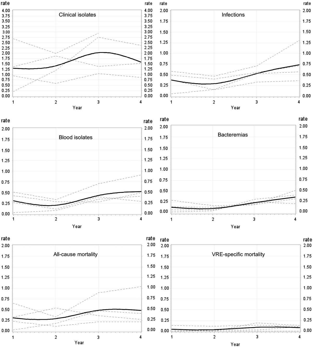
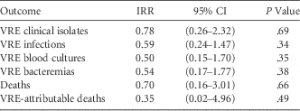
Figure 1 presents plots of hospital-specific and overall rates by year for all 6 study outcomes. Table 3 provides longitudinal analysis results by outcome. VRE control measures were not significantly associated with rates of VRE positive clinical isolates (IRR, 0.78; P=.69), infections (IRR, 0.59; P=.34), positive blood cultures (IRR, 0.50; P=.35), bacteremias (IRR, 0.54; P=.38), all-cause mortality (IRR, 0.70; P=.66), or VRE-attributable mortality (IRR, 0.35; P=.49). Although the IRR for each outcome was <1, the confidence intervals were extremely wide, and all P values were >.05.
Trends over time in annual outcome rates prior to and following cessation of VRE controls.
Rates for all six outcomes are presented. Each plot presents event rates (vertical axis) expressed as the annual number of events per 1000 patient days. Rates are presented for each individual hospital (dotted line) and for all hospitals combined (black line). VRE controls were in place for years 1 and 2 but ceased in years 3 and 4.

VRE Clinical Outcomes After Cessation of VRE Control

NOTE. VRE, vancomycin-resistant enterococci; IRR, incidence rate ratio; CI, confidence interval.
Daptomycin use was significantly higher after cessation of VRE control measures, with an increase from 83.4 DDD before the cessation of control measures to 386.3 DDD after the cessation of control measures (P=.1). Conversely, linezolid usage did not significantly change over the study period: 354.6 DDD before the cessation of control measures and 386.3 DDD after the cessation of control measures (P=.43). Nosocomial MRSA remained relatively unchanged: 6.8 per 10,000 patient days before the cessation of control measures and 5.5 per 10,000 patient days after the cessation of control measures (P=.09). C. difficile rates also remained relatively unchanged: 5.97 per 10,000 patient days before the cessation of control measures and 5.17 per 10,000 patient days after the cessation of control measures (P=.07). Compared to VSE isolates, the proportion of VRE isolates increased significantly from 4.3% to 5.9% after the control measures were stopped (P<.0001).
For the independent review of a 5% subset of cases, the Cohen’s κ statistic value was 0.60 for infections, with an overall inter-rater agreement of 82%, and Cohen’s κ statistic value was 0.53 for deaths, with an overall inter-rater agreement of 80%, indicating reasonable agreement.
DISCUSSION
To our knowledge, this is the first multicenter study to measure multiple VRE-specific patient outcomes after discontinuation of control measures. Other recent papers have demonstrated no significant clinical impact resulting from partial discontinuation of VRE control measures. In 2014, Popiel et alReference Popiel and Miller 30 evaluated the impact of discontinuation of VRE control measures in a Montreal tertiary care hospital on the incidence of VRE bacteremia, infection, and colonization. The hospital included general medicine, intensive care, and autologous bone marrow transplant populations. The authors found that both VRE colonizations and invasive infections increased over a 6–9-month period after relaxation of control measures but then decreased and remained stable over the following 6 months. The authors did not examine all-cause or VRE-attributable mortality. They concluded that adverse infection outcomes remained an uncommon event and may not be negatively impacted after cessation of control measures. More recently, Almyroudis et alReference Almyroudis, Osawa and Samonis 31 evaluated the impact of discontinuation of VRE control on VRE bacteremia in hematologic malignancy and stem cell transplant patients at a cancer center in upstate New York. In this study, the incidence of VRE bacteremia did not change during the 3-year period following the cessation of control measures. Mortality was not included as an outcome.
Our longitudinal analysis showed that cessation of VRE control measures had no statistically significant association with the risk of VRE clinical isolates, infection, blood cultures, bacteremia, all-cause mortality, or VRE-attributable mortality. Rates of all outcomes, particularly infection, positive blood cultures, and bacteremia, did appear to increase after control measures were stopped (Figure 1), but none reached statistical significance (Table 3). Notably, we have continued to follow VRE infection and bacteremia numbers beyond the end of this study period, and raw numbers for both of these outcomes have decreased to or below rates prior to cessation of control measures at all sites, similar to the findings of Popiel.Reference Popiel and Miller 30 Ongoing study will help to validate these findings.
In addition to all-cause mortality, we chose the more specific outcome of VRE-attributable mortality. We sought to minimize subjectivity in 2 ways. First, determination of infection status and attributable mortality was carried out by multiple physician reviewers at all but 1 site. Second, a subset of infections and deaths was independently reviewed by 5 physicians from varying geographic locations across Canada. Cohen’s κ analysis of these independent review results indicated a reasonable level of agreement (0.6), and inter-rater agreement was >80%. We therefore feel that subjectivity in death attribution was limited.
The proportion of VRE as compared to VSE isolates increased significantly after discontinuation of VRE control at all study sites. VRE colonization rates have been increasing across central and western Canada since the year 2000, and our findings may simply reflect this increasing colonization pressure leading to increased clinical isolates.Reference Simner, Adam and Baxter 2 , 26 In addition, the total number of VSE isolates decreased substantially from 14,100 to 10,400 after cessation of control measures, while the number of VRE isolates increased by only 18 (634 to 652); the large change in the VSE denominator had the greatest impact on the proportion. Furthermore, it is important to put the clinical impact of enterococcal bacteremias into perspective, as they represent only a small percentage of overall bacteremias in acute care hospitals, and VRE bacteremias represent an even lower percentage (4.9 and 0.5%, respectively, at 1 study site). Enterococci are also generally thought to be low-virulence pathogens, and significant infections occur more frequently in severely ill patients with multiple comorbidities.Reference Cheah, Spelman and Liew 32 , Reference Peel, Cheng, Spelman, Huysmans and Spelman 33 Indeed, enterococcal infection and bacteremia have been suggested by others to be a marker for severe underlying end-stage illness rather than a cause of mortality.Reference Vydra, Shanley and George 17 , Reference Bossaer, Hall and Garrett-Mayer 34 – Reference Theilacker, Jonas, Huebner, Bertz and Kern 36
Use of daptomycin increased significantly after VRE control measures ceased; however, use of linezolid did not. The increase in daptomycin use may reflect the relative increase in malignant hematology VRE cases after cessation of VRE control measures, as it is preferentially used in this population. Given that linezolid usage was not impacted, the reasons for the increase are more complex than simply an increase in VRE.
Whether clinical outcomes are worse with VRE than with VSE has been an ongoing question. Many early findings of more severe outcomes could plausibly be linked to the lack of availability of effective antimicrobials. A recent systematic review and meta-analysis by Prematunge et alReference Prematunge, MacDougall and Johnstone 18 compared VRE and VSE outcomes since the advent of quinupristin-dalfopristin, linezolid, and daptomycin. The authors concluded that VRE bacteremia resulted in longer hospital length of stay and increased mortality than VSE bacteremia. While the first conclusion is not surprising given challenges in discharging patients requiring isolation precautions to other facilities, and the increased incidence of other adverse events in isolated patients, the second holds less veracity. A major limitation to studies included in the Prematunge review is the use of all-cause mortality as a primary outcome measure. While commonly used to avoid challenges related to determination of disease specific mortality, all-cause mortality is particularly problematic in relation to VRE, which is inherently more common in patients who are severely ill and therefore have multiple other potential causes for proximal death.
The overall number of malignant hematology patients, as well as the number with VRE infections and VRE bacteremias, increased significantly after control measures stopped. Furthermore, the overall number of solid organ transplant patients, as well as the number of patients with VRE infections and VRE bacteremias, decreased significantly after cessation of control measures (Table 2). However, all-cause mortality and VRE specific mortality did not change in either of these populations. We are unable to explain these differences, and this result highlights the complexity of the ecology of VRE and indicates the need for further study.
We identified a number of limitations to our study. First, there were a small number of data points, with only 4 sites included. Second, our methodology for ascertaining VRE-attributable mortality introduced a level of subjectivity; however, this was attenuated through utilization of an independent audit. Importantly, no significant increase in all-cause mortality attributable to cessation of VRE control measures was detected through longitudinal analysis. Third, the follow-up period of 18 months was short, and the full impact of cessation of control measures may not have been determined during this observation period. Extended follow up may provide better information regarding the long-term impact of stopping VRE control measures.
Our findings indicate no adverse attributable clinical impact from cessation of VRE control measures across 4 large academic sites. Based on these results, we intend to continue without control measures for VRE at all of the study sites, with ongoing monitoring of clinical impact. Our data suggest that recommended best practices for the control of VRE in healthcare facilities should be re-evaluated and that measures focusing on meaningful patient-specific adverse outcomes, such as central-line–associated infection bundles and catheter-associated urinary tract infection bundles, should be explored. Further study is required regarding the optimal approaches to VRE control in high-risk populations, such as malignant hematology, and regarding the opportunity costs of VRE control.
ACKNOWLEDGMENTS
The authors thank Drs John Embil, Sarah Forgie, Mark Joffe, Jeff Powis, and Aleksandra Stefanovic for their assistance with this manuscript. The authors further thank Anita Marques for her editorial expertise, and the infection prevention and control departments at the University Health Network, the Ottawa Hospital, Kingston General Hospital and London Health Sciences Center.
Financial support: No financial support was provided relevant to this article. No funding other than local institutional support was provided for this study.
Potential conflicts of interest: All authors report no conflicts of interest relevant to this article.