Introduction
From the 14th century until the end of the 18th century, at the dawn of the industrial revolution, the organ was the embodiment of scientific and artistic universality. Tracing a legacy back to antiquity, the organ claimed a mythic status as the oldest western instrument; at the same time, it was at the forefront of technological and scientific advances. In the organ, acoustics, metallurgy, fluid dynamics, mechanics, architecture, sculpture, theology, painting, music composition, and performance come together to embody cultural collaborations and collisions. Surviving historical instruments document the social, political, scientific, and artistic impulses that yielded them.
Organs are lasting cultural monuments—the oldest continuously used organs date from the 15th century—and because of the expense of their construction, they are often objects of great civic pride, treasured for centuries. One celebrated example is the instrument in the church of St. Bavo in Haarlem, The Netherlands, constructed from 1735 to 1738 by the German builder C. Müller. This organ stands in an immensely grand and highly decorated case that advertises the city’s wealth, civic pride, and cultural patronage. Huge in scale (over 65 feet tall), with 32-foot-long pedal pipes dominating the facade, the organ was designed by Müller in conjunction with the best architects, painters, and sculptors of the day. The young Mozart played on this instrument, and today, it draws organists and audiences from all around the world. New organs inspired by instruments like the Müller organ at the St. Bavo church are now being built in Europe, the United States, and Asia, either closely based on historical models or incorporating knowledge learned from them about design, construction, and the choice and treatment of materials. Reference Snyder1,Reference Speerstra2
As multimedia artworks and models of interdisciplinarity, organs have long been a laboratory for experimentation and research, especially into the interaction of music and materials. Recently, collaborative projects in organ restoration, preservation, and construction conducted by researchers at the Göteborg Organ Art Center (GOArt) at the University of Gothenburg and the Chalmers University of Technology in Sweden, and at Cornell University and Oberlin College in the United States, have employed materials research to understand the physical environment of vulnerable historical organs and to make modern copies of surviving antiques that convey the aesthetic and material qualities of the originals.
We will describe the ways builders in the past understood the relationship between the materials from which an instrument is built, especially the metal for the pipes and the sounds it produces. We will then discuss recent scientific research initiated by efforts in preservation and restoration of these priceless monuments that opens up new insights into the behavior of organ pipe metal.
A complex materials environment
Basic organ construction has changed little over the centuries. The essential elements remain the same, as does the complexity of the materials environment. An organ may contain thousands of pipes, mostly hidden behind its facade, but it is at its simplest, a box of whistles. An array of pipes of different sizes and designs sit on a wind chest; bellows or a blower provide air to the chest. Each stop knob at the organ console represents a set of pipes of a particular timbre with a different pipe for every note on the keyboard. Pulling the stop activates a slider under that specific set of pipes, making them available as a sound source. The wind chest also contains a series of valves connected to the keyboard by a mechanical linkage. When a key is depressed, the valve is opened and air flows into the pipe for that note to create sound. Reference Thistlethwhaite and Webber3 Each pipe’s unique sound is roughly determined by its length and diameter and is honed by the pipe voicer, a craftsman who manipulates the area around the mouth of the pipe where small changes to the airflow and to patterns of turbulence have a large acoustical impact.
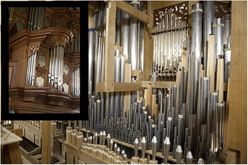
Organ pipes may be made of nearly any material—copper, paper, ivory, glass—even bamboo or plastic. Most commonly, organs contain both wood and metal pipes, with the majority made of metal. In a traditionally built organ, the pipes are situated in a complex environment of inorganic and organic materials. Figure 1 shows a new 18th century style organ at Cornell University, built at GOArt and installed in Ithaca, N.Y., under the direction of Japanese organ-builder M. Yokota. As far as possible, it uses only materials and construction techniques available to early 18th century craftsmen. The wind chests and action parts are made of wood, largely oak. The organ case is a mixture of hard and soft woods. Wind channels are made airtight with leather seals. Iron and brass are used for parts of the action—the mechanical linkages that connect the keys on the keyboard to the valves that let air into the pipes—and various kinds of glue are used throughout. These diverse materials can affect one another since they change over time, presenting a challenge to organ curators and conservators.
Interior of the Anabel Taylor Chapel Baroque Organ, Cornell University, by M. Yokota, GOArt, and Parsons Pipe Organs.

Since the Middle Ages, the most common organ pipe material other than wood has been an amalgam of tin and lead—metals that have been associated with varying degrees of sonic harshness and sweetness. The medieval theorist Adalbold of Utrecht (970–1027) ascribed particular qualities to the sounds of various metals based on their relative flexibility, writing that, “the less flexible metal makes the more violent sound, so tin is louder than lead, and silver and gold louder than tin. But aes is the most violent of all, and red aes (copper) more violent than white (brass).” Reference Barbieri4 Aes was a term used to describe copper and its alloys. Since the 14th century, organ builders have understood lead to lend a softness and vocality to the sound and tin to create a brighter, more resonant sound.
Such views were not confined to builders from hundreds of years ago. The influential 20th century organ builder C. Fisk, trained as a physicist and a pioneer in the rediscovery of historical organ-making techniques, maintained that a high proportion of lead gave the sound “a darkness, a hollowness, an astonishing agility, an ability to move as the music moves, to flit about like a freshly hatched insect; a tone with a softness about it. Yet a chorus of lead pipes has great carrying power. That bravura, that all-out quality, is the sound of lead.” Reference Fisk5 Tin-rich pipes, by comparison, “love to produce overtones, and there is something about the metal that lends itself to the production of pleasing overtones, particularly when the voicing is done in the old way.”
Lead-tin alloys continue to be the favored materials for organ pipes. The empirical sense of musicians and early acousticians that it was a metal’s flexibility that made it desirable in organ pipe construction can now be quantified with Young’s modulus. Both lead and tin are metals with low stiffness, high internal friction, and large damping capacities. The lead-tin phase diagram has limited regions of solid solubility on both ends. Microstructures in the intermediate compositional regions contain areas of tin enrichment and of lead enrichment and are affected by casting conditions. Organ pipes are manufactured as sheets by pouring molten metal mixtures onto surfaces where solidification occurs quickly. The sheets are rolled into cylinders and shaped into cones to form the body and foot of the pipe. Historically, the melt was poured onto a sand-filled bench, leading to more rapid solidification than on a modern surface of stone covered with fireproof fabric. Microscopy of lead-tin alloys following solidification at different rates has shown that faster cooling leads to a microstructure with smaller grain sizes for tin-rich and lead-rich areas. Reference Friesel and Karlsson6 In recent historically informed organ construction projects, the sand-casting method has been reproduced to manufacture alloys with microstructures as similar as possible to those of historic metals used in organ pipes. Reference Speerstra2
Another detail that emerges from close study of historical organ pipes is the complex mixture of trace elements in the alloys. For example, elemental analysis of a 1650 Swedish organ pipe showed that while the material is primarily a lead-tin mixture, bismuth, silver, arsenic, copper, indium, and antimony are present at levels of one part per thousand or less. Reference Friesel and Karlsson6 The most abundant of these, bismuth, may have been added intentionally because early metallurgists had found that it improved the flow of the melt during casting. Reference Barbieri7 The other trace elements may have been added, but more likely were part of the lead and tin ores used and remained in the alloy because of incomplete refining in early metallurgy.
The presence of trace elements is essential to the mechanical properties of an organ pipe, and particularly, its ability to resist sagging under its own considerable weight. Fisk observed, “By adding some of the impurities that come naturally in the old ‘pure’ lead of the 17th century, the metal can be made sturdy enough to stand for many years. Antimony (0.75%), copper (0.06%), bismuth (0.05%), and tin (1.0%), when added all together, will produce the desired stiffening. This explains why the lead front pipes from the Gothic and Renaissance stand without any sign of collapsing, while American common metal front pipes of the early 19th century always sagged.” Reference Fisk5 In the historic pipes, trace elements prevent creep through solid-solution hardening. Complex alloy compositions have been employed in recent historically informed instruments, frequently based upon analyses of pipes in European model instruments.
Organ pipe corrosion and conservation
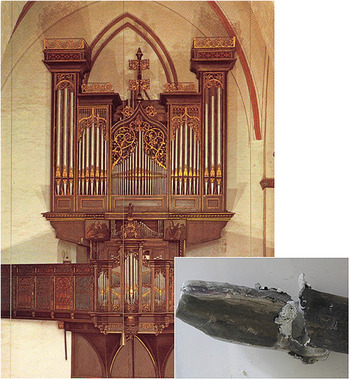
Multidisciplinary, collaborative materials research on preservation of historic organs has been motivated by the plight of organs such as that in Lübeck, Germany, completed by F. Stellwagen in 1637 ( Figure 2 ). Like many organs of its time, it incorporated pipes from an even earlier instrument, with its core dating from the 15th century. An organ of this sort—from the Baroque era, but with pipes and case still intact—is unusual; many other Baroque organs have been lost because of renovations and changes in aesthetics and, especially in Northern Europe, due to wartime bombings. Organ conservators were alarmed more than a decade ago to find significant corrosion damage in some of the Stellwagen organ’s oldest pipes. Holes rendered some pipes unable to produce sound, and earlier stages of deterioration had begun more subtly to alter the details of the sound of others. These small changes compromised the ability to access the way this instrument was meant to be heard when multiplied across the hundreds of notes in a piece.
Organ completed by F. Stellwagen in 1637 in Lübeck, Germany. Inset: Severely corrosion-damaged organ pipe. Photos by Ibo Ortgies, GOArt.

Observations of corrosion damage in the Lübeck organ and in other European organs inspired a research project with the goals of determining what was causing this corrosion, why it had been observed only recently in very old instruments, and how it could be prevented. The European Commission-funded COLLAPSE (Corrosion of Lead and Lead-Tin Alloys of Organ Pipes in Europe) Project involved organ builders and musicologists at GOArt, corrosion scientists at the Chalmers University of Technology, and metallurgists at the University of Bologna, Italy. Reference Speerstra8
Several possible causes of corrosion were initially considered. Attack by pollutants from the combustion of fossil fuels was one candidate, particularly because corrosion had only recently been observed. Local to the organ case itself were organic acids—wood acids that are released when cellulose in wood breaks down over time. It was also necessary to consider the synergistic effects of pollutants, temperature, and humidity. The placement of organs in large spaces, often without climate control, means that they are deeply affected by seasonal changes in temperature and humidity.
Another factor in deterioration of tin-rich pipes is “tin pest,” a phase transition below 13°C from the shiny, metallic beta form of tin to the powdery, nonmetallic, lower-density alpha form. Conversion to the alpha phase can cause blistering and flaking in tin-rich alloys. Reference Chiavari, Martini, Poli and Prandstraller9 For lead-rich pipes such as those in the Stellwagen organ, initial work within the COLLAPSE Project showed that organic acids released from wood are the primary cause of corrosion. Reference Niklasson, Johansson and Svensson10
Laboratory exposure experiments
Collaborative work between researchers at the Chalmers University of Technology, Cornell University, and Oberlin College addressed questions remaining after the COLLAPSE Project about the roles of humidity and alloy composition in susceptibility to acid attack. In laboratory exposure experiments, metal coupons with compositions modeling organ pipes were exposed to atmospheres of controlled pollutant content, humidity, and temperature. These exposures typically lasted a few weeks; the strength of this method was not in modeling centuries of behavior, but instead, in focusing on the initial stages of corrosion attack. In our studies, coupons containing between 1.2 at.% and 15 at.% Sn were exposed for comparison to previous work on pure lead samples. Reference Oertel, Baker, Niklasson, Johansson and Svensson11,Reference Oertel, Baker, Niklasson, Johansson, Svensson, Vandiver, Casadio, McCarthy, Tykot and Sil12 These experiments were informed by field studies on organs at a variety of geographic sites throughout Europe. The environment inside each organ case was monitored over a one-year period to measure humidity, temperature, and organic acid concentrations. Acetic acid concentrations were found in the range of 200–1500 ppb and maximum relative humidities (RHs) in the range of 74–85%. Reference Niklasson, Langer, Arrhenius, Rosell, Bergsten, Johansson and Svensson13
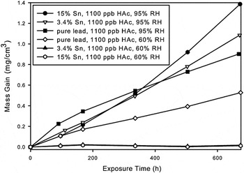
In exposure experiments, mass gains due to accumulation of corrosion products were measured over four-week time periods. At 60% RH, the presence of 3.4 at.% Sn drastically reduced the accumulation of corrosion products compared with the behavior of pure lead. At 95% RH, the extent of corrosion was greater overall, and the protective effect of tin was lost. Moreover, the extent of corrosion was enhanced with greater tin content at the higher humidity level ( Figure 3 ). Reference Oertel, Baker, Niklasson, Johansson and Svensson11
Wet mass gain curves of pure lead and 3.4 and 15 at.% Sn alloys exposed to 1100 ppb acetic acid (HAc) under 95% and 60% relative humidity (RH). Traces for 3.4% Sn and 15% Sn at 60% RH are nearly overlapping. Reference Oertel, Baker, Niklasson, Johansson and Svensson11

The outcomes of these exposure experiments suggested strategies for reduction of organ pipe corrosion. In the design of new organs, incorporation of alloys that have a small percentage of tin provide a protective effect under the humidities that are normally found in organ cases. The much-enhanced corrosion observed at 95% RH also has a practical implication, although that level of humidity does not occur naturally in organ environments. The experimental outcome shows the importance of avoiding even local over-humidification when a humidifier is used in an organ case to protect the wood and leather.
Phase identification and elemental mapping of corrosion products
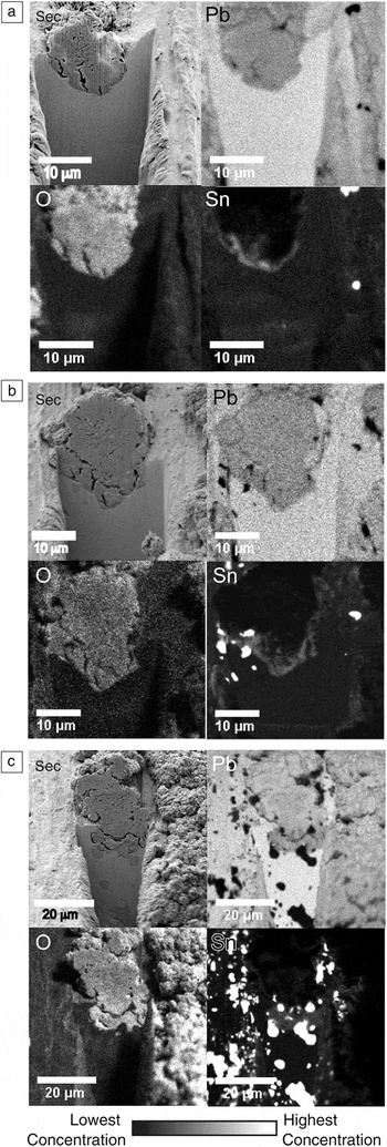
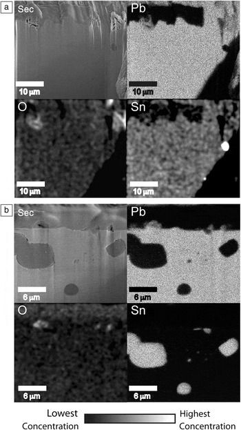
Microscale characterization of exposed lead-tin alloys was conducted to elucidate the role of each metal in the corrosion process. Scanning electron microscope (SEM) images were obtained for cross sections prepared through corrosion sites using focused ion beam milling. Wavelength-dispersive x-ray (WDX) detection was used to map the abundance of Pb, Sn, and O in the cross sections. In samples exposed to 1100 ppb acetic acid at 95% RH, lead was diluted in the corrosion crust as compared to its abundance in the bulk metal, and oxygen was more abundant in the oxide layer ( Figure 4 ). WDX maps for Sn showed tin-rich inclusions in compositions above the limit of solid solubility. More subtly, an enrichment of tin at the oxide–metal interface was observed for samples containing 3.4 and 15 at.% Sn. The mass gain data for these samples showed an enhancement of corrosion with increasing tin content (Figure 3). Under these conditions, tin participated in the oxidation process, and the images reflect its incorporation into the corrosion product layer.
Secondary electron images and wavelength-dispersive x-ray elemental maps for Pb, O, and Sn for cross sections through corrosion crusts in samples exposed to 1100 ppb acetic acid/95% relative humidities. (a) Alloy containing 3.4% Sn exposed for one week. (b) Alloy containing 3.4% Sn exposed for four weeks. (c) Alloy containing 15% Sn exposed for four weeks. Reference Oertel, Baker, Niklasson, Johansson and Svensson11

The behavior at 95% RH was compared with that at the same level of acetic acid but at 60% RH ( Figure 5 ). In cross sections of samples exposed at the lower humidity level, the oxide layer growth was less extensive. The corrosion crusts were again diluted in lead content and enhanced in oxygen content in comparison with the bulk metal. In contrast with the observation at 95% RH, there was no visible enhancement of tin around the crusts. This was consistent with the observation that a low level of tin provided a protective effect at 60% RH. At this moderate humidity, tin is suggested to reduce corrosion through formation of a nanometer-scale protective layer, which would not be visible through the cross-sectioning and SEM/WDX experiments.
Secondary-electron images and energy-dispersive x-ray elemental maps for Pb, O, and Sn for cross sections in samples exposed to 1100 ppb acetic acid/60% relative humidities for four weeks. (a) Alloy containing 3.4% Sn and (b) alloy containing 15% Sn. Platinum was deposited to protect the sample surfaces and appears as a dark area at the top of each map. Small corrosion crusts are located at (a–b) the upper left and at (b) the upper right. Reference Oertel, Baker, Niklasson, Johansson and Svensson11

Structure determination of corrosion products
Grazing incidence angle x-ray diffraction (XRD) was used to identify corrosion products on the exposed samples as well as on a historic pipe sample. The lead-based corrosion products PbO, plumbonacrite [Pb10O(OH)6(CO3)6], lead oxide acetate [Pb3O2(CH3COO)2 •0.5H2O], and lead oxide formate [Pb2O(HCOO)2] were identified following laboratory exposures. Hydrocerussite [Pb3(CO3)2(OH)2], which was found on the historic pipe sample, was not identified on samples exposed to corrosive conditions (humidity plus acetic or formic acid) over a short period of time. While the identified inorganic products were crystallographically well characterized, the structures of lead oxide acetate and lead oxide formate were unknown. Reference Kwestroo and Langereis14 We used mild hydrothermal reactions just above 100°C for ex situ synthesis and growth of diffraction-quality crystals of these materials. Reference Mauck, van den Heuvel, Hull, Zeller and Oertel15
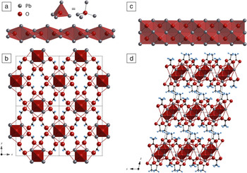
Structure determination by single-crystal XRD showed that both lead oxide carboxylates contain oxygen-centered lead tetrahedra that share edges to make one-dimensional infinite chains—single Pb2O2+ chains in the formate case and double Pb3O2 2+ chains in the acetate compound ( Figure 6 ). Linkage by the deprotonated acids leads to a three-dimensional network in the formate compound and a layered structure in the acetate compound.
Structures of the corrosion products lead oxide formate and lead oxide acetate. (a) Pb2O2+ single chain of edge-sharing Pb4O tetrahedra. (b) 3D network in lead oxide formate with Pb2O2+ chains viewed end on. (c) Pb3O2 2+ double chain of edge-sharing tetrahedra. (d) Layered structure in lead oxide acetate with Pb3O2 2+ chains viewed end on. Reference Mauck, van den Heuvel, Hull, Zeller and Oertel15

The ex situ synthesis of corrosion products enabled experiments to determine how they change upon further exposure. When exposed to a flowing atmosphere containing CO2 and water vapor, lead oxide acetate completely converted to hydrocerussite, the long-term corrosion product found on a historic organ pipe sample. Reference Niklasson, Johansson and Svensson16 Thus, synthetic chemistry provided a bridge between what was observed in four-week laboratory exposures and what eventually occurs on an organ pipe exposed to similar conditions over a much longer period of time.
Conclusions
An organ is a complex, functioning object that is integral to the cultural space in which it is situated. Understanding the behavior of any of its individual elements requires a multidisciplinary awareness of the larger context. Questions addressed during recent research on organ pipe corrosion included how to model the organ environment in the laboratory based on field data, how to reconcile outcomes from short-term exposure experiments with materials behavior over an organ’s lifetime, and how information from experiments could lead to strategies to mitigate organ pipe corrosion.
To return to the example of the Stellwagen organ in Lübeck, Germany, which introduced the problem of organ pipe corrosion, one of the sources of organic acids was new wood that had been installed as part of a relatively recent restoration campaign. This wood has now been replaced with less acid-emitting wood and a ventilation system has been added. Other emerging strategies include coating organ wood with CaO and MgO nanoparticles that act as sinks for acid Reference Giorgi, Chelazzi, Fratini, Langer, Niklasson, Rademar, Svensson and Baglioni17 and using sensing devices to alert curators when humidity or acid concentrations become too high. Reference Bergsten, Odlyha, Jakiela, Slater, Cavicchioli, de Faria, Niklasson, Svensson, Bratasz, Camuffo, della Valle, Baldini, Falciai, Mencaglia, Senesi and Theodorakopoulous18
This project demonstrates how applied research on a cultural object can lead to discoveries in fundamental materials chemistry. The reactions of lead and tin with organic acids have been elucidated on the micrometer scale at corrosion interfaces and at the angstrom scale in corrosion product crystal structures, motivating further questions on the mechanism by which high humidity leads to differential behavior of tin.
Collaborative research among scientists, musicologists, builders, and conservators brings challenges beyond those of research on new materials. This interplay of science and the humanities has advanced fundamental materials understanding while producing outcomes that preserve culturally valued historic organs and inform construction of new instruments, allowing contemporary listeners to access the sonic world of the 17th and 18th centuries.
Acknowledgments
The authors acknowledge the contributions of J.-E. Svensson and A. Niklasson Zieseniss (Chalmers University of Technology) and S. Baker (Cornell University). C.M.O. acknowledges a National Science Foundation Discovery Corps Program Grant (CHE-0412181 and CHE-0631552).

Catherine M. Oertel is associate professor of Chemistry at Oberlin College. She received a BA degree in chemistry from Oberlin College and a PhD degree in inorganic chemistry from Cornell University. She was a National Science Foundation Discovery Corps Postdoctoral Fellow at the Cornell Center for Materials Research and the Chalmers University of Technology in Göteborg, Sweden. Her current research focuses on hydrothermal synthesis and crystal chemistry of complex oxides, and is supported by a National Science Foundation CAREER Award and by the Camille and Henry Dreyfus Foundation. Oertel can be reached by email at catherine.oertel@oberlin.edu.

Annette Richards is a professor of music and university organist at Cornell University. She is a performer and scholar with specialty in 18th century music and she conducts interdisciplinary research into music, literature, and visual culture. She obtained a BA degree from the University of Oxford, UK, a PhD degree from Stanford University, and the Performer’s Diploma from the Sweelinck Conservatorium Amsterdam. Richards has been awarded prizes and fellowships from the Mellon and Alexander von Humboldt Foundations, and at the Dublin and Bruges international organ competitions. She is Executive Director of the Westfield Center for Historical Keyboard Studies. Richards can be reached by email at annette.richards@cornell.edu.