- IBD
inflammatory bowel's disease
- UC
ulcerative colitis
- CD
Crohn's disease
- DSS
dextran sodium sulphate
- MPO
myeloperoxidase
- LaBb Dahi
Lb. acidophilus and Bifido. bifidum containing probiotic Dahi
- DAI
disease activity index
Inflammatory bowel disease (IBD) comprises Crohn's disease (CD) and ulcerative colitis (UC). Both illnesses are increasingly important problems in industrialised countries and have been associated with colon cancer risk (Langholz et al. Reference Langholz, Munkholm, Davidsen and Binder1992; Gillen et al. Reference Gillen, Walmsley, Prior, Andrews and Allan1994). UC is a chronic inflammatory disease of the colon characterised by contiguous inflammation of the colonic lamina propria with subsequent injury and disruption of the mucosal barrier. Despite recent advances in the understanding of the genetics, immune and inflammatory mechanisms, as well as potential environmental factors that contribute to the disease, an exact pathogenesis remains elusive. The current therapies with aminosalicylates, steroids, and immunosuppressant therapies are limited by their side effects, poor compliance of patients and high relapse rates (Regueiro et al. Reference Regueiro, Curtis and Plevy2006; Jakobovits et al. Reference Jakobovits, Jewell and Travis2007; Lewis et al. Reference Lewis, Gelfand, Troxel, Forde, Newcomb, Kim, Margolis and Strom2008). Consequently, new alternatives for the treatment of UC are constantly being sought.
One of the latest additions to the vast therapeutic armamentarium is probiotics, defined as live microbial feed supplements, which beneficially affect the host by improving intestinal microbial balance, blocking adhesion sites on the colonocytes thus enhancing gut barrier function and improving local immune response (Fuller, Reference Fuller1989). The limitation of UC to the colon together with the microbiological impact makes it appropriate to study probiotic/prebiotic preparations. The indication for probiotics in IBD is grounded on a number of human and animal studies indicating that the enteric flora is centrally involved in the pathogenesis of CD and UC (Sartor, Reference Sartor2008). Animal and clinical studies indicate that gastrointestinal bacteria play an important role in the development of UC (Fedorak & Madsen, Reference Fedorak and Madsen2004; Rioux & Fedorak, Reference Rioux and Fedorak2006). Supplements of probiotics provide a new therapy for UC. Bibiloni et al. (Reference Bibiloni, Fedorak, Tannock, Madsen, Gionchetti, Campieri, De Simone and Sartor2005) found that VSL#3 (a probiotic mixture consisting of four strains of lactobacilli – Lb. casei, Lb. plantarum, Lb. acidophilus and Lb. delbrueckii subsp. bulgaricus; three strains of bifidobacteria – Bifido. longum, Bifido. breve and Bifido. infantis, and Streptococcus salivarius subsp. thermophilus) displays a beneficial effect on UC. It was reported that administration of Lb. casei decreases histological damage, while administration of Escherichia coli O83 and Nissle 1917 is beneficial for immunological regulation (Kokesova et al. Reference Kokesova, Frolova, Kverka, Sokol, Rossmann, Bartova and Tlaskalova-Hogenova2006).
Dahi is fermented milk widely consumed in Indian sub-continent. It is prepared using mesophilic culture of lactococci, which are not probiotic in nature. However, Dahi can be a good medium for delivery of probiotic bacteria. We have prepared a buffalo milk based probiotic LaBb Dahi by co-culturing the selected strains of Lb. acidophilus LaVK2 and Bifido. bifidum BbVK3 and Dahi starter. Studies from our laboratory have shown that this probiotic Dahi attenuates dimethylhydrazine induced gastrointestinal carcinogenesis (Rajpal & Kansal, Reference Rajpal and Kansal2008); diminishes diet induced hypercholestermia and atherogenic index in rats (Rajpal & Kansal, Reference Rajpal and Kansal2009a); stimulates the macrophage activity and confers protection against enteric infection in mice (Rajpal & Kansal, Reference Rajpal and Kansal2009b). This probiotic Dahi also reverses the age related decline in tissue antioxidant activities and improves expression of biomarkers of ageing, peroxisome proliferators activated receptors-α (PPAR-α), senescence marker protein-30 (SMP-30) and klotho (maintains calcium and phosphorus homeostasis) in hepatic and kidney tissues (Kaushal & Kansal, Reference Kaushal and Kansal2012). There is no report extending the immunoprotective effect of probiotic Dahi on UC. This study was therefore undertaken to evaluate the therapeutic potential of LaBb Dahi on DSS induced UC in mice.
Materials and Methods
Bacterial strains
Lb. acidophilus LaVK2 and Bifido. bifidum BbVK3 are our laboratory isolates with probiotic attributes tested through in vitro tests as per FAO/WHO guidelines, (2002). Lactococcus lactis ssp. cremoris NCDC-86 and Lc. lactis ssp. lactis biovar diacetylactis NCDC-60 were obtained from National Collection of Dairy Cultures, National Dairy Research Institute, Karnal, India. The lactobacilli and lactococci were propagated and maintained in MRS-broth and M17 broth (HiMedia Laboratories Pvt. Ltd., Mumbai, India) at 37 and 30 °C, respectively and were stored at 4–8 °C between transfers. Bifidobacteria were propagated and cultured under anaerobic conditions in MRS-broth (0·05% L-cysteine) at 37 °C.
Dahi preparation
Bacterial cultures were revitalised three times in reconstituted and autoclaved skim milk prior to use for preparation of fermented milk. Buffalo milk obtained from the cattle yard of the institute and standardised to 3·0% fat followed by heat treatment at 90 °C for 15 min and then cooled to 37 °C. Dahi was prepared by culturing standardised buffalo milk with Dahi starter (Lc. lactis ssp. cremoris and Lc. lactis ssp. lactis biovar diacetylactis, 1% each) at 30 °C for 8 h. The probiotic LaBb-Dahi was prepared by co-culturing standardised buffalo milk (3% fat) with Lb . acidophilus and Bifido. bifidum and Dahi starter. The final product contained lactococci, 1–2 × 10⁹ cfu/g, Lb. acidophilus, 2–20 × 107 cfu/g and Bifido. bifidum 2–20 × 107 cfu/g.
Animals
Male Swiss albino mice obtained from Small Animal House of National Dairy Research Institute, Karnal, India were grown on animal stock diet up to 2 months before start of the study. Guidelines for the care and use of animals were followed and approved by Ethical Committee of National Dairy Research Institute, Karnal, India. The animals were housed in polypropylene cages in an air conditioned room (24 ± 1 °C), provided with diet and water ad libitum. The experimental diet was comprised of bengal gram crushed (58%), wheat crushed (15%), groundnut cake crushed (10%), skim milk powder (5%), casein (4%), soybean oil (4%), vitamin mixture (0·2%), mineral mixture (4%) and choline chloride (0·2%). Vitamin and mineral mixtures were prepared and mixed according to AOAC (Reference Horowitz2005).
Experimental design
Colitis was induced by feeding dextran sodium sulphate (DSS) (M w 40 000–50 000) through drinking water (5%). Mice, divided into following four groups consisted of 12 animals each, were treated as follows: (1) fed buffalo milk for 17 d and given no DSS in water (normal control); (2) fed buffalo milk for 17 d and given DSS in water from 11 to 17 d (colitis control); (3) fed Dahi for 17 d and given DSS in water from 11 to 17 d; (4) fed LaBb Dahi for 17 d and given DSS in water from 11–17 d.
Disease activity index (DAI)
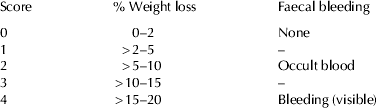
To reflect the general conditions of mice, DAI scores were determined by an investigator blind to the protocol. In the present study, weight loss and faecal bleeding were evaluated and scored individually for each animal according to Table 1. The parameters of weight loss and faecal bleeding were evaluated statistically and the score distribution was followed according to protocol developed by Hamamoto et al. (Reference Hamamoto, Maemura, Hirata, Murano, Sasaki and Katsu1999).
Disease activity scores for weight loss and faecal bleeding
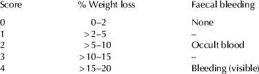
The higher the score, the more severe the symptoms. Faecal bleeding included 3 scores only: 0, 2 and 4
Tissue collection and processing
At sacrifice, the animals were examined and weighed. Their spleen and caecum were removed and weighed. The colon, from the colo-caecal to the anus was excised with its length measured, rinsed with phosphate buffer saline (PBS), pH 7·4 and cut open longitudinally. One cm length of the distal colon was fixed for histopathological examination. The other part of the colon was preserved at −20 °C for MPO assays.
Histopathological analysis
Colon of mice were excised, washed with saline and then fixed for minimum 24 h in 10% natural buffered formalin solution, pH 6·8. The tissue was processed for paraffin embedding and cut into 5-μm thick sections. The sections were stained with haematoxylin and eosin (H&E). The stained sections were examined by light microscope for evidence of colitis using the following criteria: presence of inflammatory cell infiltration, mucin depletion, presence of crypt abscesses, crypt distortion (Rosai, Reference Rosai and Rosai2004); mice fed with Dahi and LaBb Dahi were examined for histological signs of resolution.
Analysis of myeloperoxidase (MPO) activity in colonic tissue
MPO activity was measured as an indicator of neutrophil accumulation in colonic mucosa using previously described method (Bradley et al. Reference Bradley, Priebat, Christensen and Rothstein1982). The tissue were homogenized in 9 volumes of ice cold potassium phosphate buffer (50 mm, pH 6·0) containing 0·5% hexadecyl trimethyl ammonium bromide, sonicated in an ice bath for 10 s, freeze-thawed 3 times and again sonicated for 10 s. The suspension was centrifuged (40 000 g for 15 min) and the supernatant analysed for MPO assay. All reagents and tissue homogenate were brought to 25 °C before performing MPO assay. The tissue homogenate (0·1 ml) was mixed with 2·9 ml of reaction mixture (50 mm potassium phosphate buffer, pH 6·0 containing 0·167 mg/ml of o-dianisidine dihydrochloride and 0·0005% H2O2). The breakdown of H2O2 is directly proportional to oxidation of o-dianisidine dihydrochloride which is measured at 460 nm using Specord 200 double beam UV/visible spectrophotometer (Analytikjena, Germany). One unit of MPO activity was defined as degradation of one micromole of hydrogen peroxide per min at 25 °C and expressed as units/mg protein. Protein concentration was estimated using bovine serum albumin as standard (Lowry et al. Reference Lowry, Rosebrough, Farr and Randall1951).
Stool sample analysis for enzyme assays
Preparation of faecal suspension
Fresh mouse stools (0·5 g) from each group were separately collected in sterilised plastic cups, carefully suspended with 20-fold PBS in a cooled tube, and centrifuged at 250 g for 5 min. The supernatant was again centrifuged at 10 000 g for 20 min. The resulting precipitates were used as the sources for the faecal enzyme assays. All procedures were performed at 4 °C.
β-Glucuronidase assay
The activity of was assayed in faecal suspension according to the method used by Lee et al. (Reference Lee, Lee, Lee, Bae, Lee, Ahn, Lim, Huh and Kim2009). The reaction mixture (1·0 ml), consisting of 0·04 ml 2 mm p-nitrophenyl-β-D-glucuronide, 0·76 ml 0·1 m phosphate buffer (pH 7·0), and 0·2 ml faecal suspension, was incubated for 30 min at 37 °C, and the reaction was terminated by the addition of 1 ml 0·5 m NaOH. The mixture was then centrifuged at 3000 g for 10 min and the absorbance was measured at 405 nm. The activity was expressed as μmoles/h/g.
Statistical analysis
Results are expressed as mean±sd. Analysis of variance was performed using GraphPad PRISM version 5.0 statistical software package and the differences between groups were tested using Tukey–Kramer post-hoc test. Disease activity scores was evaluated statistically using Chi square and exact test Monte Carlo.
Results and discussion
UC is relapsing IBD of colon of unknown aetiology. The pathogenesis of IBD involves an interaction between genetically determined host susceptibility, deregulated immune response, and the enteric microbiota. Microflora in IBD patients becomes aberrant with normal microflora, such as bifidobacterium and lactobacillus, decreased and pathogenic or potential harmful bacteria increased, have been regarded as important pathogenic factor (Cummings et al. Reference Cummings, Macfarlane and Macfarlane2003). So the targeting of abnormal enteric microbiota to decrease the more pathogenic species and enhancing the concentration and metabolic activity of the beneficial species has tremendous potential for therapeutic benefit.
The probiotics, bifidobacteria and lactobacilli, suppress the growth of pathogens by releasing antimicrobial factors that compete with microbial pathogens for the limited number of receptors on epithelial cells (Kruis, Reference Kruis2004; Sartor, Reference Sartor2004). Several studies have demonstrated the beneficial effects of probiotics in the treatment of IBD (Campieri & Gionchetti, Reference Campieri and Gionchetti1999; Collins & Gibson, Reference Collins and Gibson1999). Madsen et al. (Reference Madsen, Doyle, Jewell, Tavernini and Fedorak1999) demonstrated that repopulation of the colonic lumen with control levels of Lactobacillus normalised the defective mucosal adherent and translocated bacterial pattern and prevents the development of colitis.
We observed no differences in body weight between the groups at allocation and at day 0 (ANOVA). In present study, significant weight loss was found at day 17 (day 7 of DSS treatment), wherein all colitis induced control animals presented a significantly lower weight compared with the normal control group. The Dahi fed DSS-induced colitis group showed a less dramatic weight loss that became significantly lower from day 3 of DSS treatment. Mice in the LaBb Dahi group maintained their body weight at its base level with non significant weight loss compared with normal control group (Fig. 1). These results are in agreement with earlier reported findings (Chapman et al. Reference Chapman, Plosker and Figgitt2007; Kamada et al. Reference Kamada, Maeda, Inoue, Hisamatsu, Okamoto, Hong, Yamada, Watanabe, Suchimoto, Ogata and Hibi2008). , When groups were compared statistically, it became evident that the LaBb Dahi group did not differ statistically from the normal control group, while the colitis control and Dahi fed group had comparatively higher disease scores than normal control group. If we can extrapolate these results to a human situation: less faecal bleeding means less blood loss, which in turn implies less haemorrhage and colonic epithelial damage, less intestinal permeability and susceptibility to infections, finally leading to less discomfort and improved conditions for the patient. The lower weight loss and faecal bleeding presented by the LaBb Dahi group could be of clinical importance.
Body weights of experimental mice expressed in mean±sd (n = 12). Differences between the groups were evaluated by ANOVA. a,b Values with different letters within groups are significantly different (P < 0·05). A,B,CValues with different letters between groups are significantly different (P < 0·05).

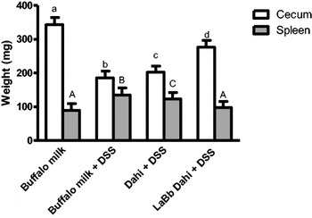
In present study, colitis control and Dahi fed group remained significantly different with respect to organ weight from the normal control group (Fig. 2). The caecal weight of these animals was significantly lighter, whereas the spleen weight was significantly elevated. No significant differences were found between the LaBb Dahi group and the normal healthy control group. The results in present context are in accordance with Herias et al. (Reference Herias, Koninkx, Vos, Huis in't Veld and van Dijk2005) who reported similar observation on protective effect of probiotic Lb. casei on organ weight. Spleen weight increase can be explained by immune activation or infection leading to hyperactivity in the spleen, as this organ is a major site of immune responses and a filter for unwanted foreign substances and aging cells. These results suggest that LaBb Dahi containing Lb. acidophilus LaVK2 and Bifido. bifidum BbVK3 may have immunoregulatory function to maintain and/or prevent intestinal disturbances.
Organ weights of experimental mice expressed in mean±sd (n = 12). Differences between the groups were evaluated by ANOVA. a,b,c,dValues with different letters are significantly different (P < 0·05).

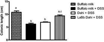
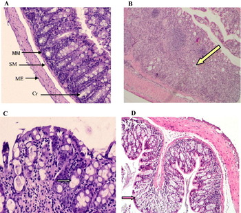
Animals orally administered DSS develop colitis with features similar to symptomatic and histological findings in human IBD. The inflammatory response provoked by DSS is considered to reproduce many of the macroscopic and immunological hallmarks of clinical colitis. We found significant reduction in colonic length of the colitis control and Dahi groups suggesting an acute damage to colon caused by UC (Fig. 3). However, on feeding LaBb dahi, we found non-significant reduction in colonic length as compared with normal control group, representing protective effect of probiotics on the DSS induced colitis. In the present study, histopathological analysis of colonic tissue of colitis control mice showed extensive accumulation of neutrophils, disruption of surface epithelium, loss of goblet cells, complete distortion and loss of crypts (Fig. 4). These changes were less severe in colonic tissue of Dahi fed mice which showed moderate loss of crypts, crypt abscess and infiltration of inflammatory cells. However, histological analysis of the colonic tissue of colitis induced mice fed with LaBb Dahi ameliorated the severity of inflammation, declined accumulation of neutrophils, presumably reflecting attenuation of the tissue disruption and crypt loss.
Length of colon of experimental mice on day 17, expressed in mean±sd (n = 12). Differences between the groups were evaluated by ANOVA. a,b,cValues with different letters are significantly different (P < 0·05).

Longitudinal section of colonic tissue stained with haematoxylin and eosin. (a) Mice fed with buffalo milk having no colitis induced (200×). Micrograph shows normal mucosa (MM), submucosa (SM), muscularis externa (ME) and crypt (Cr). (b) Mice fed with buffalo milk with colitis induced (200×). Micrograph shows severe infiltration of inflammatory cells and complete loss of crypts ( ). (c) Mice fed with Dahi having colitis induced (400×). Micrograph shows neutrophil infiltration, crypt loss and crypt abscess (
 ). (d) Mice fed with LaBb Dahi having colitis induced (100×). Micrograph shows mild inflammation, integrated surface epithelium (
 ) and attenuated crypt cell loss.

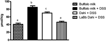
The results of present study revealed that the level of harmful intestinal enzyme β-glucuronidase which produces carcinogenic and cytotoxic metabolites, was significantly elevated in colitis control and Dahi group compared with normal control group (Fig. 5). The feeding of LaBb dahi was able to inhibit the enzyme activity of β-glucuronidase in colonic tissue of colitis induced mice. These results suggest that LaBb Dahi containing Lb. acidophilus LaVK2 and Bifido. bifidum BbVK3 may inhibit the growth of harmful intestinal enzymes. These results are in accordance with a previous study which reported that probiotic Lb. suntoryeus HY7801 inhibits the activity of β-glucuronidase in UC-induced mice (Lee et al. Reference Lee, Lee, Lee, Bae, Lee, Ahn, Lim, Huh and Kim2009).
Effect of feeding LaBb Dahi on glucoronidase activity. Values are mean±sd (n = 12); a,b,c,dValues with different letters are significantly different (P < 0·05).

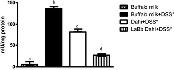
The macroscopic injury caused by DSS challenge was accompanied by a substantial increase in the levels of MPO, an index of neutrophil infiltration, in the colonic tissue of mice that was confirmed by the histological analysis. The results of our study indicated that MPO activity in colonic tissue of colitis control group increased 24 fold of that in normal control group (Fig. 6). The treatment of colitis induced mice with Dahi decreased MPO activity by about 40%. However, the treatment with LaBb Dahi was more efficacious in reducing MPO activity, resulting 80% decline in MPO activity compared with colitis control group, suggesting that LaBb Dahi can relieve the symptoms of experimental colitis by decreasing the infiltration of neutrophils. These observations extend and corroborate with previous studies which reported increased MPO activity, in the colonic mucosa of UC and that decreased after probiotic treatment (Shibolet et al. Reference Shibolet, Karmeli, Eliakim, Swennen, Brigidi, Gionchetti, Campieri, Morgenstern and Rachmilewitz2002; Hegazy & El-Bedewy, Reference Hegazy and El-Bedewy2010).
Effect of feeding Dahi and LaBb Dahi on myeloperoxidase activity in colonic tissue of colitis induced mice.*DSS, dextran sodium sulphate; Values are mean±sd for 6 animals; a,b,c,dValues with different letters are significantly different (P < 0·001).

In conclusion, probiotic Dahi preparation containing Lb. acidophilus LaVK2 and Bifido. bifidum BbVK3 (LaBb-Dahi) developed in our laboratory attenuated the UC related histological damage, body weight loss, sharp rise in MPO activity, β-glucuronidase activity and thus conferred therapeutic effect on DSS induced colitis in mice. Thus probiotic Dahi as adjunctive treatment to conventional therapy may provide a simple and attractive way to treat ulcerative colitis.
The authors thankfully acknowledge the Indian Council of Agricultural Research (ICAR) and Department of Science and Technology (DST), New Delhi for awarding fellowship grants to authors Sagar R. Jadhav and Umesh Kr. Shandilya, respectively.