Introduction
The development of embryo technologies has long challenged scientists to find better strategies and apply them in an efficient and low-cost manner for the purposes of improving animal production (Meirelles et al., Reference Meirelles, Chiaratti, Ferreira, Meirelles and Bressan2013). Although widely used in bovine, the in vitro production of embryos (IVP) continues to have limiting factors. Currently, in vitro maturation (IVM) and fertilisation (IVF) rates are relatively high at 90 and 80%, respectively (Lonergan & Fair, Reference Lonergan and Fair2014); however, the lower rates of embryonic development are generally attributed to the inability of oocytes to support cell multiplication to the blastocyst stage (Camargo et al., Reference Camargo, Viana, Sá, Ferreira, Ramos and Vale Filho2006; Meirelles et al., Reference Meirelles, Chiaratti, Ferreira, Meirelles and Bressan2013).
Thus, the selection of competent oocytes remains a bottleneck in the IVP of mammalian embryos and, to improve blastocyst rate, studies have been conducted to attempt to identify a more precise selection method for competent oocytes. Different indirect approaches have been employed using tools such as molecular biology, fluorescent stains, metabolomics and proteomics (reviewed by Bukowska et al., Reference Bukowska, Kempisty, Piotrowska, Walczak, Sniadek, Dziuban, Brussowm, Jaskowski and Nowicki2011). These techniques, despite great accuracy, are often harmful and usually cause cell death or are prohibitively expensive for commercial applications. Therefore, the morphological selection of oocytes is still commonly used in most IVP laboratories.
However, a promising technique was described by Ericsson et al. (Reference Ericsson, Boice, Funahashi and Day1993) based on the use of brilliant cresyl blue (BCB) stain to determine glucose-6-phosphate dehydrogenase (G6PDH) activity in porcine immature oocytes. Immature oocytes synthesise a variety of enzymes, including G6PDH, and this protein, which is a component of the pentose phosphate cycle, is active in the growing oocyte but is relatively less active in oocytes that have completed growth (Janowski et al., Reference Janowski, Salilew-Wondim, Torner, Tesfaye, Ghanem, Tomek, El-Sayed, Schellander and Hölker2012; Gutnisky et al., Reference Gutnisky, Dalvit, Thompson and Cetica2013).
G6PDH activity degrades the BCB stain, allowing for the identification of oocytes with (BCB–: described as immature oocyte) or without (BCB+: described as competent) enzyme activity (BCB– test) according to cytoplasm colour (for review see Opiela & Katska-Ksiazkiewicz, Reference Opiela and Katska-Ksiazkiewicz2013). Different studies in other species have shown the efficiency of the BCB– test to identify competent oocytes for IVM in goats (Rodríguez-González et al., Reference Rodríguez-González, López-Béjar, Izquierdo and Paramio2003), cows (Pujol et al., Reference Pujol, López-Béjar and Paramio2004; Alm et al., Reference Alm, Torner, Lohrke, Viergutz, Ghoneim and Kanitz2005, Silva et al., Reference Silva, Rodriguez, Galuppo, Arruda and Rodrigues2011), buffalo (Manjunatha et al., Reference Manjunatha, Gupta, Devaraj, Ravindra and Nandi2007), dogs (Rodrigues et al., Reference Rodrigues, Rodriguez, Silva, Cavalcante, Feltrin and Rodrigues2009), rats (Alcoba et al., Reference Alcoba, Braga, Sandi-Monroy, Proença, Lopes and Oliveira2011) and sheep (Wang et al., Reference Wang, Lin, Huang, Wang, Zhao and Chen2012).
The population of bovine oocytes from slaughterhouse-derived ovaries is not homogeneous (Ferreira et al., Reference Ferreira, Vireque, Adona, Meirelles, Ferriani and Navarro2009; Boni, Reference Boni2012), but the proportion of BCB+ and BCB– oocytes following morphological pre-selection (grade 1 and 2 oocytes) ranges from 57.9 to 66.5% BCB+ (Pujol et al., Reference Pujol, López-Béjar and Paramio2004; Alm et al., Reference Alm, Torner, Lohrke, Viergutz, Ghoneim and Kanitz2005, Silva et al., Reference Silva, Rodriguez, Galuppo, Arruda and Rodrigues2011; Janowski et al., Reference Janowski, Salilew-Wondim, Torner, Tesfaye, Ghanem, Tomek, El-Sayed, Schellander and Hölker2012; Castaneda et al., Reference Castaneda, Kaye, Pantaleon, Phillips, Norman, Fry and D´Occhio2013; Mirshamsi et al., Reference Mirshamsi, Karami Shabankareh, Ahmadi-Hamedani, Soltani, Hajarian and Abdolmohammadi2013).
Silva et al. (Reference Silva, Rodriguez, Galuppo, Arruda and Rodrigues2011) have described different patterns of nuclear and cytoplasmic maturation in bovine oocytes during IVM between BCB+ and BCB– oocytes. The BCB+ oocytes undergoing IVM show a synchronous nuclear and cytoplasmic maturation. In contrast, BCB– oocytes that undergo IVM show an asynchronous pattern of nuclear and cytoplasmic maturation.
Usually, oocyte competence is defined as the ability to properly progress through maturation, cleave following fertilisation and support embryonic development to the blastocyst stage (Brevini-Gandolfi & Gandolfi, Reference Brevini-Gandolfi and Gandolfi2001; Blanco et al., Reference Blanco, Demyda, Moreno Millán and Genero2011). Many factors can affect the competence of female gametes including ovarian physiological status, follicular environment, actions of hormones or growth factors, oocyte diameter and culture media (Camargo et al., Reference Camargo, Viana, Sá, Ferreira, Ramos and Vale Filho2006).
Based on these observations, we hypothesised that the presence of BCB– oocytes within the same environment during IVM (within the same medium drop) would impair the competence of BCB+ oocytes to undergo IVM, IVF and IVC.
Materials and methods
Chemicals and media
Media for IVM, IVF and IVC were obtained from Biotecnologia Animal® (Brasília, Brazil). Fetal calf serum (FCS) was purchased from Nutricell® (Campinas, SP, Brazil). Modified phosphate-buffered saline (PBS) and BCB stain solutions were assembled in our laboratory using an ultrapure water system (Millipore, Bedford, MA, USA) and chemicals were purchased from Sigma (St. Louis, MO, USA) unless specifically stated otherwise.
Oocyte collection
Bovine ovaries were obtained from a local slaughterhouse and transported to the laboratory in PBS within 3 h of slaughter and maintained at 32–34°C. Once in the laboratory, the ovaries were washed in PBS and COCs were aspirated from follicles (3–8 mm in diameter) using an 18-gauge needle attached to a 10-ml syringe. After 20 min (to allow sedimentation to occur), the pellet containing COCs was re-suspended in PBS containing 0.4% BSA (mPBS) and examined under a stereomicroscope (×15 magnification) to recover COCs. Only COCs with a compact cumulus mass with more than three layers were selected for the experiment (morphological pre-selection – grades 1 and 2; according to Leibfried & First, Reference Leibfried and First1979).
After morphological selection, COCs were grouped randomly into the designed control and experimental groups.
Brilliant cresyl blue test and in vitro maturation
COCs were exposed to 26 μM BCB in mPBS for 90 min at 38.5ºC in an incubator with atmospheric air and saturated relative humidity. Afterwards, oocytes were washed three times in mPBS (without BCB stain), observed using a stereomicroscope (×40 magnification) with incident light and then selected based on their cytoplasm colouration: BCB+ oocytes had a blue cytoplasm colour, and BCB– oocytes had a colourless cytoplasm. After being classified according to the BCB test, COCs were distributed randomly into the experimental groups. The COCs within all groups were washed three times in IVM medium (Bio Biotecnologia Animal®, Brasília, Brazil) containing 10% FCS and cultured in drops containing 3 μl of IVM medium per oocyte under mineral oil during 24 h in an incubator at 38.5°C with 5% CO2 in atmospheric air and saturated relative humidity.
In vitro fertilisation (IVF)
Straw (0.5 ml) containing bovine semen that was previously IVP tested was thawed and immediately evaluated for sperm progressive motility and vigour. Next, spermatozoa were selected using the swim-up procedure. Briefly, semen was divided into two parts (approximately 240 μl), and each part was gently placed on the bottom of a Falcon tube (15 ml) containing 1 ml Sperm Selection Medium (SSM; Bio Biotecnologia Animal®, Brasilia, Brazil) containing 0.6% BSA. After 1 h, 900 μl of the supernatant medium from both tubes were mixed and washed in 1 ml SSM and then centrifuged at 300 g for 10 min. The pellet was collected and diluted in IVF medium (Bio Biotecnologia Animal®, Brasilia, Brazil) containing 0.6% BSA and was exposed to heparin–hypotaurine–epinephrine solution (HHE) for 15 min in an incubator at 38.5°C with 5% CO2 in air and saturated relative humidity to induce sperm capacitation. While the spermatozoa were capacitated, the COCs were washed three times in IVF medium (Bio Biotecnologia Animal®, Brasília, Brazil) containing 0.6% BSA and were placed into IVF medium drops (80 μl) under mineral oil based on each experimental group. In total, 1 × 10⁶ spermatozoa were added to the IVF medium drops containing the COCs and co-cultured (D0) for 22 h at 38.5ºC in atmosphere containing 5% of CO2 in air and saturated relative humidity.
In vitro culture (IVC)
After IVF, presumptive zygotes were stripped of cumulus cells, washed individually into 100 μL of IVC medium (Biotecnologia Animal®, Brasilia, Brazil) containing 10% FBS, and then transferred to holes of the Well-the-Well culture system (WOW) for 7 days at 38.5ºC in a controlled atmosphere (5% CO2, 5% O2, 90% N2) and saturated humidity. The cleavage rate was observed 44 h following IVF, and all 2-cell embryos were cultured until they reached the blastocyst stage (D7).
Experimental design
First experiment (three replicates)
The aim of this experiment was to observe if oocytes directly submitted to IVM show similar cleavage and blastocyst rates as those obtained with the oocytes maintained under the same in vitro conditions as the oocytes that undergo the BCB test. Selected COCs were submitted to IVM after been morphologically selected and randomly grouped into three groups: (1) directly submitted to IVM; (2) oocytes submitted to BCB test without the addition of BCB stain (BCB control group); and (3) submitted to BCB test.
Second experiment (six replicates)
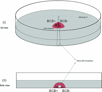
The aim of this experiment was to determine the cleavage and blastocyst rates obtained from BCB+ oocytes undergoing IVM in the presence (paracrine effect) of BCB– oocytes at ratio of 10:1. For this purpose after morphological selection COCs were first divided into two groups: directly undergo IVM and submitted to the BCB test before IVM. After the BCB test, the COCs were classified as either BCB+ (blue cytoplasm) or BCB– (colourless cytoplasm), and finally distributed randomly into four experimental groups: two groups matured in different IVM drops, BCB+ and BCB–, and two paracrine groups BCB+ and BCB– matured within the same IVM drop medium but separated by a heat-sealed glass microcapillary, at a 10:1 (BCB+:BCB–) ratio, (Fig. 1).
Scheme for IVM in which BCB+ oocytes were maintained separate from BCB– oocytes using the same medium drop. To create a closed glass microcapillary, the extremities were closed by flame; the microcapillary was sterilised by flame and then gently placed into the medium drop (80 μl) under mineral oil. This procedure allowed us to divide the drop environment into two compartments (I) while maintaining a common medium between them via the top of the drop (II).

Statistical analysis
Data analysis was performed by using Statistical Analysis Software 9.1 (SAS Institute, Cary, NC). The proportions of BCB+ and BCB– oocytes were expressed as 95% confidence intervals. Cleavage data were tested by the chi-squared test (P = 0.05) and blastocyst data were tested using Fisher's test (P = 0.05).
Results
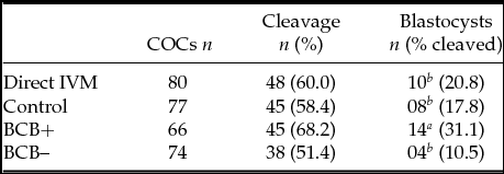
During the first experiment, we observed that oocytes submitted directly to IVM showed similar (P > 0.05) cleavage and embryonic development rates to the blastocyst stage as the results obtained with the BCB control group oocytes (Table 1). Therefore, we did not replicate this group in the subsequent experiment, which provided additional oocytes for the BCB test.
Influence of the BCB test on the further development of bovine oocytes
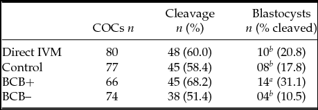
a,bP ≤ 0.05.
In the following steps after morphological selection, 805 COCs were exposed to BCB stain, and 543 (67.5%) maintained the blue stain within cytoplasm (BCB+); 262 (32.5%) were classified as having colourless cytoplasm (BCB–).
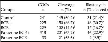
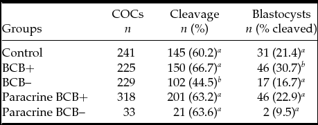
Table 2 shows the capacities of oocytes after IVF to achieve cleavage and to promote the development of the embryo to the blastocyst stage. Regarding the cleavage rate, only the BCB– group (102/229; 44.5%) was less than the other experimental groups. The BCB+ oocytes submitted to IVM alone without BCB– paracrine effects produced the greatest number of blastocysts (46/150; 30.7%). Between the other oocyte groups, there were no differences in the capacity to reach the blastocyst stage after IVF and IVC.
Cleavage and embryonic development rates obtained from in vitro matured oocytes according to the BCB test
a,bP ≤ 0.05.
Discussion
The results of our first experiment showed that the oocytes submitted directly to IVM or maintained in the same conditions as the oocytes submitted to BCB test without the addition of BCB stain (control group) did not differ in their ability to support embryonic development to the blastocyst stage (Table 1). This observation is in agreement with the data published by Alm et al. (Reference Alm, Torner, Lohrke, Viergutz, Ghoneim and Kanitz2005), which also showed no differences in the capacity of bovine oocytes from directly in vitro matured versus holding groups to progress through IVM, IVF and IVC.
The quality of oocytes from slaughterhouse-derived ovaries is extremely variable (Boni, Reference Boni2012), especially due to follicular status (Vassena et al., Reference Vassena, Mapletoft, Allodi, Singh and Adams2003), oocyte diameter (Fair, Reference Fair2010) and other factors (reviewed by Boni, Reference Boni2012). However, when the BCB test is employed, the proportion of BCB+ and BCB– oocytes is relatively consistent at approximately 60% and 40%, respectively, independent of bovine donor age (calves or adults) (Pujol et al., Reference Pujol, López-Béjar and Paramio2004; Alm et al., Reference Alm, Torner, Lohrke, Viergutz, Ghoneim and Kanitz2005; Janowski et al., Reference Janowski, Salilew-Wondim, Torner, Tesfaye, Ghanem, Tomek, El-Sayed, Schellander and Hölker2012; Castaneda et al., Reference Castaneda, Kaye, Pantaleon, Phillips, Norman, Fry and D´Occhio2013; Mirshamsi et al., Reference Mirshamsi, Karami Shabankareh, Ahmadi-Hamedani, Soltani, Hajarian and Abdolmohammadi2013).
In a previous study conducted in our laboratory, Silva et al. (Reference Silva, Rodriguez, Galuppo, Arruda and Rodrigues2011) observed that 65% of the oocytes selected for IVM were BCB+, which is similar to the percentages obtained in our experiment (67% BCB+ oocytes); this ratio was similar to the result described by Pujol et al. (Reference Pujol, López-Béjar and Paramio2004). Pujol et al. (Reference Pujol, López-Béjar and Paramio2004) also correlated the data obtained from the BCB test with oocyte morphology and observed that grades 1 and 2 oocytes had a greater proportion of BCB+ (78.6 and 66.2%, respectively) than BCB– oocytes.
Alm et al. (Reference Alm, Torner, Lohrke, Viergutz, Ghoneim and Kanitz2005) suggested that the oocyte retrieval technique (slicing versus aspiration) could be an important factor in determining the percentage of BCB+ oocytes; the slicing technique leads to the collection of more growing and consequently more BCB– oocytes. However, these authors compared their data (in bovine) to the oocyte retrieval procedure in swine, and this was not confirmed by our data or the literature (Pujol et al., Reference Pujol, López-Béjar and Paramio2004; Silva et al., Reference Silva, Rodriguez, Galuppo, Arruda and Rodrigues2011; Janowski et al., Reference Janowski, Salilew-Wondim, Torner, Tesfaye, Ghanem, Tomek, El-Sayed, Schellander and Hölker2012; Castaneda et al., Reference Castaneda, Kaye, Pantaleon, Phillips, Norman, Fry and D´Occhio2013; Mirshamsi et al., Reference Mirshamsi, Karami Shabankareh, Ahmadi-Hamedani, Soltani, Hajarian and Abdolmohammadi2013).
When we artificially increased the percentage of BCB+ oocytes in an IVM drop from 60–90% in an attempt to induce competence in BCB– oocytes through a paracrine effect, we observed that the cleavage rates of BCB+ and BCB– oocytes were similar. When we observed the BCB– oocyte group submitted to IVM without the presence of BCB+ (paracrine effect), the cleavage rate was reduced (Table 2). Different from this observation, the BCB– oocyte group in one of our previous experiments (Silva et al., Reference Silva, Rodriguez, Galuppo, Arruda and Rodrigues2011) showed similar cleavage rate as the control and BCB+ oocyte groups.
The interactions between oocytes and other cells have been widely studied, especially between somatic cells and gametes, for example by oocyte-secreted factors (Suttom et al., Reference Suttom, Cetica, Beconi, Kind, Gilchrist and Thompson2003; Hussein et al., Reference Hussein, Thompson and Gilchrist2006). Undefined factors secreted by BCB+ oocytes could positively affect the cleavage rate of BCB– oocytes maturing within the same drop. The decreased cleavage rates of BCB– oocytes maturing alone could be explained by a reduction in the activity of maturation-promoting factor (MPF) detected in this oocyte class (Catalá et al., Reference Catalá, Izquierdo, Uzbekova, Morató, Roura, Romaguera, Papillier and Paramio2011). According to the data of Salamone et al. (Reference Salamone, Damiani, Fissore, Robl and Duby2001), the acquisition of meiotic and possibly developmental competence by bovine oocytes might be related to changes in the quantity or phosphorylation states of ooplasmic kinases, among these by MPF and mitogen-activated protein kinase (MAPK); however, MAPK has not been studied in oocytes classified using the BCB test.
In observing the capacity of oocytes (BCB+ or BCB–) to promote embryo development until the blastocyst stage, our data are similar to the data described by different authors (Silva et al., Reference Silva, Rodriguez, Galuppo, Arruda and Rodrigues2011, Janowski et al., Reference Janowski, Salilew-Wondim, Torner, Tesfaye, Ghanem, Tomek, El-Sayed, Schellander and Hölker2012; Mirshamsi et al., Reference Mirshamsi, Karami Shabankareh, Ahmadi-Hamedani, Soltani, Hajarian and Abdolmohammadi2013). Looking at the rate of embryonic development, the BCB+ oocytes submitted to IVM alone showed the greatest blastocyst number, which differs from the BCB+ oocytes that were submitted to IVM at a ratio of 10:1 within the same environment as BCB– oocytes (Table 2). The presence of BCB– oocytes together with BCB+ during IVM (control and paracrine groups) probably has reduced the capacity of BCB+ oocytes to achieve the blastocyst stage during IVC. These data suggest possible different negative effects from BCB– oocytes that impair the developmental capacity of BCB+ oocytes to support embryo development to the blastocyst stage. Interesting is the observation that blastocyst development rate obtained in the control group was not different from the results obtained with the paracrine BCB+, paracrine BCB– and BCB– oocytes (Table 2). Different from that observed regarding cleavage rates, here the presence of BCB+ oocytes during IVM had no effect over the capacity of BCB– oocytes to promote embryo development until blastocyst stage. In our view the presence or absence of BCB+ in the same IVC system during maturation as the BCB– has no effect over BCB– oocytes' capacity to undergo embryo development after cleavage until blastocyst stage. It is possible that an apoptotic factor could be involved, whose roles in oocyte maturation have been controversial and is usually described as a negative factor (Opiela et al., Reference Opiela, Katska-Ksiazkiewicz, Lipinski, Słomski, Bzowska and Rynska2008). These authors also have pointed out that oocytes submitted to BCB staining show the tendency to apoptosis. Moreover these apoptotic-related genes at the protein level did not confirm this conclusion. However, this pathway also plays an important role in influencing oocyte developmental competence in the surrounding follicular cells (Warzych et al., Reference Warzych, Pers-Kamczyc, Krzywak, Dudzinska and Lechniak2013) and in stimulating the oocytes to acquire developmental competence (Janowski et al., Reference Janowski, Salilew-Wondim, Torner, Tesfaye, Ghanem, Tomek, El-Sayed, Schellander and Hölker2012).
The capacity of oocytes to acquire competence can be affected only when a threshold of apoptosis (a still undefined apoptotic index) is surpassed (Zeuner et al., Reference Zeuner, Müller, Reguszynski and Jewgenow2003). Recently Warzych et al. (Reference Warzych, Pers-Kamczyc, Krzywak, Dudzinska and Lechniak2013) showed that there is no relationship between the morphology of COCs and the apoptotic index of cumulus cells.
Opiela et al. (Reference Opiela, Katska-Ksiazkiewicz, Lipinski, Słomski, Bzowska and Rynska2008) carried out an experiment to determine the relationship between apoptotic (Bax), anti-apoptotic (Bcl-2) genes and G6PDH activity. The authors were not able to identify a relationship between the activity of G6PDH and the expression of the Bcl-2 or Bax proteins in immature and mature oocytes. In contrast, Janowski et al. (Reference Janowski, Salilew-Wondim, Torner, Tesfaye, Ghanem, Tomek, El-Sayed, Schellander and Hölker2012), in studying granulosa cell gene expression, observed that competent oocytes show an increased degree of late apoptotic events compared to non-competent oocytes. Taking both of these observations into account, our data suggest a possible apoptotic cascade that occurs in non-competent oocytes could induce the same phenomenon in competent oocytes, inhibiting their further developmental capacity.
Briefly, BCB– oocytes could be helped by BCB+ in their progression through MIV, FIV and the first embryo cell cycles (cleavage rate). However, the presence of BCB– oocytes during IVM may have affected the embryonic developmental capacity of BCB+ oocytes. Our results suggest that the BCB test is an efficient tool for selecting competent oocytes and that it can be used to increase the number of blastocysts produced by IVP, to separately submit BCB+ and BCB– oocytes to IVM and to avoid negative in vitro interactions between competent and non-competent oocytes. However, Bols et al. (Reference Bols, Jorssen, Goovaerts, Langbeen and Leroy2012) reported that, although double COC screening (morphological pre-selection and BCB test) resulted in a substantial increase in the number of blastocysts, the final absolute number of blastocysts did not increase.
In conclusion, submitting BCB+ oocytes that were separated from BCB– oocytes to IVM increases the rate of embryonic development to the blastocyst stage when compared to the control group, BCB– oocyte group, BCB+ paracrine group and BCB– paracrine group. The presence of non-competent oocytes during IVM, even in low proportion (1:10), reduces the capacity of competent oocytes to undergo embryo development and achieve blastocyst stage during IVC.
Acknowledgements
The authors thank the National Council for Scientific and Technological Development (CNPq), the Coordination for the Improvement of Higher Education Personnel (CAPES) for the provided grant no. 308505/2009–3 and fellowships and the Biotecnologia Animal Company for kindly providing the culture media.