Introduction
Germinal cells play key roles in heredity, the differentiation of these cells requires complex and specific structural architecture dynamics of their genetic material.
One particular aspect of amphibian oogenesis is the formation of the lampbrush chromosomal phenotype during the major phase of oocyte growth and differentiation (Macgregor, Reference Macgregor1993). Lampbrush chromosomes are considered uniquely valuable tools in the study of development of their two major characteristics: (i) they are transient structures that are formed during an prolonged diplotene of the first meiotic division; and (ii) their most functional conspicuous feature is widespread RNA transcription, which take place on hundreds, and in some cases thousands, of loops that are laterally projected from the main axis of the bivalent at short intervals along its entire length. Therefore, lampbrush chromosomes are good experimental models for the study of chromosome organization and gene expression during meiotic prophase (Macgregor, Reference Macgregor1993).
The early diplotene nucleus is also characterized by the formation of a large number of extrachromosomal nucleoli (up to 1500, in Xenopus), which further are distributed beneath the nuclear membrane (Barsacchi et al., Reference Barsacchi, Bussoti and Mancino1970; Steopoe et al., Reference Steopoe, Gavrilă, Mihăescu and Comşa1985). These nucleoli are the morphological expression of a phenomenon known as specific ribosomal gene amplification (Brown & Dawid, Reference Brown and Dawid1970), an event controlled by certain chromatin changes that trigger local decondensation and remodelling in order for the nuclear machinery to progress through the signalling and transcription processes (Grant, Reference Grant2001). An emerging theme in the field of chromatin research has relied on the role of histone post-translational modifications in nuclear function regulation.
One of the most important landmarks in the chromatin field during the last two decades has been the dynamic view, which states that histones are not merely passive structural players but rather they have an important functional role in the modulation of gene expression (Grunstein, Reference Grunstein1990; Wolfe, Reference Wolffe1992). Research over the past decade has revealed that their chromatin structure is far more than a static carrier of the genetic information encoded in their DNA, as it actively and dynamically mediates regulation of processes that require protein access to DNA, i.e. transcription, recombination and replication. Emerging evidence indicates that epigenetic factors control and regulate most nuclear processes. These factors alter chromatin structure by covalent DNA modifications, covalent histone modifications and nucleosome repatterning. These changes seem to be interpreted by proteins that recognize a particular modification and facilitate the appropriate downstream biological effect (Turner, Reference Turner1991, Reference Turner1993; Ausio, Reference Ausio1992; Bird, Reference Bird1996; Strahl & Allis, Reference Strahl and Allis2000; Jenuwein & Allis, Reference Jenuwein and Allis2001; Lusser, Reference Lusser2002; Jaenish & Bird, Reference Jaenish and Bird2003; Santoro, Reference Santoro2005).
The nucleolus, the site of ribosomal precursor synthesis and ribosome assembly, is an example of an essential nuclear organelle (Peng & Karpen, Reference Peng and Karpen2007). In somatic cells, the nucleolar organizing region (NOR) contains tandemly repeated ribosomal RNA genes (rDNA), and is embedded in heterochromatin in most eukaryotes. Single rDNA genes inserted into euchromatin are able to form mini-nucleoli through a self-assembly process that is probably initiated by rDNA transcription (Karpen et al., Reference Karpen, Schaefer and Laird1998). The genes that encode rRNA are tandemly repeated at one or few chromosomal loci in eukaryotes. The number of rRNA gene repeats varies greatly among organisms, ranging from less than 100 to more than 10 000 (Long & Dawid, Reference Long and Dawid1980). One might imagine that the presence of large numbers of rRNA gene copies reflects a demand for higher levels of rRNA synthesis during rapid growth of early embryo. In somatic cells only a fraction of these repeats is used for rRNA synthesis at any given time. In metabolically active human or mouse cells, approximately half of the 400 rRNA gene copies are transcriptionally active, and the other half are silent (Conconi et al., Reference Conconi, Widmer, Koller and Sojo1989; Santoro et al., Reference Santoro, Li and Grummt2002; Lawrence et al., Reference Lawrence, Early, Pontes, Silva, Chen, Neves, Viegas and Pikaard2004).
Active and silent genes are distinct from one another with respect to their chromatin configurations; active genes are located in euchromatin, whereas silent genes are usually located in heterochromatin (Santoro et al., Reference Santoro, Li and Grummt2002). Recent studies have established that epigenetic mechanisms are involved in marking the transcriptional state of rRNA genes in human and mouse somatic cells, which is transmitted to daughter cells in order to assure a correct chromatin conformation dynamics. The promoter of active rRNA genes is free of CpG methylation and is associated with acetylated histones, an opposite pattern being predominant among silent genes (Lawrence et al., Reference Lawrence, Early, Pontes, Silva, Chen, Neves, Viegas and Pikaard2004). The epigenetic mechanism of rRNA gene silencing was extensively studied for the nucleolar dominance phenomenon (Reeder, Reference Reeder1985; Lawrence et al., Reference Lawrence, Early, Pontes, Silva, Chen, Neves, Viegas and Pikaard2004; Santoro & Grummt, Reference Santoro and Grummt2005; McStay, Reference McStay2006), which is common in plant and animal hybrids, whereby rRNA genes inherited by one parent are transcribed and those from the other parent are silent.
In contrast to rRNA genes from somatic cells, epigenetic networks that mediate transcriptional state of rDNA from gametes have been poorly studied. The aim of this study was to investigate amphibian oocyte chromatin remodelling process from genome level to 28S–5.8S–18S ribosomal gene promoter. This approach (using complex methodology that included a variation on the chromatin immunoprecipitation assay (RE-ChIP), a transmission electron microscopy technique, immunohistochemistry and the electrophoretic analysis of histones) explained a relationship between chromatin structure and DNA function, thereby suggesting certain aspects of epigenetic regulation of the gene expression and molecular signalling during oogenesis.
Materials and Methods
Animals
Female newts (Triturus cristatus) were obtained from Bucharest Botanical Garden Pool, University of Bucharest. Animals were sacrificed in accordance with stipulations of European Council law (86/609/CEE/24.11.2004) regarding the protection of animals used for experimental and scientific aims.
Acridine orange (AO) staining of oocyte nucleoli
A small piece of ovary was placed in a mixture of five parts 0.1 M KCl and one part 0.1M NaCl. This mixture contained these two cations in the ratio that was found within the oocytes nucleus. The cell was enucleated and the nucleus was transferred to a flat-bottomed well slide, previously filled with chromosome isolation medium (five parts 0.1 M KCl to one part 0.1M NaCl, with formaldehyde added to a final concentration of 0.5%). After spreading the nuclear contents, the samples were fixed with a mixture of methanol and acetic acid 3 : 1 (v/v), and after 10 min washed and stained with AO 0.01%. Acridine orange interacts with DNA and RNA by intercalation or electrostatic attraction respectively. DNA-intercalated AO fluoresces green and RNA electrostatically bound to AO fluoresces red. The samples were examined with an Olympus fluorescence microscope.
Electron microscopy
Vitellogenic oocytes were collected and enucleated directly in 2.5% glutaraldehyde in 0.1 M sodium cacodylate buffer. Electron microscopy technique was performed as described previously (Burlibaşa & Gavrilă, Reference Burlibasa and Gavrila2005). The samples were examined with a Philips 201 electron microscope.
Extraction of oocyte histones
For extraction of histones from previtellogenic and vitellogenic oocytes of T. cristatus, frozen oocytes from a single animal were resuspended in approximately 0.5 ml of 0.15 M NaCl, 10 mM Tris–HCl pH 7.5 buffer containing 0.2 mM phenylmethylsulfonylfluoride (PMSF). The samples were then disrupted in this buffer using an homogeniser. The homogenate was spun down for 10 min at 16 000 g at 4 °C, in a centrifuge. The pellets were re-suspended in buffer (0.5 ml 0.15 M NaCl, 10 mM Tris–HCl, 0.2 mM PMSF, 0.5% Triton X-100) and were homogenized and centrifuged for 10 min at 16 000 g at 4 °C. This step was usually repeated twice. The final pellets were resuspended in approximately 0.8 ml of 0.4 N HCl, homogenized and kept at 4 °C with occasional stirring. The HCl extracts were next spun down at 16 000 g in the Eppendorf microfuge for 10 min. The supernatants thus obtained were then precipitated with 5 volumes of acetone for 1–2 h at –20 °C. The tubes were centrifuged using the above procedure and the pellets were dried and then re-suspended in distilled water.
Electrophoretic fractionation of histones
Sodium dodecyl sulfate-polyacrylamide gel electrophoresis (SDS-PAGE) was carried out in 12.5% slab gels, pH 6.8, with 1.5 M Tris–HCl running buffer, pH 8.8, according to the Laemmli system. Unfractionated histones (1 mg/ml) were dissolved in the tray buffer, which contained 0.125 M Tris–HCl, 4% SDS, 20% v/v glycerol, 0.2 M DTT and 0.02% bromophenol blue, pH 6.8. The gel was run at a constant voltage 10 V/cm. After electrophoresis, gels were stained with 0.025% (w/v) Coomassie blue in 40% (v/v) methanol/7% (v/v) acetic acid and distained in 40% (v/v) methanol/7% acetic acid (v/v). Histones were fractionated comparatively with a lysine-rich histone marker (f1 fraction – Sigma), an arginine-rich histone fraction (f3 fraction – Sigma) and a molecular weight marker (Sigma) ranging between 11.2 kDa and 66 kDa. The gels were scanned with Jencons-PLS UVB Bioimaging System.
Immunohistochemistry
Acetylated histone H4 labelling was performed using rabbit polyclonal antibody against the penta-acetylated peptide corresponding to histone H4 (Upstate Biotechnology). Fragments of newt ovary were fixed in Bouin solution dehydrated in ethanol, cleared in toluene and embedded in paraffin. Immunohistochemistry was performed on 7-μm thick sections as described previously (Zarnescu, Reference Zărnescu2007).
Chromatin immunoprecipitation (ChIP) and RE-ChIP assays
The following antibodies were used in this study: (1) rabbit anti-histone H4, hyperacetylated (Penta) polyclonal antibody (Upstate Biotechnology NY); (2) anti-ubiquityl-histone H2A, clone E6C5, mouse IgM, (Upstate Biotechnology NY); and (3) anti-5′-methyl cytosine, sheep IgG (Biomol).
ChIP studies were performed using a Quick ChIP Kit (Imgenex) according to a variant adapted by us from the Quick ChIP Kit user manual. All the steps were performed at 4 °C. Immature (previtellogenic) and vitellogenic oocytes (defolliculated previously) were incubated for 10 min with 1% formaldehyde by gentle agitation. Cross-linking was stopped by the addition of 0.125 M glycine for 10 min with gentle agitation at room temperature. The cells were then washed with phosphate-buffered saline (PBS), scrapped off in PBS and centrifugated at 1000 g for 5 min. The cell pellet was resuspended in lysis buffer containing SDS, a mixture of proteinase inhibitors (10× PIC) (Quick ChIP kit) and 10× PMSF and then further incubated for 10 min on ice. The cell extract was collected by centrifugation at 1000 g for 10 min, resuspended in washing buffer A and homogenized with an equal volume of washing buffer B. The mixture was then centrifuged at 1000 g for 15 min. The cell pellet was resuspended in NUC buffer (15 mM HEPES pH 7.5, 60 mM KCl, 15 mM NaCl, 0.13 mM sucrose, 0.15 mM β-mercaptoethanol, 0.15 mM spermine, 0.5 mM spermidine) and then CaCl2 (3 mM final concentration) and 50 UI/ml MNase (micrococcal nuclease) (Biomol) were added. The reaction was stopped with an equal volume of Buffer X (90 mM HEPES pH 7.9, 220 mM NaCl, 10 mM EDTA, 2% Triton X-100, 0.2% SDS, 0.5 mM PMSF) containing 10 μl 10× PIC. After centrifugation at 16 000 g, the supernatant was collected and mixed with dilution buffer and 10 μl of 10× PIC (Quick ChIP kit). An aliquot was used for A260 measurements. Cross-linked extracts (10 A260 units) were resuspended in 75 μl salmon sperm DNA/protein G Agarose (Quick ChIP kit), early blocked with bovine serum albumin (BSA) (Sigma) and were incubated for 30 min at 4 °C by agitation. After centrifugation at 1000 g for 2 min, the supernatant was collected and aliquots of 10 μl were stored at 4 °C. After centrifugation at 1000 g for 2 min, the supernatant was collected and immunoprecipitated with anti-histone H4 hyperacetylated (Upstate Biotechnology) by mixing overnight at 4 °C. The immunocomplexes were recovered with the addition of 60 μl salmon sperm DNA/protein G agarose and subsequent incubation for 1 h at 4 °C with agitation. The complexes were washed with buffer A and centrifuged at 1000 g for 1 min. The pellets were resuspended and washed with buffer B, centrifuged and the procedure repeated for washing with buffer C (Quick ChIP kit) and buffer D (Quick ChIP kit). The protein–DNA complexes were then eluted by incubation with 250 μl of elution buffer (50 mM NaHCO3 and 1% SDS) for 15 min at 65 °C. The beads were incubated with an equal volume of 10 mM DTT for 30 min at 37 °C, and then centrifuged at 1200 g for 1 min and the supernatant was transferred into a new tube. The samples were diluted 40 times with elution buffer and 10% of the sample was kept as an input sample (positive control for first antigen presence) and processed by adding the second antibody, anti-ubiquityl-histone H2A, clone E6C5, respectively anti-5′-methyl cytosine. The protein–DNA complexes were then eluted by incubation in 250 μl of elution buffer (50 mM NaHCO3 and 1% SDS) for 15 min at 65 °C, then centrifuged at 1200 g for 1 min and the supernatant was transferred into a new tube. After centrifugation at 1000 g for 5 min, the supernatant was collected and incubated with 10 μg/ml RNase A for 1 h at 42 °C. Then NaCl was added to mixture to a final concentration of 200 mM and incubated at 65 °C to reverse cross-linking. The proteins were then digested with 200 μg/ml proteinase K for 2 h at 50 °C. The DNA was recovered by phenol/chloroform extraction and ethanol precipitation. PCR amplification of a region of 544 bp from ribosomal genes promoter −85 to +395 has been performed using primers having sequences chosen from NCBI accession numbers X02995 and X98876 (forward 5′-ATTAAGGTCCGCCGGACT-3′ and reverse 5′-GCCCTCGCTCGGATAAAA-3′) and AmpliTaq polymerase (Biomol), in a Perkin Elmer thermocycler. The following PCR programme was set up: initial denaturation 94 °C for 5 min, 25 cycles × (94 °C/60 s, 53 °C/60 s, 72 °C/60 s) and final elongation for 7 min at 72 °C. Two control samples: positive control (input) sample – for the presence of H4 hyperacetylated and negative control sample were run through each step of the ChIP procedure without antibody. PCR products were run on an agarose 1% gel.
Results
Acridine orange staining of vitellogenic oocyte nucleoli
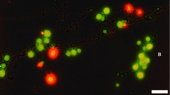
The multinucleolar state of the vitellogenic oocytes nucleus was evidenced by spreading isolated nuclei using a saline medium technique and acridine orange staining. The nucleolar organizer region (NOR) is the main structure that is involved in the synthesis of high-molecular-weight rRNAs and in the assembly of ribosomes. Different nucleoli have different fluorescent colours in accordance with their composition (DNA – yellow/green; RNA – red/orange). Fluorescent staining revealed a ‘string of beads’ structural pattern for the extrachromosomal nucleoli (Fig 1).
Acridine orange staining of nucleoli isolated in saline solution from Triturus cristatus fully grown oocytes (red – RNA, yellow – proteins). Extrachromosomal nucleoli of newt oocytes are arranged in ‘beaded rings’ (bar: 5 μm).

Ultrastructural analysis of a lampbrush loop
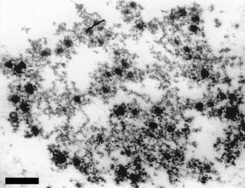
We have studied electronmicroscopically nuclei that were isolated from vitellogenic oocytes. The multiple ribonucleoproteins (RNP) complexes are distributed at the lampbrush loops (Fig. 2). Fine sections were visualized using TEM, these showed that the lateral loops are actively transcribed and their profiles are associated with the richness of RNP particles, representing the transcription products.
Electron micrograph of a nucleus from Triturus cristatus oocyte. A putative folded transcriptionally active lampbrush loop is evidenced. Multiple RNP particles are randomly distributed over almost whole length of the lampbrush loop (bar: 0.5 μm).

Electrophoretic fractionation of previtellogenic and vitellogenic oocytes histones
Previous literature had suggested that linker histones and their variants might play a specific role in transcription regulation (Zlatanova, Reference Zlatanova1990), therefore the histone content pattern during oogenesis was studied. Histones from previtellogenic and vitellogenic oocytes were isolated and fractionated.
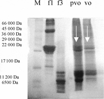
The electrophoregram indicated the presence of nucleosomal core histones (H2A, H2B, H3 and H4), both in previtellogenic and vitellogenic oocytes, and were seen as four bands that exhibited the same electrophoretic mobility as the f3 fraction (Fig. 3).
Triturus cristatus: 12.5% SDS-polyacrylamide gel electrophoresis of histones from previtellogenic (pvo) and vitellogenic (vo) oocytes. M, molecular weight marker; f1, lysine rich histone marker (H1); f3, arginine rich histone fraction (nucleosomal core histones). The electrophoregram indicates the presence of four variants of linker histone H1 (pvo line) in previtellogenic oocytes and two variants of linker histone (vo line) in vitellogenic oocytes.

In previtellogenic oocytes, the electrophoretic pattern indicated the presence of four variants of H1 histone (pvo line), whereas, for vitellogenic oocytes, isolated histones indicated the presence of two variants of H1 histone (vo line). These results suggest a clear variation in the linker histone pattern in previtellogenic and vitellogenic oocytes. Stages III and IV of vitellogenic oocytes are cytological correspondents of the lampbrush chromosome phenotype involved in amplification of the ribosomal genes and of their high transcriptional activity.
Immunohistochemical investigations of the hyperacetylation of histone H4 in vitellogenic oocytes
The aim of the next set of experiments was to investigate the relationships between H4 hyperacetylation and the specific regions of transcriptional activity within chromatin of newt oocytes. A study of the immunohistochemical distribution of H4 hyperacetylated in vitellogenic oocytes (stages II–IV) and oocyte stages, correlated with the lampbrush phenotype, was performed.
Immunodetection of hyperacetylated histone H4 (tri- and tetra-acetylated forms of histone H4) on histological preparations of crested newt ovary revealed a characteristic nuclear staining.
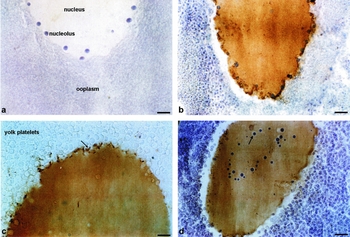
In the oocyte nucleus there are numerous extrachromosomal nucleoli with preferential distribution at the nuclear periphery (Fig 4a, b). From such extrachromosomal multiple nucleoli, smaller nucleolar bodies are released and that are ready to pass through the nuclear envelope into the cytoplasm (Fig. 4c).
Immunohistochemistry for hyperacetylated H4 from vitellogenic oocytes of Triturus cristatus (a, b, d – counterstained with Harris' haematoxylin). (a) Negative control for immunohistochemical reactions; early vitellogenic oocytes; numerous extrachromosomal nucleoli with a preferential distribution at the periphery of the nucleus are evidenced. (c–d) Immunostaining for H4 hyperacetylated revealed a homogeneous nucleoplasm pattern with a massive concentration of signals for highly acetylated H4 at the periphery of the nucleus. (b, c) Some peripheral blocks of hyperacetylated H4 are detached and the invagination of the nuclear membrane becomes evident. (d) Nucleoli exhibit a characteristic form as ‘beaded rings’. Immunostaining was lacking in extrachromosomal nucleoli and in some perinucleolar regions (bars: 25 μm).

Immunoperoxidase staining of hyperacetylated H4 resulted in a homogeneous nuclear pattern with a massive concentration of signals for hyperacetylated H4 at the periphery of the nucleus, in close vicinity to the internal surface of the nuclear envelope (Fig. 4b, c). These peripheric blocks showed a high immunoreactivity for hyperacetylated H4. In some sections, multiple extrachromosomal nucleoli exhibited a characteristic form known as ‘beaded rings’ (Fig. 4d). Immunostaining failed in multiple extrachromosomal nucleoli. Another peculiar aspect of the immunostaining pattern was the lack of a signal for highly acetylated isoforms of histone H4 in certain perinucleolar regions (Fig. 4d).
In Fig. 4c, a detachment at the level of these peripheral blocks of hyperacetylated H4 histone to the oocyte cytoplasm is evident. Interestingly, this event is mediated by the detachment of the H4 hyperacetylated blocks and concomitant nuclear envelope invagination.
RE-ChIP assay for studying the epigenetic modifications at the rRNA gene promoter
To our knowledge the epigenetic modifications at the level of ribosomal genes promoter in amphibian oocytes has not been reported previously. For this approach, we considered it appropriate to analyse the changes in chromatin structure at the promoter region of ribosomal cistron employing the methods for the detection of certain epigenetic modifications (H4 hyperacetylation, H2A-ubiquitination and cytosine methylation).
In order to investigate the epigenetic repertoire at the level of ribosomal cistron promoter, a variation on the immunoprecipitation ChIP assay, RE-ChIP, was used.
The ChIP assay was employed to evaluate the association of histone and non-histone proteins with a specific DNA region. This technique involves crosslinking of proteins with DNA, fragmentation and preparation of soluble chromatin, followed by immunoprecipitation with an antibody that recognizes the protein of interest. The segment of the genome that is associated with the protein is then identified by PCR amplification of the DNA in the immunoprecipitate. Re-ChIP involves sequential chromatin immunoprecipitations with two antibodies in order to study the simultaneous presence of two proteins or different histone modifications in the genome sequence of interest.
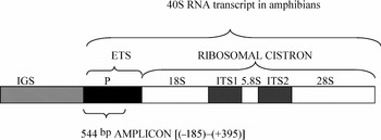
Because ribosomal genes are organized in tandem repeats (i.e. many copies of ribosomal genes exist in a genome) and in order to study the simultaneous presence of two epigenetic modifications at the same promotor region, this variation of the ChIP assay was used. In this study, the segment of genome analysed for association with certain epigenetic modifications (H4 hyperacetylation, H2A-ubiquitination and cytosine methylation) is represented by a sequence found 544 bp from the ribosomal cistron (18S–5.8S–28) promoter. The amplicon also contains a small fragment from the external transcribed spacer (ETS) (Fig. 5). The amphibian rRNA gene promotor is an oligo dT promotor that contains a conserved 5′-GCTTTTTTG-3′ sequence.
Amphibian ribosomal cistron organization. ITS, internal transcribed spacer; ETS, external transcribed spacer; IGS, intergenic spacer; P, promoter.

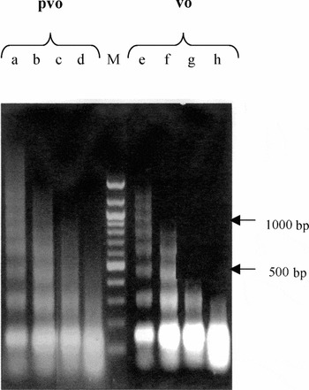
In order to ensure accurate results in the RE-ChIP assays, chromatin was fragmented by MNase incubation for several time periods, thus obtaining fragments with a range of 300–1000 bp. We tested four different MNase (50 UI/ml) incubation times (5, 10, 15 and 30 min) for both previtellogenic and vitellogenic oocytes. We choose the length of chromatin fragments to be less then 1000 bp (confirmed by electrophoretic analysis) at 15 min MNase incubation time for previtellogenic oocytes and, respectively, we choose a 10 min MNase incubation time for vitellogenic oocytes (Fig. 6). The electrophoresis pattern indicates different levels of chromatin sensitivity to MNase in previtellogenic and vitellogenic oocytes depending on its level of condensation.
Micrococcal nuclease digest (50 UI/ml) of the chromatin from previtellogenic (pvo) and vitellogenic (vo) oocytes. Various MNase incubation times were tested (a) 5 min; (b) 10 min; (c) 15 min; (d) 30 min; (e) 5 min; (f) 10 min; (g) 15 min; and (h) 30 min). Samples (c) and (f) were used in RE-ChIP experiments (M, marker 100 bp, Roth).

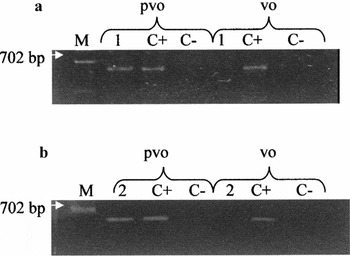
In the first RE-ChIP experiment (hacH4 + 5MeC), the electrophoregram indicated the simultaneous presence of both epigenetic modifications (high acetylated H4 forms and 5′-methyl cytosine) in previtellogenic oocytes at equivalent levels in the rRNA promoter region. For vitellogenic oocytes, only highly acetylated H4 forms were present. The Re-ChIP procedure, combined with PCR analysis, showed a negative result for cytosine methylation for the sequence of interest (Fig. 7a).
RE-ChIP experiments. (a) PCR analysis of hacH4 + 5MeC immunoprecipitates from previtellogenic (pvo) and vitellogenic (vo) oocytes. M, molecular weight marker BstE II λ; 1, hacH4 + 5MeC, (C+), positive control (input sample); (C−), negative control (without antibodies); (b) PCR analysis of hacH4 + uH2A immunoprecipitates from previtellogenic (pvo) and vitellogenic (vo) oocytes. 2, hacH4 + uH2A; (C+), positive control (input sample); (C−), negative control (without antibodies).

The second experiment was designated to study the simultaneous presence of high acetylated forms of histone H4 and ubiquitinylation of H2A (hacH4 + uH2A), using the same comparative study of a negative control and positive (input) sample for hyperacetylated H4.
Interestingly, PCR analysis gave a positive result for the RE-ChIP assay in previtellogenic oocytes and a negative result for both antibody immunoprecipitations in vitellogenic oocytes (Fig. 7b). These results indicate the simultaneous presence of highly acetylated H4 and ubiquityl-H2A at the ribosomal gene promoter region, in previtellogenic oocytes. For vitellogenic oocytes only highly acetylated forms of histone H4 were detected.
Taken together, our results suggest that previtellogenic oocytes have a different repertoire of epigenetic modifications at the level of the ribosomal gene promoter region as compared with vitellogenic oocytes.
Discussion
During amphibian oocytes development, chromatin remodelling factors act towards the decondensation of chromatin fibre; this process enables the access of transcription factors or signalling molecules to DNA embedded in the chromatin structure. Chromosomes that adopt a lampbrush phenotype are involved in extensive transcription activity. Chromatin structure plays an important role in the control of gene expression by limiting the accessibility of sequence-specific binding proteins to DNA. The histone components of chromatin play an integral role in this regulation machinery. Several years ago it was suggested that linker histones may play a more specific role in transcription regulation by acting at the level of critically placed individual nucleosomes, rather than by fibre compaction generally (Zlatanova, Reference Zlatanova1990; Brown et al., Reference Brown, Gunjan, Alexander and Sittman1997; Gunjan & Brown, Reference Gunjan and Brown1999). H1 was often supposed to function as a general repressor of transcription by stabilizing higher order chromatin structure (Carruthers et al., Reference Carruthers, Bednary, Woodcock and Hansen1998). Linker histones have also been demonstrated to reject factor binding directly (Juan et al., Reference Juan, Utley, Adams, Vetesse-Dadey and Workman1994), in order to limit nucleosome mobility (Ura et al., Reference Ura, Hayes and Wolffe1995) and to reduce the transient dynamic exposure of DNA on the nucleosome surface (Polach & Widom, 1995). All of these activities are expected to have repressive effects on transcription. The fact that linker histones are actually involved in transcriptional regulation of individual genes was first shown unambiguously in two systems: (i) the selective transcription of oocyte- and somatic-type 5S rRNA genes in early Xenopus development (Kandolf, Reference Kandolf1994); and (ii) the activation and repression of transcription of certain genes in Tetrahymena knockouts for histone H1 (Shen et al., Reference Shen and Gorovsky1996).
In our study, 12.5% SDS-PAGE electrophoresis results regarding linker histones from previtellogenic and vitellogenic oocytes revealed an interesting dynamics of chromatin H1 variant content during oocyte development. Previtellogenic oocytes were shown to contain more linker histone variants than mature oocytes, the latter being involved in an extensive transcriptional activity. These results suggest a hypothesis in which oocyte differentiation is marked by a gene expression regulation process through specific dynamics of histone H1 variants. Moreover, we also suggest that these H1 variants have a functional significance during amphibian oocytes differentiation.
Most of our knowledge of transcriptional regulation comes from studies in somatic cell, in which specific chromatin remodelling events are directly coupled with transcriptional activation and silencing (Jenuwein et al., Reference Jenuwein and Allis2001). One can ask: ‘Are the same events operating in egg cells?’
Oogenesis is a critical event in the formation of female gametes, highly specialized cells that transfer the maternal genomic information to the next generation. During this process, the gene expression pattern changes dramatically, concomitant to genome remodelling, while genomic information is maintained. There are also many meiosis-specific events other than the reprogramming of gene expression in oocytes, e.g., homologous chromosomes recombination and successive divisions without DNA replication, asymmetric cell division. All these events are accompanied by meiotic specific modifications in chromatin structure and some of these might be controlled by epigenetic modifications including histone acetylation and DNA methylation.
One of the extensively studied epigenetic modification is histone acetylation. The acetylation of nucleosome core histones is thought to play an important role in various functions. The biological significance of histone acetylation was first suggested after the discovery that the amount of acetylation is correlated with the level of transcription (Allfrey et al., Reference Allfrey, Faulkner and Mirsky1964). At the beginning of this century, the reversible acetylation of core histones had been recognized as a major mechanism by which chromatin-mediated gene regulation is affected (Gunjan et al., Reference Gunjan, Sittman and Brown2001). Some reports provide direct evidence that the acetylation status of core histones has a causal relationship to gene activity (Wang et al., Reference Wang, Liu and Berger1998). It has been demonstrated that many of the histone-acetyltransferases (HATs) that acetylate core histones are components of transcriptional activator or coactivator complexes and are specifically targeted to genes in order to activate transcription (Utley et al., Reference Utley, Ikeda, Grant, Cote, Steger, Eberharter, John and Workman1998). On the other hand, transcriptionally silent chromatin is often hypo-acetylated and histone-deacetylases (HDACs) have been shown to be components of transcriptional corepressors and silencers that are also targeted to the appropriate DNA sequences (Ayer, Reference Ayer1999).
Certain studies on H4 acetylation (Sommerville et al., Reference Sommerville, Baird and Turner1993; Kim et al., Reference Kim, Liu, Tazaki, Nagata and Aoki2003) and its turnover (Magnaghi-Jaulin & Jaulin, Reference Magnaghi-Jaulin and Jaulin2006) in oocyte chromatin have been reported previously. Lampbrush chromosomes from previtellogenic (immature) and vitellogenic (mature) oocytes of the amphibian Triturus cristatus have been used to examine the role of histone acetylation in transcription by indirect immunofluorescence with antisera to H4 that was acetylated at specific lysine residues (lysines 5, 8, 12 and 16) (Sommerville et al., Reference Sommerville, Baird and Turner1993). This work suggested that H4 acetyl groups turn over more rapidly in mature than in immature oocytes and that histone hyperacetylation proceeds, and possibly induces, loops formation and transcriptional activation.
Our immunohistochemical investigations on the hyperacetylation of the histone H4 in amphibian mature oocytes are the first report that attempts to correlate the pattern of high acetylated H4 immunostaining with nuclear architecture in these specialized cell types.
Whereas a homogeneous H4 hyperacetylated reaction in the nucleus of the vitellogenic oocyte, a rather massive concentration of the hyperacetylated H4 was noticed towards the nucleus periphery. The occurrence of a H4 hyperacetylated homogeneous signal in the vitellogenic oocyte nucleus can be explained as a requirement of this chemical modification for the extensive transcription revealed in the nucleus of this type of oocytes. Many ‘housekeeping’ genes that are involved in establishing embryo segments and ribosomal genes are abundantly transcribed and even amplified in oogenesis.
The occurrence of the concentrated H4 hyperacetylated signal in the form of blocks at the periphery of the nucleus may be interpreted as follows: firstly as a reflection of the level of rDNA axes of extrachromosomal nucleoli involved in intense rRNA transcription, H4 hyperacetylated histone is accumulated; or secondly, as ribonucleoproteins that detach from extrachromosomal nucleoli are exported from the nucleus to the cytoplasm, H4 hyperacetylated histone remains at the periphery of the nucleus in order to be recycled and/or degraded. A peculiar aspect of the immunostaining pattern was the lack of signal for hacH4 in extrachromosomal nucleoli and in perinucleolar regions.
Other experiments performed on plants have shown that perinucleolar regions and nucleoli during S phase generally do not contain hyperacetylated forms of H4 (Jasencakova et al., Reference Jasencakova, Meister, Walter, Turner and Schubert2000) and are transcriptionally inactive (Shaw et al., Reference Shaw, Highett, Beven and Jordan1995). In vitellogenic oocytes, rDNA axes of extrachromosomal nucleoli are involved in an intense transcription activity, thus we interpreted these findings because of abundant deposition of ribonucleoproteins particles (RNP) at the nucleolar organizing region, covering the rDNA axes.
Chromatin immunoprecipitation experiments have shown that histone H4 is hyperacetylated in the promoter regions of active genes (Shestakova et al., Reference Shertakova, Bandu, Doly and Bonnefoy2001; Zhao et al., Reference Zhao, Herrera-Diaz and Gross2005; Workman, Reference Workman2006). Recently, it has been demonstrated that distinct histone modification patterns function as recognition codes for the recruitment of different transcription factors upon transcriptional activation (Agalioti et al., Reference Agalioti, Chen and Thanos2002). Thus the histone code may function as an epigenetic marker that is directly associated with transcriptional activation (Jenuwein & Allis, Reference Jenuwein and Allis2001).
Our RE-ChIP experiments performed for the first time on chromatin from previtellogenic and vitellogenic oocytes revealed that histone H4 at the level of rRNA genes promoter remains in a highly acetylated state during oocyte differentiation. However, significant dynamics of other chromatin epigenetic changes, such as H2A ubiquitinylation and cytosine methylation, are occurring concomitantly.
We noticed multiple epigenetic modifications at the rRNA gene promoter regions in previtellogenic oocytes. Thus, at the same copy level of ribosomal cistron promoter we found, using two RE-ChIP experiments, the presence of highly acetylated histone H4 and 5MeC, respectively; highly acetylated H4; and H2A ubiquitinylated. In this case it is very difficult to make any correlation with the transcriptional state of that gene. In previtellogenic oocytes, ribosomal genes are actively transcribed, but this activity has not been extensively observed in vitellogenic oocytes. Before yolk genesis, at the beginning of oocytes development, these genes may be marked for some important and intensive functional state.
It is possible that our experiment corresponded to the moment of oogenesis, which is needed to mark ribosomal genes for future transcriptional activity. These hypotheses need further investigation, including the study of other components of the concerted chromatin conformation dynamics. As we had previously shown, some researchers have demonstrated that H4 is hyperacetylated in the promoter of active genes (Kuo et al., Reference Kuo, Zhou, Jambeck, Churchill and Allis1998; Shestakova et al., Reference Shertakova, Bandu, Doly and Bonnefoy2001).
Ubiquitinylation is a chemical modification involved in protein degradation via a proteasome pathway. Nevertheless, ubiquitinylation is not limited to degraded proteins, some studies have shown that this modification is also involved in rDNA repair, cell cycle control, cellular response to stress and histone code (Baarends et al., Reference Baarends, Hoogerbrugge, Roest, Jan Vreeburg, Hoeijnakers and Grootegoed1999).
Immunohistochemical studies of meiotic male mouse cells evidenced that the amount of H2A-ubiquitinylated (uH2A) reaches its maximum during the pachytene stage of meiotic prophase. In early pachytene, uH2A is concentrated in the condensed and transcriptional inactive XY chromatin of the sex body. By mid-pachytene, uH2A is detected throughout the whole nucleus, but is subsequently limited to the sex body of late pachytene spermatocytes (Baarends et al., Reference Baarends, Hoogerbrugge, Roest, Jan Vreeburg, Hoeijnakers and Grootegoed1999). At present, the function of histone ubiquitinylation is not clear. There are some indications that this histone modification results in destabilization of the H2A–H2B dimer, which may facilitate dimer displacement and DNA transcription (Li et al., 1993). Our results support the hypothesis that uH2A may be a signal for H2A–H2B dimer displacement at the nucleosomal level. It might be possible that uH2A and hyperacetylated H4 at the level of our sequence of interest (544 bp) represent modifications with marking roles for the future functional state of the promoter.
Much work had been performed on the silenced gene promoter, which has revealed strong evidence for the presence of 5'-methyl cytosine at CpG dinucleotides (Bird, Reference Bird1996, Reference Bird2002; Nan et al., Reference Nan, Ng, Johnson, Laherty, Turner, Eisenman and Bird1998; Bird & Wolffe, Reference Bird and Wolffe1999; Lawrence et al., Reference Lawrence, Early, Pontes, Silva, Chen, Neves, Viegas and Pikaard2004). Methylation of cytosine bases located 5′ to a guanosine in a CpG dinucleotide is an epigenetic marker known to be involved in gene transcriptional repression (Jones & Baylin, Reference Jones and Baylin2002). Yet, until recently, the role of cytosine methylation in rRNA gene regulation was unclear (Grummt & Pikaard, 2002). Methylated ribosomal genes are transcribed efficiently when injected into oocyte nuclei or when transcribed in vitro (Macleod & Bird, Reference Macleod and Bird1983; Pennock & Reeder, Reference Pennock and Reeder1984), suggesting that DNA methylation is not sufficient for transcriptional repression.
Our intriguing result that indicated a hyperacetylated and, in the same time, a methylated promoter, might be explained by the fact that the ribosomal cistron could be inactive at our moment of investigation, but it might start the preparation for its future intense transcriptional activity. H4 hyperacetylation of the ribosomal gene promotor might be required to keep chromatin in a potentially relaxed or accessible state in which transcription can be induced as required.
It is obvious that the described chromatin dynamic changes could be ascribed to a specific complex epigenetic code for gamete differentiation. Consequently, chromatin may adopt an ‘open’ or a ‘closed’ conformation depending upon the functional state of that domain.
The lack of 5′-methyl cytosine and ubiquityl–H2A at the rRNA gene promoter region in vitellogenic oocytes allows us to conclude that, during oogenesis, a dynamic of epigenetic modification takes place. Nucleosomal core histones are targets for many post-translational modifications. All these epigenetic modifications act in a synergistic way to modulate DNA-binding protein accessibility, and might represent signals for the recruitment of transcription complexes.
Whereas the functional correlations between histone variants/histone modifications/DNA methylation appear to become increasing clear, in contrast the structural modifications of chromatin remain to be defined in most instances. An interesting possibility that has increasing experimental support is that some variants and modifications might operate synergistically. Moreover, it is possible that most histone variants and modifications operate in vivo in a concerted way, affecting either an individual histone or different histones.
Previously, the role of a single epigenetic modification was studied (Kaplan et al., Reference Kaplan, Bauer, Morrison, Langan and Fosman1984, Ausio & van Holde, Reference Ausio and van Holde1986, Garcia-Ramirez et al., Reference Garcia-Ramirez, Rocchini and Ausio1995). Taken together, the results of our present study have revealed that a dynamic epigenetic network mediates the transcriptional state of rDNA, by interplay of histone post-translational modification, linker histone variants, DNA methylation and other chromatin ATP-dependent remodelling complexes, in order to establish the complex functional state of rRNA genes.