Prothymosin alpha: features and biological role
Prothymosin alpha (PTMA) is a small peptide belonging to the thymosin family. It was isolated for the first time from rat thymus in 1984 as an immunogenetic factor (Haritos et al., Reference Haritos, Goodall and Horecker1984a), but soon detected in a variety of mammalian tissues (Haritos et al., Reference Haritos, Tsolas and Horecker1984b; Clinton et al., Reference Clinton, Frangou-Lazaridis, Panneerselvam and Horecker1989). PTMA shows a very peculiar structure, definitely correlated to its functional promiscuity in that it lacks aromatic amino acids, cysteines and methionines, and approximately the 50% of its primary structure is represented by glutamic and aspartic residues (Frangou-Lazaridis et al., Reference Frangou-Lazaridis, Clinton, Goodall and Horecker1988). For these reasons, PTMA can be considered as one of the most acidic proteins present in nature, with an isoelectric point of 3.5. The negative charges present in this protein define the physiological adoption of a random-coil conformation with no secondary or tertiary structure (Gast et al., Reference Gast, Damaschun, Eckert, Schulze-Forster, Maurer, Müller-Frohne, Zirwer, Czarnecki and Damaschun1995). These features make PTMA able to interact with many cationic molecules and, potentially, with membranes and receptors. The absence of a tertiary structure does not represent a functional limit, as some proteins are able to fold themselves during interaction with other molecules, acquiring transitional conformations that are biologically active.
PTMA localization is extended, consistent with its structural flexibility and it has been confirmed in the nucleus, cytoplasm and extracellular compartment (Enkemann et al., Reference Enkemann, Ward and Berger2000b; Mosoian, Reference Mosoian2011). In fact, a bipartite nuclear localization signal has been identified in the C-terminal region, phylogenetically preserved.
At a genomic level, Ptma has been well characterized in human: it is coded by a family made of seven members (Eschenfeldt et al., Reference Eschenfeldt, Manrow, Krug and Berger1989; Rubtsov & Vartapetian, Reference Rubtsov and Vartapetian1995). One of these, localized on chromosome 2 (in Homo sapiens; Szabo et al., Reference Szabo, Panneerselvam, Clinton, Frangou-Lazaridis, Weksler, Whittington, Macera, Grzeschik, Selvakumar and Horecker1993) has exons, introns and standard regulatory signals, while the others are devoid of introns (Eschenfeldt et al., Reference Eschenfeldt, Manrow, Krug and Berger1989; Rubtsov & Vartapetian, Reference Rubtsov and Vartapetian1995) and probably are pseudogenes (Manrow et al., Reference Manrow, Leone, Krug, Eschenfeldt and Berger1992). Ptma transcripts have been revealed in all the analyzed tissues: brain, heart, liver, muscles, spleen, intestine, lung, cerebellum and testis (Clinton et al., Reference Clinton, Frangou-Lazaridis, Panneerselvam and Horecker1989) and, overall, the Ptma gene is one of the most expressed loci, together with heat shock protein 90, the light chain of myosin and ribosomal proteins (Adams et al., Reference Adams, Kerlavage, Fleischmann, Fuldner, Bult, Lee, Kirkness, Weinstock, Gocayne and White1995).
The peculiar structure, extended distribution and association with several molecular partners have soon suggested that PTMA could perform roles those go beyond its immunogenic nature. Among the multiple functions that have been associated with PTMA are H1 histone interaction and chromatin remodelling (Karetsou et al., Reference Karetsou, Sandaltzopoulos, Frangou-Lazaridis, Lai, Tsolas, Becker and Papamarcaki1998; Ueda et al., Reference Ueda, Matsunaga and Halder2012), cell death (Enkemann et al., Reference Enkemann, Wang, Trumbore and Berger2000a; Jiang et al., Reference Jiang, Kim, Shu, Zhao, Zhang, Kofron, Donnelly, Burns, Ng, Rosenberg and Wang2003; Malicet et al., Reference Malicet, Giroux, Vasseur, Dagorn, Neira and Iovanna2006; Ueda, Reference Ueda2009), transcriptional regulation (Martini et al., Reference Martini, Delage-Mourroux, Kraichely and Katzenellenbogen2000; Karetsou et al., Reference Karetsou, Kretsovali, Murphy, Tsolas and Papamarcaki2002; Martini & Katzenellenbogen, Reference Martini and Katzenellenbogen2003) cancer development (Domínguez et al., Reference Domínguez, Magdalena, Cancio, Rosón, Paredes, Loidi, Zalvide, Fraga, Forteza and Regueiro1993; Tsitsiloni et al., Reference Tsitsiloni, Stiakakis, Koutselinis, Gogas, Markopoulos, Yialouris, Bekris, Panoussopoulos, Kiortsis and Voelter1993; Wu et al., Reference Wu, Habib, Mitry, Reitsma, van, Deventer and Chamuleau1997; Skopeliti et al., Reference Skopeliti, Voutsas, Klimentzou, Tsiatas, Beck, Bamias, Moraki, Livaniou, Neagu, Voelter and Tsitsilonis2006; Zhang et al., Reference Zhang, Cui, Lu, Lu, Jiang, Chen, Zhang, Jin, Peng and Tang2014) and, as already alluded to, immunity (Pan et al., Reference Pan, Haritos, Wideman, Komiyama, Chang, Stein, Salvin and Horecker1986; Baxevanis et al., Reference Baxevanis, Thanos, Reclos, Anastasopoulos, Tsokos, Papamatheakis and Papamichail1992; Voutsas et al., Reference Voutsas, Baxevanis, Gritzapis, Missitzis, Stathopoulos, Archodakis, Banis, Voelter and Papamichail2000). Furthermore, recent studies have highlighted its action as an important anti-apoptotic and cell proliferation mediator that could eventually become a target for therapeutic approaches in diseases such as cancer, inflammation and sepsis (Iannou et al., Reference Iannou, Samara, Livaniou, Derhovanessian and Tsitsilonis2012; Samara et al., Reference Samara, Karachaliou, Ioannou, Papaioannou, Voutsas, Zikos, Pirmettis, Papadopoulos, Kalbacher, Livaniou, Tsitsilonis and Voelter2017). It has been proposed that the considerable number of processes in which this small peptide is involved may be due to its molecular mimicry, suggesting that there might not be a univocal definition describing its physiological role (Piñeiro et al., Reference Piñeiro, Cordero and Nogueira2000; Wang & Pan, Reference Wang and Pan2007).
PTMA and the spermatogenesis
The first studies
Reproduction is one of the processes for which a role for the molecule has been suggested, and in particular in male gametogenesis. The first studies conducted in the 1990s were focused on the testis of the mammal Rattus norvegicus. Dosil and colleagues (Reference Dosil, Freire and Gómez-Márquez1990) found that PTMA was mainly expressed during the first 2 weeks of age, when the testis cell population is mostly represented by self-renewing type A spermatogonia (SPG) and by proliferating immature Sertoli cells (SC), and decreases with the onset of puberty. Coherently, a further paper reported that the protein is localized in the nucleus and cytoplasm of SPG at the initial stages (preleptotene, leptotene and zygotene) primary spermatocytes (SPC) of the first meiotic division and disappears in other stages (Rosón et al., Reference Rosón, Gallego, García-Caballero, Heimer, Felix and Domínguez1990). These results were then in agreement as both authors found that PTMA was expressed during the proliferative phase of spermatogenesis.
PTMA in non-mammals vertebrate
Before 2002, PTMA was found only in mammal tissues, and Trumbore et al. (Reference Trumbore, Manrow and Berger1998) failed to find its presence in yeast and other organisms ranging from bacteria to amphibians, therefore they considered the presence of the gene in animals other than mammals to be highly unlikely. In 2002, PTMA was detected for the first time in a non-mammalian vertebrate, the amphibian Pelophylax esculentus, and in particular in the Harderian gland, liver, muscle brain, kidney and at a very high levels in testis (Aniello et al., Reference Aniello, De, Rienzo, Ferrara, Palmiero and Minucci2002; Ferrara et al., Reference Ferrara, Izzo, Liguori, d'Istria, Aniello and Minucci2009). Mindful that P. esculentus is a seasonal breeder with a spermatogenic cycle regulated by endocrine and environmental factors (Rastogi & Iela, Reference Rastogi, Iela and Dallai1992), the authors focused their interest on the temporal and spatial localization of the ptma transcript in frog testis during the reproductive cycle. They found that it is always present during the seasons, but shows an usual distribution. In spring, concomitant with the high proliferative activity of SPG and when SPC I and II are rare, spermatids (SPT) are totally absent and ptma is at its lower level. In contrast, it is expressed at its highest level in September–October when SPG are at their lowest proliferative activity and the testis presents the highest number of SPC and SPT nests (Aniello et al., Reference Aniello, De, Rienzo, Ferrara, Palmiero and Minucci2002).
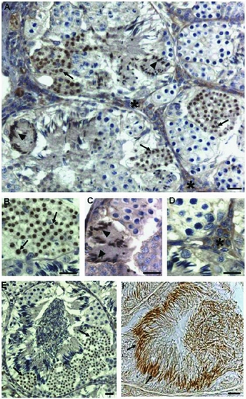
These data were confirmed and completed in a further paper in which, for the first time, the protein was found in the nuclei of GC from SPC II to elongating SPT, and in the cytoplasm of interstitial cells (Fig. 1). It is worthy noting that the localization pattern in GC mirrors deleted expression of frog Ptma compared with that of the corresponding transcript. This pattern is not unusual for a seasonal breeder in which, along with the slow progression of spermatogenesis, a transcript can be synthetized and stored in a specific phase to be translated later (Ferrara et al., Reference Ferrara, Izzo, Liguori, d'Istria, Aniello and Minucci2009).
Localization of Ptma in Pelophylax esculentus testis. In September, Ptma was present in the GC nuclei from SPCII (arrows, A, B, E) to elongating SPT (arrowheads, A, C) and in the cytoplasm of interstitial LC (asterisks, A, D). In April, (F) Ptma was also present in the nuclei of SPZ (arrows); the arrowhead indicates a SPG. Scale bar represent 20 µm (Ferrara et al., Reference Ferrara, Izzo, Liguori, d'Istria, Aniello and Minucci2009).

With the aim of expanding the comparative knowledge, PTMA localization has been studied in the cartilaginous fish Torpedo marmorata (Prisco et al., Reference Prisco, Donizetti, Aniello, Locascio, Del, Giudice, Agnese, Angelini and Andreuccetti2009) and in the teleost Danio rerio (Pariante et al., Reference Pariante, Dotolo, Venditti, Ferrara, Donizetti, Aniello and Minucci2016).
Torpedo marmorata presents a cystic organization (Prisco et al., Reference Prisco, Liguoro, D'Onghia, Ricchiari, Andreuccetti and Angelini2002) and a peculiar distribution of androgen-producing cells in the testis (Prisco et al., Reference Prisco, Liguoro, Ricchiari, Del, Giudice, Angelini and Andreuccetti2008). Every cyst is characterized by the presence of only one germ cell stage (Stanley, Reference Stanley1966; Prisco et al., Reference Prisco, Liguoro, D'Onghia, Ricchiari, Andreuccetti and Angelini2002) and, after spermiation, the SC within a cyst are removed by apoptosis (Prisco et al., Reference Prisco, Liguoro, Comitato, Cardone, D'Onghia, Ricchiari, Angelini and Andreuccetti2003). Leydig cells are active only when they are located among cysts containing meiotic and differentiating germ cells. In the regions containing cysts with SPG or mature SPZ, androgen synthesis is sustained by SC as well as by SPG (Prisco et al., Reference Prisco, Liguoro, Comitato, Cardone, D'Onghia, Ricchiari, Angelini and Andreuccetti2003, Reference Prisco, Liguoro, Ricchiari, Del, Giudice, Angelini and Andreuccetti2008).
PTMA transcript and protein are expressed in a stage-dependent manner during Torpedo spermatogenesis. Indeed, mRNA is present in SPC II and round SPT and the protein is localized in SPC II, round and elongating SPT and SPZ, whereas both mRNA and protein expression were absent in SPG and SPC I. These findings demonstrate that PTMA synthesis takes place in SPC II and probably continues in round SPT, suggesting that it is probably involved in spermiogenesis.
Furthermore, both mRNA and protein were present at the level of some Leydig cell groups located among cysts containing meiotic and differentiating male germ cells suggesting its possible involvement in the synthesis of androgen.
The teleost Danio rerio represents a very common vertebrate model, in which two paralog genes have been described and characterized during embryogenesis (ptmaa and ptmab), this suggests that a gene duplication event occurred in the teleost fish lineage after their separation from tetrapods (Donizetti et al., Reference Donizetti, Liccardo, Esposito, Del, Gaudio, Locascio, Ferrara, Minucci and Aniello2008).
The male zebrafish, as commonly found in other ‘early’ teleosts, develops anastomosing tubular gonads, in which the germinal compartment branches in loops or tubules that form a connected network (Grier, Reference Grier, Russell and Griswold1993). The tubules are formed by clusters of progressing germ cells enclosed by the cytoplasm of a somatic SC, defining the spermatocysts that line the lumina, where the SPZ are released (Billard, Reference Billard and Lamming1990; Pudney, Reference Pudney, Payne, Hardy and Russell1996; Parenti & Grier, Reference Parenti and Grier2004; Schulz et al., Reference Schulz, de França, Lareyre, Le, Gac, Chiarini-Garcia, Nobrega and Miura2010; Huszno & Klag, Reference Huszno and Klag2012).
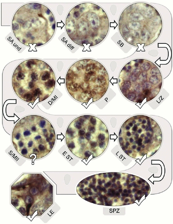
The localization of Ptma in the testis of Danio rerio approximately matches the pattern previously highlighted in the other species, with a clear predilection for meiotic and post-meiotic germ cells (Fig. 2). Its absence in SPG suggests that it does not participate in the proliferation of staminal/mitotic phases, while its presence in primary SPC and in SPT supports its possible role during meiosis and/or during the subsequent stages of SPT differentiation into mature SPZ. Moreover, the protein has also been detected in the gametes and, in particular, in the head of luminal SPZ.
A schematic illustration of the progression of the germinal line in the tubules, with pictures of selected cysts that describe the pattern of Ptma sub-localization in germ cells during zebrafish spermatogenesis (for review of the germ cell types see Schulz et al., Reference Schulz, de França, Lareyre, Le, Gac, Chiarini-Garcia, Nobrega and Miura2010). Ptma-positive cell types are marked with a ‘V’, the negative types with an ‘X’. Non-stained secondary SPC are marked by a ‘?’. SA and SA diff: undifferentiated and differentiated (A) SPG; SB: (B) SPG. L/Z: leptotene/zygotene primary SPC; P: pachytene primary SPC; D/MI: diplotene/metaphase I primary SPC; S/MII: secondary/metaphase II SPC; EST: early SPT; LST: late SPT; SPZ: spermatozoa; LE: interstitial Leydig cells (Pariante et al., Reference Pariante, Dotolo, Venditti, Ferrara, Donizetti, Aniello and Minucci2016).

PTMA in mammals testis and spermatozoa
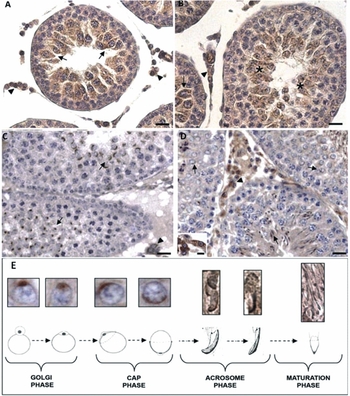
As described before, some non-mammals vertebrates represent an useful model to study testis structure and the spermatogenesis, due to the cystic architecture of the male gonad. This organ has separated nests of synchronous GC, each enveloped by Sertoli cell cytoplasm, and the appearance of different GC at various periods of the annual reproductive cycle. In contrast, in mammals, testis spermatogenesis occurs continuously, and all GC types are represented at the same time. Therefore, to minimize this difference, a study on PTMA localization in a mammal model was conducted that covered the first wave of spermatogenesis and, in particular, three representative points: 27 days post-partum (dpp), when meiosis has just been completed for the first time, 35 dpp, while spermiogenesis was occurring, and 60 dpp, when the first wave of spermatogenesis had definitely ended (Ferrara et al., Reference Ferrara, Izzo, Pariante, Donizetti, d'Istria, Aniello and Minucci2010). The authors observed that the ptma transcript was always localized in the same GC types, from pachytene I SPC to newly formed round SPT. The protein followed this localization only at 27 dpp, while it was almost absent in the meiotic SPC and was evident in differentiating SPT in the subsequent stages (Fig. 3). In particular, their results demonstrated, for the first time, the presence of PTMA in association with the developing acrosome system. During the Golgi phase, it was detected at the level of the forming acrosomic vesicle and, later in this phase, it was retained as the vesicle attached to the nuclei of the round SPT. In the cap phase the signal delineated the acrosome as it gradually spread and capped the apical region of the SPT nucleus. The signal persisted throughout the acrosome phase in the elongating acrosome capped nuclei. At the end of the maturation phase, PTMA was still detected in the apical region of the SPZ head and, finally, was still present in the apical region of the head of epididymal SPZ (eSPZ) (Fig. 3).
PTMA localization in rat testis at 27 (A, B), 35 (C) and 60 (D) dpp. It is present in LC (arrowheads in A, B), in pachytene I SPC (arrows in A, B) and in newly formed SPT (asterisks in B). It is also localized in differentiating SPT (arrows in C, D). PTMA localization in the head of differentiating rat SPT in comparison with schematic representations of acrosome system biogenesis during the different phases of spermiogenesis (E). Scale bars represent 20 µm in (A–D), and 5 µm in the inset (Ferrara et al., Reference Ferrara, Izzo, Pariante, Donizetti, d'Istria, Aniello and Minucci2010).

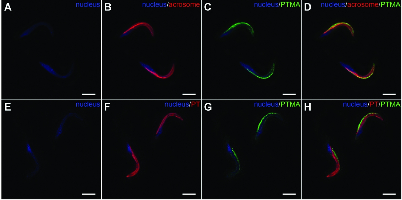
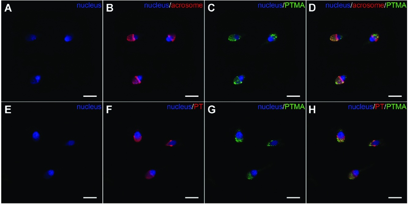
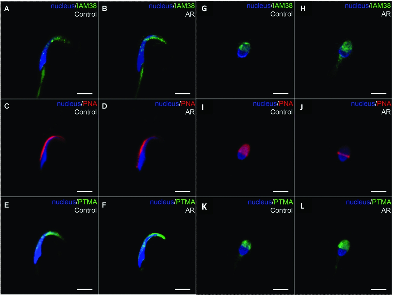
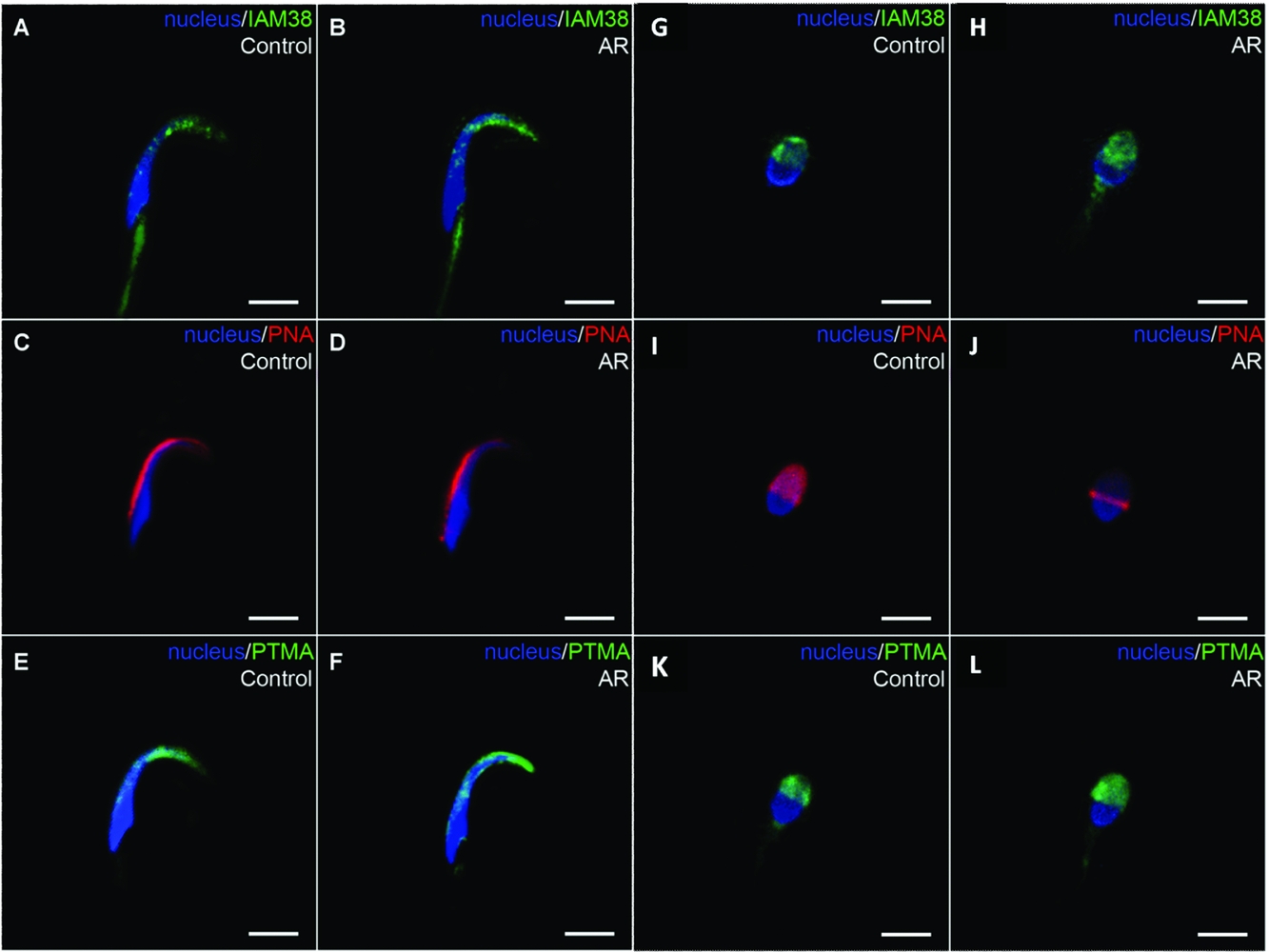
The presence of the protein in the mature SPZ has led to improved efforts to better understand its role in the mammalian sperm head (Ferrara et al., Reference Ferrara, Pariante, Di, Matteo, Serino, Oko and Minucci2013). The sperm head shows a peculiar and complex morphology, which comprises: (1) the nucleus, which contains the paternal genome; (2) the acrosome, which plays a crucial role in the early phases of fertilization; and (3) the cytoplasmic layers, which consist of the residual cytoplasm surrounded and packed by a complex membrane system (Toshimori, Reference Toshimori2009). The acrosome is bordered by the outer acrosomal membrane (OAM) underlying the plasma membrane (PM) and the inner acrosomal membrane (IAM) overlying the nuclear envelope (NE). Among the cytoplasmic layers, the perinuclear theca (PT) is of particular interest. Its apical region, sandwiched between the IAM and the NE, contains molecules that act in the acrosomal assembly during spermiogenesis and in the maintenance of nucleus–acrosome contact in the mature SPZ. Its caudal region, located between the PM and the NE, is involved in sperm–egg interactions during fertilization, such as egg activation (Oko & Sutovsky, Reference Oko and Sutovsky2009). With experiments of co-localization, the association of PTMA with the acrosome has been confirmed. This is also a suitable environment for such an acidic protein while not related with the PT, which is located closer to the nucleus (Fig. 4). The analysis was been extended to human SPZ and, for the first time, confirmed its localization in the acrosome, from the equatorial segment to the anterior-most part of the latter (Fig. 5). Encouraged by these results, the authors carried out an in vitro acrosome reaction that, by fusion of the OAM and the PM over the anterior region, led the SPZ to release acrosome soluble contents and to expose its matrix compartment and the IAM (Manandhar & Toshimori, Reference Manandhar and Toshimori2001; Miranda et al., Reference Miranda, Allaire, Sosnik and Visconti2009). Their data showed that the PTMA signal is retained in the apical region of the reacted SPZ, thus demonstrating that the peptide is associated with the IAM rather than with the soluble compartment (Fig. 6).
PTMA (green, C, D, G, H) co-localization with either the acrosome (red, B, D) or the PT (red, F, H) on rat eSPZ. PTMA is definitely associated with the acrosome while its signal is distinctly dorsal to the PT. The nucleus is marked in blue (A–H). Scale bars represent 5 µm (Ferrara et al., 2011).

PTMA (green, C, D, G, H) co-localization with either the acrosome (red, B, D) or the PT (red, F, H) on human SPZ. The data show that PTMA co-localizes with the acrosome and not with the PT, in human as in the rat. The nucleus is marked in blue (A–H). Scale bars represent 5 µm (Ferrara et al., 2011).

Analysis on rat (A–F) and human (G–L) SPZ after the induction of acrosome reaction (B, D, F, H, J, L) and on control SPZ (A, C, E, G, I, K). IAM is retained (green, A, B, G, H), while acrosome staining decreases (red, C, D, I, J). PTMA is easily detectable both in control and in reacted SPZ (green, E, F, K, L). Scale bars represent 5 µm (Ferrara et al., 2011).
Possible involvements of PTMA in spermatogenesis
To date, the effective role of PTMA in the vertebrate testis remains unclear, but some hypotheses can be made.
The most extensive studies referring to the presence of this protein in the cytoplasmic compartment suggest an antiapoptotic function that negatively regulates caspase-9/3 activation by inhibiting apoptosome formation (Jiang et al., Reference Jiang, Kim, Shu, Zhao, Zhang, Kofron, Donnelly, Burns, Ng, Rosenberg and Wang2003). Moreover, the presence of several overlapping caspase-3 recognition sites makes PTMA itself a well designed substrate of caspase-3 (Evstafieva et al., Reference Evstafieva, Belov, Kalkum, Chichkova, Bogdanov, Agol and Vartapetian2000). These sites are highly conserved from lower vertebrates to mammals. It has been proposed that the cleavage by caspase-3 might abolish the inhibitory effect of PTMA on apoptosome formation (Hannappel & Huff, Reference Hannappel and Huff2003). Although in mammalian testis the fully differentiated LC rarely proliferates and rarely undergoes apoptosis, it has been hypothesized that the presence of survival proteins might actively suppress the apoptotic signal cascade, suggesting an antiapoptotic role of PTMA in LC. Furthermore, both nuclear localization and the ability to bind the linker histone H1 in vitro has lent support to the hypothesis that PTMA can act as a chromatin-remodelling protein (Gómez-Márquez & Rodríguez, Reference Gómez-Márquez and Rodríguez1998; Karetsou et al., Reference Karetsou, Sandaltzopoulos, Frangou-Lazaridis, Lai, Tsolas, Becker and Papamarcaki1998; Rodríguez et al., Reference Rodríguez, Viñuela, Alvarez-Fernández, Buceta, Vidal, Domínguez and Gómez-Márquez1998; Gómez-Márquez, Reference Gómez-Márquez2007). This is a major event that occurs during spermatogenesis. In mammals, from the meiotic prophase onwards, somatic linker histones H1a and H1b are replaced at first by testis-specific subtype H1t (Doenecke et al., Reference Doenecke, Drabent, Bode, Bramlage, Franke, Gavenis, Kosciessa and Witt1997), then by transition nuclear proteins TNP1 and TNP2 (Yu et al., Reference Yu, Eriksson and Stillman2000), and finally by protamines PRM1 and PRM2 (Cho et al., Reference Cho, Willis, Goulding, Jung-Ha, Choi, Hecht and Eddy2001). Bearing this in mind, it could be assumed that PTMA might be involved in the central event of chromatin condensation in the nuclei of SPC and SPT. In addition, the presence of PTMA in SPZ heads, its association with the IAM and its preservation after the acrosome reaction might also suggest a role after fertilization, probably when, following the penetration into the ooplasm, the highly packed sperm chromatin undergoes decondensation. This hypothesis seems to be supported by the reported efficiency of PTMA in decondensing human sperm chromatin (Karetsou et al., Reference Karetsou, Martic, Tavoulari, Christoforidis, Wilm, Gruss and Papamarcaki2004).
Closing remarks
In this review, we tried to describe the conservation of PTMA distribution in the testis and SPZ of all the analyzed species, and have provided an evolutionary perspective and a better comprehension of basal mechanisms of reproductive activity. Furthermore, the presence of PTMA in the SPZ head and its retention and exposure on the IAM after the acrosome reaction, provides evidence for involvement of PTMA in fertilization. Further studies could provide information on PTMA as a possible marker of a correct physiology of testis and male gametes. It is clear that this small peptide is involved in spermatogenetic events and, the results obtained so far represent a starting point to understand and define the effective role of PTMA in gamete physiology and vertebrate reproduction.
Acknowledgements
This work was supported by grants from ‘Ricerca di Ateneo’, Università degli Studi della Campania ‘Luigi Vanvitelli’.