INTRODUCTION
Leukoaraiosis refers to neuroimaging findings of white matter disease, seen as hyperintense lesions on T2 magnetic resonance imaging (MRI) sequences. These may be circumscribed or confluent, typically found in periventricular or subcortical regions (Smith et al., Reference Smith, Saposnik, Biessels, Doubal, Fornage, Gorelick and Seshadri2017) and is often presumed to be of vascular origin (Wardlaw et al., Reference Wardlaw, Smith and Dichgans2019). It is a common finding in older adults, especially those with vascular risk factors (Abraham et al., Reference Abraham, Wolfson, Moscufo, Guttmann, Kaplan and White2016; Jeerakathil et al., Reference Jeerakathil, Wolf, Beiser, Massaro, Seshadri, D’Agostino and DeCarli2004). The extent to which leukoaraiosis results in cognitive impairment remains unclear; it has been hypothesized that these effects are attributable to coincident neurodegeneration (e.g., Ross et al., Reference Ross, Hansel, Orbelo and Monnot2005; Frisoni et al., Reference Frisoni, Galluzzi, Pantoni and Filippi2007) or only confer modest but robust effects on cognition, particularly attention and executive functioning (Kloppenborg et al. Reference Campbell and Pike2014).
Leukoencephalopathies are diseases characterized by especially severe leukoaraiosis. These are diseases of white matter that are acquired following toxic exposure or infections (Filley et al., Reference Filley, McConnell and Anderson2017), are inherited forms involving small vessel ischemia (Vanderver et al., Reference Vanderver, Prust, Tonduti, Mochel, Hussey, Helman and Rodriguez2015), or result from congenital disorders of myelin development or maintenance (Köhler et al., Reference Köhler, Curiel and Vanderver2018). Leukoencephalopathies are typically functionally impairing and often progressive. The inherited forms are called “leukodystrophies,” implying congenital etiologies regardless of age of symptom onset (Köhler, et al., Reference Köhler, Curiel and Vanderver2018). In these cases, functional impairments are common, including dementia, emotional disturbance, motor incoordination, autonomic dysfunction, and other neurologic symptoms like chronic headaches and seizures (Di Donato et al., Reference Di Donato, Bianchi, De Stefano, Dichgans, Dotti, Duering and Markus2017; Hageman et al., Reference Hageman, Gabreels, De Jong, Gabreels-Festen, Van den Berg, Van Oost and Wevers1995; Joutel et al., Reference Joutel, Corpechot, Ducros and Vahedi1996; Nozaki et al., Reference Nozaki, Nishizawa and Onodera2014).
Remarkably, there are rare cases where neuropsychological functioning remains preserved despite severe leukoaraoisis (e.g., Duning, Kugel, & Knecht, Reference Duning, Kugel and Knecht2005). Such findings are illustrations of the concepts of cognitive or brain reserve. Described in the white paper by Stern et al., Reference Stern, Arenaza-Urquijo, Bartrés-Faz, Belleville, Cantilon and Chetelat2018, cognitive reserve is defined as “the adaptability (i.e., efficiency, capacity, flexibility)… of cognitive processes that helps to explain differential susceptibility of cognitive abilities or day-to-day function to brain aging, pathology, or insult.” In contrast, brain reserve is conceptualized as more static “neurobiological capital,” wherein greater brain integrity confers an advantage prior to the onset of disease or injury. Although both likely have shared mechanisms, cognitive reserve is often estimated using socio-behavioral proxies (e.g., IQ, occupational complexity, educational attainment, literacy, socioeconomic status, access to healthcare) or functional neuroimaging, whereas brain reserve is typically measured in vivo via neuroimaging-based measures of brain structure including cortical and regional volumetry and metrics of white matter microstructure. Current terminology precludes a specific method of assaying brain reserve (Stern et al., Reference Stern, Arenaza-Urquijo, Bartrés-Faz, Belleville, Cantilon and Chetelat2018), as the knowledge base on the extent to which these measures are totemic of the concept of brain reserve versus indicators of disease is still evolving. Nonetheless, advances in structural neuroimaging as applied to unusual cases may illuminate future innovations of its measurement.
In this paper, we present a case study to demonstrate how an advanced diffusion MRI technique may identify potential structural mechanisms (i.e., “brain reserve”) supporting a patient’s cognitive resilience in the face of leukoaraiosis, so severe and extensive as to prompt workup for leukoencephalopathy. The technique, fiber ball imaging (FBI; Jensen et al., Reference Jensen, Glenn and Helpern2016; Moss et al., Reference Moss, McKinnon, Glenn, Helpern and Jensen2019), is a single-shell, high b-value diffusion MRI technique that can be implemented on 3-T MRI systems. It is distinguished from more conventional methods (e.g., diffusion tensor imaging [DTI]) in that it suppresses the extra-axonal signal such that axonal integrity can be independently imaged, which is the hypothesized mechanism for the preservation of this patient’s cognitive functioning. In particular, FBI enables white matter fiber tractography that is unaffected by water in the extra-axonal space. In this way, FBI allows for an assessment of structural connectivity that is not confounded by microstructural changes external to axons, such as edema, gliosis, and myelin loss. Here, we provide data from the patient and a matched control with which to compare and contrast FBI results.
METHODS
For this case study, the patient completed the following over the course of 1 month: a neurological consultation, a neuropsychological evaluation, and a brain MRI on a research-dedicated 3-T Siemens Prismafit scanner with a 32-channel head coil, acquiring T1-weighted magnetization-prepared rapid gradient-echo sequence, T2-weighted fluid-attenuated inversion recovery sequence, T2-weighted susceptibility-weighted imaging, DTI, and FBI. Total scan time was 1 hr and 50 s (see Supplementary Material for acquisition parameters and image analysis procedures). Her images were reviewed by a neuroradiologist, who had the patient’s scans from 1, 3, and 6 years prior for comparison.
Prior clinical and research data are also summarized here. Specifically, clinical evaluations from neurology, neuroradiology, and neuropsychology were reviewed from 6 years prior (when the participant was 68 years of age), and neuropsychological test results from another research study was reviewed from 3 years prior (when the participant was 71 years of age) in which an author (A. B.) was the principal investigator. This case study was approved by the Institutional Review Board (IRB) at the Medical University of South Carolina. The control FBI data were obtained from a separate IRB-approved study. Both were compensated for their participation in research.
RESULTS
Case Description
Presenting Problem
The patient is a 74-year-old Caucasian woman who denied any clinically significant cognitive decline other than greater difficulty recalling names that has worsened over the years. She denied any other memory, language, visuospatial, or executive functioning difficulties. Her neurologic complaints included balance difficulties confounded by pelvic pain which began several years prior and was treated via physical therapy. She occasionally stumbles and falls, with the last major fall 3 years prior to this study. There were two prior episodes of dizziness/vertigo. She endorsed occasional dull frontal headaches relieved by ibuprofen. She denied photophobia or phonophobia but had a baseline sensitivity to bright moving lights and loud noises. She also endorsed fatigue, hearing loss, urge incontinence, and constipation. Her husband concurred with her report, adding that he has noticed a subtle “mumbling” quality to her speech which has been stable over time. He otherwise denied any cognitive issues and confirmed that she remains independent in all routine activities.
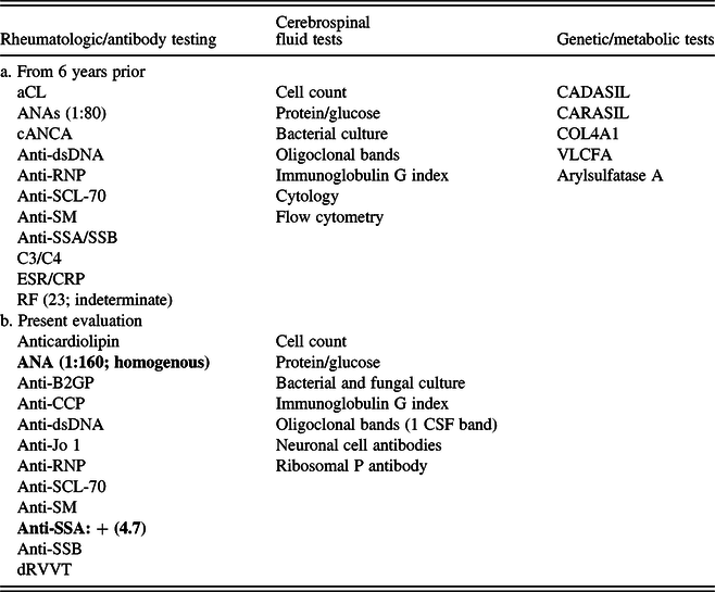
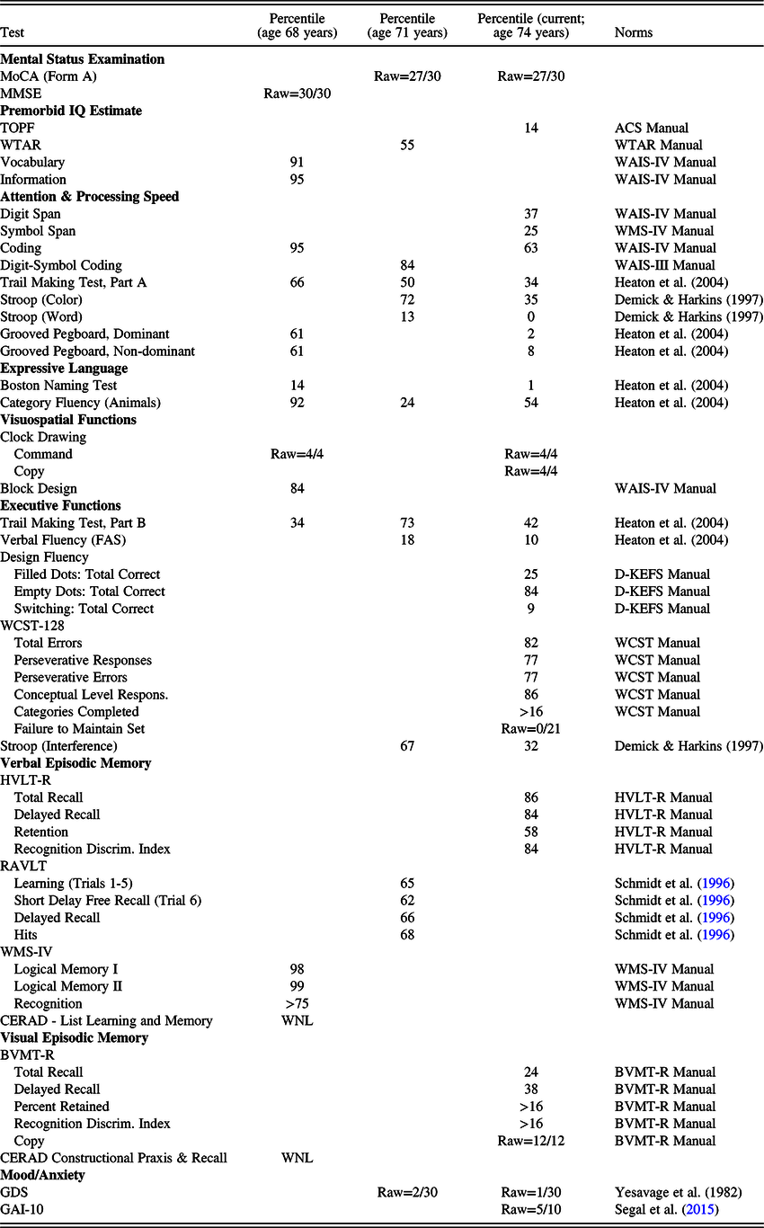
Her brain MRI abnormalities were first noted 6 years prior at the age of 68 years, as she had experienced acute onset of dizziness and vertigo and was admitted to a hospital for possible stroke (she previously had an episode of vertigo, although this had resolved after 3–4 days with meclizine). Brain MRI was negative for acute stroke but showed extensive leukoaraiosis of bilateral subcortical white matter and increased T2 signal in the pons, with no enhancement. Neurology subsequently ordered an extensive workup including computed tomography angiogram (normal), and metabolic, autoimmune, and genetic testing, all of which were negative (see Table 1.a). A neuropsychological evaluation at the time identified normal performance overall except for a select weakness in confrontation naming, consistent with her self-report (see Table 2, column 1).
Summary of the patient’s past laboratory and genetic testing

Note: Positive findings are in bold. All other findings are in negative or equivocal. ACE = angiotensin converting enzyme; ANAs = antinuclear antibodies; cANCA = anti-neutrophilic cytoplasmic antibodies; Anti-B2GP = anti-beta 2 glycoprotein; aCL = anti-cardiolipin; Anti-CCP = anti-cyclic citrullinated peptide; Anti-dsDNA = anti-double stranded DNA; Anti-RNP = anti-U1 ribonucleoprotein; Anti-SCL-70 = anti-topoisomerase (scleroderma); Anti-SM = anti-Smith; Anti-SSA = anti-Sjögren’s syndrome-related antigen A; Anti-SSB = anti-Sjögren’s syndrome-related antigen type B; C3/C4 = complement proteins C3 and C4; CADASIL = cerebral autosomal dominant arteriopathy with subcortical infarcts and leukoencephalopathy; CARASIL = cerebral autosomal recessive arteriopathy with subcortical infarcts and leukoencephalopathy; COL4A1 = collagen type IV alpha 1; CSF = cerebrospinal fluid; dRVVT = dilute Russell viper venom time; ESR/CRP = erythrocyte sedimentation rate and C-reactive protein; RF = rheumatoid factor; VLCFA = very long-chain fatty acids.
Summary of the patient’s neuropsychological test results by cognitive domain and assessment period

Notes: MoCA = Montreal Cognitive Assessment; MMSE = Mini Mental State Exam; WAIS-IV = Wechsler Adult Intelligence Scale – Fourth Ed.; WMS-IV = Wechsler Memory Scale – Fourth Ed.; D-KEFS = Delis–Kaplan Executive Function System; WCST-128 = Wisconsin Card Sorting Test 128 Card Version; CERAD = Consortium to Establish a Registry for Alzheimer’s Disease; HVLT-R = Hopkins Verbal Learning Test – Revised; RAVLT = Rey Auditory Verbal Learning Test; BVMT-R = Brief Visuospatial Memory Test – Revised; GDS = Geriatric Depression Scale; GAI-10 = Geriatric Anxiety Inventory – 10 item
Personal and Family Medical History
Relevant diagnoses included obstructive sleep apnea (managed with Continuous positive airway pressure (CPAP) therapy), mild depression, hypertension, and hyperlipidemia. The patient saw ophthalmology for dry eye syndrome, glaucoma, and cataracts. She had a past history of uveitis. She reported ulcers in her mouth for the previous 1–2 years, diagnosed as lichen planus. Surgical history included hysterectomy, oophorectomy for a noncancerous cyst, and knee replacement. She had severe measles with high fevers at the age of 6 years, prompting concerns about encephalitis but this was never diagnosed; she had no neurologic sequelae and returned to school without issue. She denied any history of anoxic episodes, known toxic exposures, or head injuries.
Current medications included a calcium channel blocker, aspirin, a statin, a selective serotonin reuptake inhibitor, a beta-3 adrenergic agonist, anastrozole for prophylaxis, an antihistamine, nasal spray for rhinitis, a steroidal nasal spray, a topical corticosteroid, eye drops (brimonidine, cyclosporine, and patanol), a stimulant laxative, a stool softener, vitamins and an omega 3 fatty acid supplement, as well as ibuprofen as needed. She endorsed good medication compliance overall and per her records she demonstrated a clear pattern of consistent engagement with various healthcare providers.
Her family medical history is noteworthy for neurological and psychiatric conditions. Her father had a history of alcoholism, depression, and dementia of unspecified etiology which reportedly first developed at the age of 59 years. Her mother had a stroke at the age of 95 years but was previously living independently. One sibling had a history of alcoholism and bipolar disorder with onset of cognitive decline and walking difficulties around the age of 60–70 years, currently diagnosed with vascular dementia and is no longer able to ambulate independently. Two other siblings reportedly have no neurologic or cognitive impairments.
Psychosocial History
The patient obtained her doctorate (20 years of education) in a humanities field and has a premorbid IQ that is estimated to be in the superior range (Table 2, column 1), suggesting high cognitive reserve. She had trouble with reading and with foreign languages in school but was never diagnosed with a learning disability or ever received educational remediation. She is married and has no biological children. She has never smoked. In her 30s, she drank alcohol excessively until her late 40s (2–4 glasses of wine/daily) and has only drunk wine occasionally since. She denies any history of substance abuse.
Clinical Examinations
Neurology
On mental status exam, the patient was alert and fully oriented with normal attention, memory, and fund of knowledge. Evaluation of language identified mild anomia with frequent phonemic paraphasic errors. She was able to describe the objects shown and correctly identified objects given multiple choice options. Propositional speech was normal and fluent. She could read irregular words but was impaired when reading nonwords. On frontal executive motor tasks, fist-edge-palm was normal. She had impaired crossed response inhibition (5 errors out of 10 trials). The remainder of her neurologic exam was normal with the exception of right eye ptosis (present since childhood), 3+ reflexes in all extremities and bilateral crossed adductors (downgoing plantar reflex bilaterally), decreased sensation to pinprick and temperature in her feet and lower calves, a mild intention tremor (L > R) on finger to nose testing, and an impaired tandem gait.
Neuroradiology
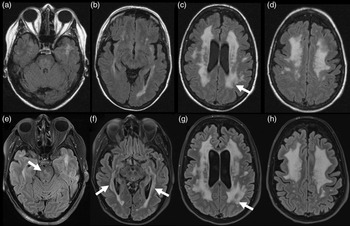
A board-certified neuroradiologist reviewed all of the patient’s MRI studies and described the following results. There was extensive scattered and confluent T2 prolongation, present in subcortical and periventricular white matter of both hemispheres including the temporal lobes as well as in the brain stem, nonspecific in appearance and distribution but most likely represent the sequelae of severe small vessel ischemic disease (Figure 1). These results were stable compared to images obtained 1 and 3 years prior, and only slightly progressed compared to exam 6 years prior, particularly in the peritrigonal region. Mild interval cerebral volume loss was noted compared to the study 6 years prior, but with no significant changes in the degree of volume loss over the last 3 years. In the left lateral precentral gyrus, an 8-mm area of T1 prolongation was observed in the subcortical white matter, stable since the first exam conducted 6 years prior. This was considered to be of uncertain etiology but may represent an area of encephalomalacia surrounded by gliotic changes. Her exam was otherwise unremarkable.
Axial T2 FLAIR images of the patient 6 years earlier at the age of 68 years (Panels A–D) and during the present evaluation at the age of 74 years (Panels E–H). Images of the patient at the age of 74 years (Panels E–H) revealed extensive confluent white matter T2 prolongation, present in the subcortical and periventricular white matter of both cerebral hemispheres including the temporal lobes (see arrows at Panel F), as well as in the brain stem (see arrow at Panel E), nonspecific in appearance and distribution but deemed most likely to represent the sequelae of severe small vessel ischemic disease. Findings were only slightly progressed compared to images obtained 6 years earlier at the age of 68 years, especially in the parietal regions (see arrows at Panels C and G). Mild interval cerebral volume loss was noted when compared to the prior examination.

Neuropsychology
Comprehensive testing revealed intact cognition with minimal deficits to correlate with her severe imaging results (Table 2) and three noteworthy findings. First, fine motor dexterity and coordination were in the impaired/borderline ranges bilaterally. This finding suggests non-lateralized slowing, although her overall presentation and intact performance on other speeded tasks argue against globally diminished processing speed. Second, confrontation naming was impaired, but she correctly identified all missed items with phonemic or multiple choice cues. This finding suggests compromised lexical access with preserved semantic representations, which is consistent with her weaker performance on letter relative to semantic fluency. It is possible that these findings are consistent with deficits in rapid automatized naming seen in developmental dyslexia (Wolf & Bowers, Reference Wolf and Bowers1999). Indeed, her performance on the word but not the color condition was impaired on the Stroop, and her ability to pronounce words with irregular grapheme-to-phoneme translation was low average, belying her high educational attainment. There was no evidence for clinically significant depression or diminished effort that may better account for her performance. Third, compared to her prior neuropsychological results for clinical and other research purposes 3 and 6 years previously, her current performance was remarkably consistent except for: a 1 standard deviation decline on confrontation naming, 2.5–3 standard deviation decline in fine motor dexterity bilaterally, and 1 standard deviation decline in aspects of processing speed, as above (i.e., coding and word reading speed).
Rheumatology
Due to her history of mouth ulcers and uveitis, she was also evaluated by rheumatology. A laboratory evaluation (Table 1.b) was significant for positive antinuclear antibodies and Sjogren’s syndrome antibody (Anti-SSA). Given the combination of these results with her presenting symptoms, she was diagnosed with Sjogren’s and started on hydroxychloroquine, with reported benefit. She was subsequently re-evaluated clinically by neurology and completed a lumbar puncture to assess for evidence of inflammation or autoimmunity. The results of this were negative (Table 1.b) and conservative management has since been maintained.
Advanced Diffusion MRI: Identifying a Potential Mechanism for Brain Reserve
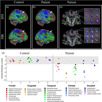
Figure 2 depicts the results of tractography analyses to provide a qualitative depiction of the patient’s white matter tracts using conventional (i.e., DTI) and advanced (i.e., FBI) diffusion MRI, vis-à-vis results from a well-matched cognitively unimpaired control (i.e., female, aged 77 years, 18 years of education). Figure 2A highlights FBI’s ability to resolve crossing fibers in the frontal lobe of the control in an area with complex fiber orientations from well-validated fiber bundles (i.e., genu of the corpus callosum, anterior corona radiata). Similarly, the patient’s FBI tractography resolves more fibers in this region (Figure 2B), suggesting a possible mechanism supporting the patient’s cognitive resilience as particularly evident in her overall intact executive functions relative to the severity of her leukoaraiosis in the frontal lobes. FBI is able to resolve complex white matter architecture via multi-directional diffusion orientation distribution functions (Figure 2C) and assesses diffusion within axons (i.e., via suppressing extra-axonal water), thereby revealing white matter tracts that are likely present and unresolved using DTI.
DTI and FBI tractography (A–C) and cortical thickness normed z-scores (D) of the control and the patient. (A and B) The sagittal slices include tracts with anterior-to-posterior (green), inferior-to-superior (blue), and left-to-right (red/magenta) directional orientations. Compared to DTI, FBI is better able to resolve crossing fibers particularly in the frontal regions (see tracts indicated by white arrows in A, particularly the red/magenta tracts using the FBI data). Compared to DTI, FBI resolves the potential presence of intact axons in the frontal regions (see tracts indicated by white arrows in B, particularly the green and red/magenta tracts using the FBI data). (C) This figure shows coronal slices of DTI and FBI fiber orientation distribution functions (ODFs) with a zoomed-in 3 × 3 matrix of voxels highlighting FBI’s ability to generate ODFs with multiple directions, thereby resolving crossing fibers. (D) This figure depicts the normed z-scores (y-axis) of the cortical thickness regions of interest within the F = Frontal, C = Caudate, T = Temporal, P = Parietal, and O = Occipital lobes (x-axes) for both the control and the patient, showing more aberrant cortical thickness measurements in the patient than the control across most regions. DTI = diffusion tensor imaging; FBI = fiber ball imaging.

Figure 2D depicts the cortical thickness measurements of lobar regions of interest segmented using FreeSurfer. FreeSurfer outputs were converted to z-scores using a large normative database accounting for age, sex, and estimated total intracranial volume (Potvin et al., Reference Potvin, Dieumegarde and Duchesne2017) to provide a qualitative comparison of individualized deviation scores for each region of interest for both the control and patient (cf. Supplementary Material). Compared to the control, the majority of the patient’s cortical thickness measurements were below the normative range (i.e., z = ±2.0), providing less support for cortical compensation and the relevance of cortical thickness as a measure of brain reserve in this particular case.
DISCUSSION
This case study illustrates cognitive resilience in the presence of severe leukoaraiosis and demonstrates how an advanced diffusion MRI method (i.e., FBI) can identify a potential mechanism of brain reserve. That is, as illustrated by tractography in both a control and the patient, FBI resolves white matter tracts with complex orientations and in areas where axonal architecture may remain intact despite alterations in the extra-axonal space (i.e., due to gliosis, myelin rarefaction, etc.). While advanced techniques such as this are still under development, individual case studies serve as powerful examples of their future potential to ascertain resilience in vivo. These findings have the following implications with regard to the patient, the concept of brain reserve, and the potential clinical use of advanced diffusion MRI.
This case illustrates the startling finding of intact functioning despite severe brain disease. While this patient demonstrated some declines over time (i.e., in processing speed and confrontation naming) that may reflect advancing vascular or rheumatologic disease, her overall intact abilities are remarkable given her severe leukoaraiosis. In contrast to conventional structural imaging, data from FBI suggest intact axonal architecture, revealing white matter that is not too discrepant from that of a similarly cognitively intact control. As implied by her cognitive resilience, these findings also suggest a favorable prognosis given her brain reserve.
This study adds to the literature by illustrating how an advanced diffusion MRI technique can yield other measures of brain reserve, specifically indicators of axonal architecture. However, the broader field of diffusion MRI has yet to achieve consensus regarding acquisition and analysis parameters for clinical use. Many developers agree that neuroscience is best served by going beyond DTI, but as to which advanced diffusion MRI technique to use remains up for debate (Le Bihan, Reference Le Bihan2013). It is especially important to analyze these data judiciously given the potential sources of bias and error endemic to all diffusion techniques from DTI to advanced ones like FBI (Jones & Cercignani, Reference Jones and Cercignani2010), including when subjecting these data to tractography analyses (Campbell & Pike, Reference Campbell and Pike2014). Achieving consensus on how best to acquire, analyze, and validate diffusion data will be critical for its eventual application as a measure of brain reserve.
This case study has some limitations. First, tractography is inherently a qualitative technique that serves as an in vivo estimate of white matter architecture. Only postmortem validation could verify our assertions. We also only had FBI data for one time point. Having multiple acquisitions and comparing their progression (ideally vis-à-vis a control’s progression) would strengthen the argument that her sustained cognitive resilience is mediated by the stability of her brain reserve. Another valuable extension of this work would be to examine the FBI results in another matched patient with similarly severe leukoaraiosis but with severe cognitive deficits. If the preserved axonal architecture highlighted by the FBI findings in our patient were absent in this cognitively impaired leukoaraiosis “control,” then we would have more compelling evidence supporting FBI as a brain reserve marker. At present, these data represent a preliminary demonstration of how an advanced technique resolves axonal architecture in a patient whose neuropsychological functioning belies what can be inferred from conventional techniques (i.e., DTI, cortical thickness).
Furthermore, the patient’s neurologic diagnosis remains unknown. As best we can ascertain, she has a rheumatologic condition, and although white matter lesions can be seen in Sjogren’s syndrome, the severity of her leukoaraiosis would be very atypical (Morgen et al., Reference Morgen, McFarland and Pillemer2004; McCoy & Baer, Reference McCoy and Baer2017). It is also possible that vascular disease, such as hypertension and hyperlipidemia, are the main drivers of her white matter pathology. However, while data from a large observational study (i.e., UK Biobank) suggest that hypertension is associated with diffusion MRI findings indicating alterations in axonal structure (Suzuki et al., Reference Suzuki, Gao, Bai, Evangelou, Glocker, O’Regan and Matthews2017), abnormal cholesterol was not shown to be uniquely associated with any MRI marker (Cox et al., Reference Cox, Lyall, Ritchie, Bastin, Harris, Buchanan and Deary2019). It is perhaps more likely that these risk factors are additive and synergistic in their impact (Cox et al., Reference Cox, Lyall, Ritchie, Bastin, Harris, Buchanan and Deary2019; Maillard et al., Reference Maillard, Carmichael, Reed, Mungas and DeCarli2015), and it remains difficult to ascertain these effects on an individual patient basis, particularly in this case where medication and treatment compliance was high and perhaps contributing to her reserve. Given the absence of a known etiology, we are unable to more definitively comment on the suspected pathology underlying her imaging findings, which is likely complicated given her history (i.e., possible childhood encephalitis, remote history of alcohol use disorder).
The extent to which FBI findings are both sensitive and specific to this patient’s unique set of circumstances or to a particular disease remains unknown. Nonetheless, this study serves to illustrate how an advanced technique such as FBI may improve upon the characterization of white matter integrity. Indeed, Figure 2 demonstrates better resolution of fiber architecture in both the control and the patient compared to DTI, which has significant implications for the investigation of white matter integrity as a method of quantifying brain reserve. A recent study demonstrated that greater occupational complexity (an index of cognitive reserve) in earlier adulthood was associated with better white matter microstructure and cognitive performance in mid-life, suggesting that reserve may attenuate cognitive and brain decline (Kaup et al., Reference Kaup, Xia, Launer, Sidney, Nasrallah, Erus and Yaffe2018). Importantly, this study did not find an association between occupational complexity with gray matter volume (another investigational measure of brain reserve), introducing the hypothesis that reserve may be conferred via white matter-mediated processes (e.g., axonal and myelin maintenance and plasticity) rather than changes to neuronal cell bodies or synaptic density approximated in vivo via gray matter morphometry (including cortical thickness, such as in this study). Utilizing advanced diffusion approaches in similar investigations may further explore this possibility given the greater fidelity with which it characterizes white matter microstructure, particularly if contrasted with other candidate measures of brain reserve. Future validation studies would ideally be longitudinal in design, given the importance of disentangling innate neurobiological capital (“brain reserve”) from resilience to age-related or pathological changes that may be modifiable (“brain maintenance”) (Nyberg et al., Reference Nyberg, Lövdén, Riklund, Lindenberger and Bäckman2012; Stern et al., Reference Stern, Arenaza-Urquijo, Bartrés-Faz, Belleville, Cantilon and Chetelat2018). In addition, future applications of diffusion MRI must be mindful of the fact that this technique is inherently an indirect estimation, and work integrating physical, numerical, and histological validation strategies are necessary to substantiate the biological inferences made in clinical studies (Dyrby et al., Reference Dyrby, Innocenti, Bech and Lundell2018; Fieremans & Lee, Reference Fieremans and Lee2018).
Despite these limitations, this study adds to the literature by introducing a clinical application of an advanced technique that may illuminate mechanisms of brain reserve. The current findings suggest that reserve may not only be implied based on discrepant cognitive and neuroimaging findings, but could potentially be quantified using advanced diffusion MRI. This project has also offered our patient some insight into her diagnostic conundrum as well as some hope, given that her previous diagnosis (i.e., leukoencephalopathy) had a far worse prognosis. Future work remains necessary to verify the application of this novel technique across individuals and populations and to further develop the construct of brain reserve for clinical use.
ACKNOWLEDGMENTS
We would like to acknowledge the early clinical workup and professional input provided by our colleagues Drs. Tanya Turan, Mark Wagner, Gisele Matheus, and Melissa Cunningham. We would also like to acknowledge Kayti Keith for her assistance with plotting the data. We especially would like to thank our patient, who generously gave her time and effort so that we could make this meaningful contribution to her care and to science. This project was supported by the Rare Disease Foundation Microgrant and the National Institutes of Health (K23 AG044434 and R01 AG054159).
CONFLICT OF INTEREST
The authors have nothing to disclose.
SUPPLEMENTARY MATERIAL
To view supplementary material for this article, please visit https://doi.org/10.1017/S1355617720000569