Introduction
Giant cell tumour of the tendon sheath (GCTTS) is the second most common tumour of the hand after ganglion cysts. Reference Coroneos, O’Sullivan, Ferguson, Chung and Anastakis1 It is a slow growing, painless benign lesion of soft tissues. The tumour affects individuals between the age of 30 and 50 years old and is seen more often in women with the most common site being fingers. These benign tumours have the capacity for local recurrence as they tend to spread to the neighbouring synovium rendering complete excision often tricky. Definitive surgery with wide excision margins is the treatment of choice. Up to 45% of cases recur locally after excision Reference Ozben and Coskun2 hence post-operative radiotherapy (PORT) has been tried in the past to reduce recurrences and was found promising. Reference Coroneos, O’Sullivan, Ferguson, Chung and Anastakis1,Reference Kotwal, Gupta and Malhotra3
With the use of PORT, there are reports of decreased range of motion of the affected joint, sensory changes and nail changes. Reference Steen, Hayward, Novak, Anastakis and McCabe4 These secondary effects are directly correlated to the dose received to the skin, phalanges, nail bed etc. There are various choices of radiotherapy technique available which includes external beam radiotherapy (EBRT) using photons or electrons and surface mould brachytherapy. Among these, surface mould brachytherapy is the least used modality, possibly due to less technical proficiency.
In this paper, we present a case of GCTTS of left-hand index finger and illustrate a surface mould brachytherapy technique. We will also compare this dosimetrically with conventional EBRT using photons and electrons. Good dosimetry may result in reduced secondary effects of radiotherapy.
Case Presentation
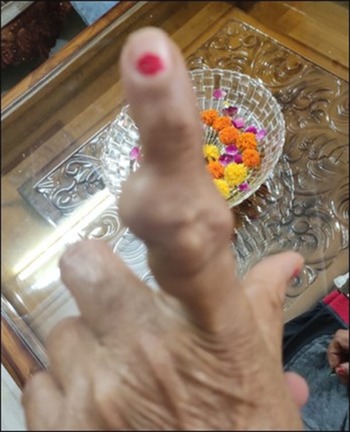
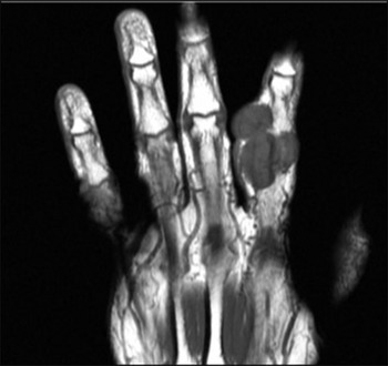
A 66-year female presented with a slow-growing swelling on dorso-lateral surface of the left index finger for 1 year. It was associated with mild-to-moderate pain and restriction of finger movement. On clinical examination a multi-lobulated swelling of 40 × 35 mm at dorso-lateral aspect of left index finger found which was partially mobile was found (Figure 1). An MRI of the left hand suggested a lobulated cystic swelling 35 × 30 × 34 mm around proximal inter-phalangeal joint of left index finger not eroding underlying bone (Figure 2). The patient underwent wide local excision of swelling and histopathology showed a proliferation of fibroblast and histiocytes with collagen in between the cells interspersed with scattered giant cells suggesting GCTTS with unknown margin status. After excision of the tumour, the patient was considered for radiation therapy in view of the high risk of recurrence after surgery, due to unknown margin status.
Showing index presentation of GCTTS of the left index finger.

Showing MRI GCTTS of the left index finger.

Materials and Methods
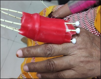
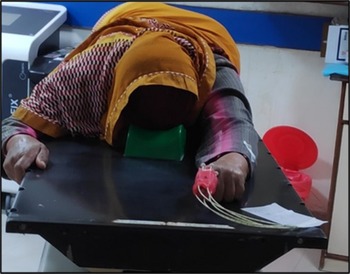
A customised thermoplastic thumb mould was made with a thermoplastic sheath such that the finger could easily slide through it. A 5-mm thick paraffin wax bolus was wrapped around the mould. The basic rationale was to increase the gap between the applicator needles and the skin thus avoiding a high dose to the skin at the time of treatment. Five applicator needles each of 8-cm length were placed at a uniform spacing of 1 cm each on the thermoplastic mould (Figure 3). These applicators were tagged for the purpose of identification at the time of planning and treatment. A planning CT scan (Philips Brilliance Big Bore 16 slice CT scanner) was taken in a prone position with left arm extended, fist closed and index finger extended (Figure 4), slice thickness of 3 mm. CT scan was imported in Oncentra® brachy planning system (Elekta, Stockholm, Sweden). The pre-operative MRI fused with the planning CT scan and clinical target volume delineated by taking into account surgical scars. A margin of 2 mm superior-inferiorly and 1 mm laterally and anteriorly were added to create planning treatment volume (PTV) to account for any possible movement of the mould during multiple sessions of brachytherapy. Organs at risk (OARs) such as skin, phalangeal bone and nail bed were delineated. A treatment plan with a prescription dose (Dp) of 30 Gy in 10 fractions to PTV was generated for the 192-Ir HDR Microselectron® digital brachytherapy afterloading platform (Elekta, Stockholm, Sweden) (Figure 5). Treatment was given for 5 days, 2 fractions per day 6 hours apart.
Showing five-catheter surface mould brachytherapy of GCTTS of the left index finger.

Showing planning CT with surface mould brachytherapy.

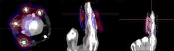
Showing surface mould brachytherapy dose distribution in axial, sagittal and coronal sections.

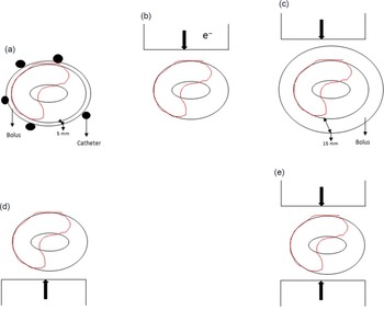
Another planning CT was taken, this time without the surface mould, in the same treatment position and the PTV was defined with the same parameters as before. A digitally reconstructed radiograph (DRR) based on 2D planning and using 10 MeV electron and 6 MV photon beams, several treatment plans were generated in Monaco® treatment planning system (Elekta, Stockholm, Sweden). Three different plans for 6 MV photons were generated, one with a bolus, the other two were without bolus (single posterior field and two anterior–posterior fields). Thus, adopting all the standard techniques that could be used to deliver PORT to the index finger (Figure 6). The same Dp of 30 Gy in 10 fractions was prescribed to PTV. The surface mould brachytherapy using 192-HDR, 10 MeV electron and multiple 6 MV photons treatment plans were compared for dosimetric superiority on the parameters of dose coverage to PTV, dose to OARs, Homogeneity index (HI), Conformity index (CI) as shown in Table 1.
Showing beam arrangements, the red structure is PTV. (a) Surface mould brachytherapy, (b) Direct 10 MeV electron, (c) DRR 6 MV photon with bolus, (d) DRR 6 MV photon without bolus PA and (e) DRR 6 MV photon without bolus AP-PA.

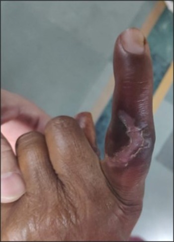
After 6 weeks of completion of surface mould brachytherapy treatment.

Showing dosimetric parameters of (1) Surface mould brachytherapy, (2) Direct 10 MeV electron, (3) DRR 6 MV photons with bolus anterior, (4) DRR 6 MV photons without bolus posterior field and (5) DRR 6 MV photons without bolus anterior–posterior. PTV-D95% is the percentage of the prescribed dose (% of Dp) received by 95% of volume, Dmax to the skin is the maximum percentage of the prescribed dose (% of Dp) received to 0·1 cc volume of skin, Dmax to phalanges is the percentage of prescribed dose (% of Dp) received to 0·1 cc volume of phalanges, Dmax to nail bed is the percentage of prescribed dose (% of Dp) received to 0·1 cc volume of nail bed, Heterogeneity index (HI) is dose received by 5% of PTV volume (D5) divided by is dose received by 95% of PTV volume (D95), Conformity index (CI) is volume covered by 100% dose isoline of Dp (VDp) divided by the volume of PTV.

Results
The DRR 6-MV photon treatment plan with a bolus plan provided the least dose to skin (106%) and phalanges (103%). It has HI (1·06) closest to 1, whereas CI of all plans was similar. The dose coverage was adequate in all plans. The second-best plan dosimetrically was the surface mould brachytherapy.
Discussion
GCTTS is interchangeably called as pigmented villonodular synovitis. It is a benign tendon sheath swelling, although various case series have shown local recurrences ranging from 7 to 45%. The risk of recurrence is more in (a) incomplete excision; (b) the presence of mitotic figures on histological examination; (c) involvement of bone or (d) site of lesion. Reference Ozben and Coskun2,Reference Di Grazia, Succi, Fraggetta and Perrotta5 Multiple local recurrences can lead to multiple surgeries which further leads to an increase in morbidity such as decrease range of motion, reduced sensation and amputation.
Post-operatively radiotherapy has shown to be very promising in reducing recurrences in various case series. PORT could demonstrate a local control rate of up to 100% when using total RT doses of 20–50 Gy. Reference Coroneos, O’Sullivan, Ferguson, Chung and Anastakis1,Reference Kotwal, Gupta and Malhotra3,Reference O’Sullivan, Cummings and Catton6 Some authors have reported no long-term effects of radiotherapy, but some have shown concerns and reported secondary effects of radiation mainly late toxicity. Reference Steen, Hayward, Novak, Anastakis and McCabe4 Sensory changes, joint stiffness, nail changes, pigmentation changes, decreased range of movements, skin atrophy, dryness, hair loss and itching were reported in decreasing order. Although no fracture of the digit, bony pain and ulceration were reported. Reference Steen, Hayward, Novak, Anastakis and McCabe4 As these are deterministic effects, these toxicities are directly proportional to radiation doses received to the organ at risk structures such as skin, nail bed and phalangeal bone. The radiotherapy technique which can decrease the doses to these OARs may decrease the secondary effects of radiation. In anatomical locations such as the thumb, radiation treatment with conventional photon and electron beams may result in excessive exit dose to the nail bed. Electron treatment may result in an inhomogeneous dose distribution potentially under-dosing the target volume. The surface mould brachytherapy radiation delivery technique is an attractive alternative technique for treating tumours located at such sites. It is the least adopted technique and only one case report is available. Reference Goda, Patil, Krishnappan and Elangovan7 The possible reason for this could be a lack of technical proficiency.
PTV-D95% represents the dose coverage to PTV. Usually, D95 above 95% is considered adequate. In all the three modalities, we were able to achieve adequate dose coverage. Skin, phalanges and nail bed are the OARs. The HI is an informative parameter to analyse the dose distribution uniformity in the target volume. The ideal value of HI is 1—the closer the value to 1, the more uniform the distribution of dose. Having a uniform dose distribution reduces the risk of secondary effects of radiation. Reference Kataria, Sharma, Subramani, Karrthick and Bisht8 The CI analyses the coverage of target volume by prescribed dose. CI equal to 1 is an ideal scenario where dose coverage to the target volume is 100% of the prescribed dose. Greater than 1 shows that the irradiated volume is greater than the target volume and includes healthy tissues whereas less than 1 shows the target volume is not completely covered. Reference Feuvret, Noël, Mazeron and Bey9 Better dosimetry correlates to a reduced risk of secondary effects. In our case, the DRR plan of 6 MV photons with a bolus provided the most homogenous and conformal plan with a reduced dose to the skin and phalanges. Except dose to nail bed, it is the best plan followed by surface mould brachytherapy. Perhaps, increasing the wax bolus thickness of surface mould brachytherapy would have increased the distance between source and finger, and may have resulted in a reduced dose to the skin and phalanges.
Our patient, treated by surface mould brachytherapy, developed grade II CTCAE V5 radiation dermatitis in week 3 post-treatment (Supplementary Figure S8), progressing to a grade III reaction in week 4 following completion of radiotherapy (Supplementary Figure S9). The patient was managed conservatively and skin reaction started healing by week 6G (Figure 7). The patient also complained of pain and dryness of the skin, which was managed conservatively and at week 6 the patient had minimal pain.
Conclusion
Surface mould brachytherapy is feasible in index finger GCTCS to deliver PORT. In our case, the treatment plan for EBRT using 6 MV photons with bolus was dosimetrically superior to the treatment plan of surface mould brachytherapy, electron and photon without bolus.
Funding
The authors received no financial support for the research, authorship, and/or publication of this article.
Acknowledgements
None.
Ethical Approval
Ethical approval is not required at our institution for publishing an anonymous case.
Statement of Human and Animal Rights
All procedures followed were in accordance with the ethical standards of the responsible committee on human experimentation (institutional and national) and with the Helsinki Declaration of 1975, as revised in 2008.
Statement of Informed Consent
Informed consent was obtained from patient for being included in the study. Additional informed consent was obtained for identifying information included in the article.
Declaration of Conflicting Interests
The authors declared no potential conflicts of interest with respect to the research, authorship, and/or publication of this article.
Supplementary material
To view supplementary material for this article, please visit https://doi.org/10.1017/S1460396920000965.