Introduction
Working memory (WM) deficit, a core feature of cognitive impairment in schizophrenia, appears to be a persistent phenomenon that is closely related to poor long-term outcome (Coyle, Reference Coyle2006; Minzenberg & Carter, Reference Minzenberg and Carter2012), but is often resistant to anti-psychotic drugs (MacDonald & Schulz, Reference MacDonald and Schulz2009). The lack of a coherent insight into the neuropathological mechanism of WM deficits has been a critical hurdle in the development of remedial treatments (Lett et al. Reference Lett, Voineskos, Kennedy, Levine and Daskalakis2014). For example, conflicting results, both prefrontal hypoactivity (Barch et al. Reference Barch, Carter, Braver, Sabb, MacDonald, Noll and Cohen2001) and hyperactivity (Manoach et al. Reference Manoach, Gollub, Benson, Searl, Goff, Halpern, Saper and Rauch2000) during WM tasks, have been observed in patients with schizophrenia when compared with healthy controls. Recent functional magnetic resonance imaging (fMRI) experiments in healthy subjects suggest that the construct of WM not only arises from the activation of local brain regions responsible for high-order cognitive functions (e.g. prefrontal cortex; PFC), but also critically depends on the integration of large-scale brain networks (Sala-Llonch et al. Reference Sala-Llonch, Pena-Gomez, Arenaza-Urquijo, Vidal-Pineiro, Bargallo, Junque and Bartres-Faz2012; Liang et al. Reference Liang, Zou, He and Yang2015). Thus, investigation on the dynamic interaction of large-scale brain networks during WM processing in schizophrenia may help to provide further insight into the mechanism of WM deficits in this severe mental disorder.
Of the major brain networks regarding to WM deficits in schizophrenia, an external attention system (EAS) and a default mode network (DMN) have drawn the most intensive attention in this field (Whitfield-Gabrieli et al. Reference Whitfield-Gabrieli, Thermenos, Milanovic, Tsuang, Faraone, McCarley, Shenton, Green, Nieto-Castanon and LaViolette2009; Metzak et al. Reference Metzak, Riley, Wang, Whitman, Ngan and Woodward2012; Anticevic et al. Reference Anticevic, Repovs and Barch2013; Fryer et al. Reference Fryer, Woods, Kiehl, Calhoun, Pearlson, Roach, Ford, Srihari, McGlashan and Mathalon2013). The EAS, encompassing the dorsal prefrontal and parietal cortex, dorsal cingulate cortex and anterior insula, is often activated when performing external attention-demanding tasks, and has been directly related to top-down attentional and cognitive control (Fornito et al. Reference Fornito, Harrison, Zalesky and Simons2012). In contrast, the DMN, mainly anchored at the midline brain regions, typically shows decreased activation during external cognitive processing, but increased activation during internal cognitive processing (e.g. self-referential thinking and autobiographical memory) (Buckner et al. Reference Buckner, Andrews-Hanna and Schacter2008). In schizophrenia, robust evidence exists for both abnormal activation and functional dysconnectivity within the EAS during WM tasks (Kim et al. Reference Kim, Tura, Potkin, Fallon, Manoach, Calhoun, FBIRN and Turner2010; Barch & Ceaser, Reference Barch and Ceaser2012; Kyriakopoulos et al. Reference Kyriakopoulos, Dima, Roiser, Corrigall, Barker and Frangou2012; Anticevic et al. Reference Anticevic, Repovs and Barch2013), providing an important lead to the neural basis of WM deficits. Recently, hyperactivity (i.e. non-suppression) and hypo-connectivity in the DMN have also been observed in patients with schizophrenia during WM tasks (Whitfield-Gabrieli et al. Reference Whitfield-Gabrieli, Thermenos, Milanovic, Tsuang, Faraone, McCarley, Shenton, Green, Nieto-Castanon and LaViolette2009; Anticevic et al. Reference Anticevic, Repovs and Barch2013; de Leeuw et al. Reference de Leeuw, Kahn, Zandbelt, Widschwendter and Vink2013). This hyperactivity of the DMN is proposed to reflect an inability of patients to reallocate the attentional resources away from introspectively oriented mental activity and towards the demands posed by external stimuli when performing cognitive tasks (Whitfield-Gabrieli et al. Reference Whitfield-Gabrieli, Thermenos, Milanovic, Tsuang, Faraone, McCarley, Shenton, Green, Nieto-Castanon and LaViolette2009). Considering the specific roles of both networks in attention control related to WM performance, one might anticipate that an abnormal dynamic interaction between the EAS and DMN could be a crucial neuropathological mechanism underlying WM deficits in schizophrenia.
Traditionally, resting-state fMRI studies indicate that the default network is anticorrelated with the task-positive network (i.e. EAS) (Fox et al. Reference Fox, Zhang, Snyder and Raichle2009). Recent evidence, however, suggests that these two networks may not always ‘compete’ (negatively correlated), but indeed ‘cooperate’ (positively correlated), during certain cognitive processes including attention (Spreng et al. Reference Spreng, Stevens, Chamberlain, Gilmore and Schacter2010), WM (Bluhm et al. Reference Bluhm, Clark, McFarlane, Moores, Shaw and Lanius2011) and recollection tasks (Fornito et al. Reference Fornito, Harrison, Zalesky and Simons2012). This suggests that certain task demands may reconfigure the dynamic interactions between these networks from an intrinsic state of competition into a task-dependent cooperation. Most importantly, both the negative and positive correlations between the EAS–DMN networks relate to better cognitive performance in healthy subjects (Eichele et al. Reference Eichele, Debener, Calhoun, Specht, Engel, Hugdahl, von Cramon and Ullsperger2008; Kelly et al. Reference Kelly, Uddin, Biswal, Castellanos and Milham2008; Leech et al. Reference Leech, Kamourieh, Beckmann and Sharp2011; Fornito et al. Reference Fornito, Harrison, Zalesky and Simons2012; Sala-Llonch et al. Reference Sala-Llonch, Pena-Gomez, Arenaza-Urquijo, Vidal-Pineiro, Bargallo, Junque and Bartres-Faz2012; Wen et al. Reference Wen, Liu, Yao and Ding2013). Thus on one hand, the greater anti-correlation between the two networks facilitates the attention to be shifted effectively from the introspective mental activity to extrospective processing (Fox et al. Reference Fox, Zhang, Snyder and Raichle2009; Sala-Llonch et al. Reference Sala-Llonch, Pena-Gomez, Arenaza-Urquijo, Vidal-Pineiro, Bargallo, Junque and Bartres-Faz2012). On the other hand, the DMN (especially the posterior cingulate cortex; PCC) functions as a transitional module (or ‘hub’) in the brain (Margulies et al. Reference Margulies, Vincent, Kelly, Lohmann, Uddin, Biswal, Villringer, Castellanos, Milham and Petrides2009; Fornito et al. Reference Fornito, Harrison, Zalesky and Simons2012; Leech et al. Reference Leech, Braga and Sharp2012), to integrate with other modules such as the EAS to support goal-directed cognitive processing. Therefore, optimal cognitive performance may rely on both ‘competition’ and ‘cooperation’ between the DMN and EAS, depending on the demands placed. In schizophrenia, despite the consistent demonstration of disrupted interaction between large-scale brain networks during resting state (Mannell et al. Reference Mannell, Franco, Calhoun, Canive, Thoma and Mayer2010; Liu et al. Reference Liu, Kaneko, Ouyang, Li, Hao, Chen, Jiang, Zhou and Liu2012), it is unclear whether and how this disruption influences the WM deficits seen in patients.
Recently, fMRI studies applying Granger causality analysis (GCA) in healthy subjects found that the DMN, in particular the PCC node, influences EAS activity during both resting-state (Uddin et al. Reference Uddin, Kelly, Biswal, Castellanos and Milham2009) and unconscious information processing (De Pisapia et al. Reference De Pisapia, Turatto, Lin, Jovicich and Caramazza2012). Our prior work adopting GCA has revealed a breakdown of the salience-execution loop at rest (disrupted reciprocal causal connectivity between insula and dorsal PFC) in schizophrenia (Palaniyappan et al. Reference Palaniyappan, Simmonite, White, Liddle and Liddle2013). Employing a well-established preprocessing pipeline for GCA in fMRI data, we studied the putative ‘competitive’ and ‘cooperative’ interactions between the EAS and DMN during an n-back WM task in patients with schizophrenia (SCZ) and healthy controls (HC). We expected to see reduced DMN suppression during the WM task in SCZ, and hypothesized that: (1) the dynamic interactions between the EAS and DMN would be disrupted in patients; (2) and both DMN suppression and disrupted EAS–DMN interaction would relate to WM performance in both groups and to clinically observed attention impairment in SCZ.
Method
Participants
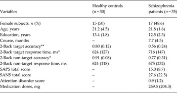
Using the Structured Clinical Interview for DSM-IV – patient version (SCID-P) (First et al. Reference First, Spitzer and Gibbon1996), 35 first-episode SCZ patients were recruited from in-patient and out-patient units of the Department of Psychiatry, the Second Xiangya Hospital of Central South University, Changsha, Hunan province, People's Republic of China. The inclusion criteria were: (a) age between 18 and 45 years; (b) Han Chinese ethnicity; (c) 9 years of education or above; (d) right-handed by a determination of hand preference; (e) meeting DSM-IV criteria for schizophrenia; (f) total duration of illness ⩽18 months. The exclusion criteria were: (a) any contra-indications to MRI scanning; (b) substance-related disorders; (c) recorded loss of consciousness (duration >5 min); (d) chronic neurological disorders or severe physical diseases. All the patients were receiving second-generation antipsychotic drugs, and all the medication doses were converted to chlorpromazine equivalence (50–1000 mg/day; Table 1). The clinical symptoms of the SCZ patients were evaluated by two experienced psychiatrists using the Scale for the Assessment of Positive Symptoms (Andreasen, Reference Andreasen1984) and Scale for the Assessment of Negative Symptoms (Andreasen, Reference Andreasen1983), in which the attention disorder subscale of the SANS was used to evaluate the attentional impairment in SCZ patients (Table 1).
Sociodemographic and behavioural variables of healthy controls and schizophrenia patients

Data are given as mean (standard deviation) unless otherwise indicated.
* p < 0.05, ** p < 0.01.
We recruited 30 HC from a community sample in Changsha city. The inclusion and exclusion criteria were the same as those of the patient group except that the HC did not meet the DSM-IV diagnostic criteria of any psychiatric disorders when interviewed using the SCID non-patient version. All participants gave voluntary informed consent for participation in the study, with procedures approved by the ethics committee of the Second Xiangya Hospital of Central South University.
Experimental design, fMRI data acquisition and analysis
The details of the task design, image acquisition protocol, preprocessing steps for the image data , and general linear model for brain activity analysis are presented in the online Supplementary Text, section S.1, S.2 and S.3, and also described elsewhere (Wu et al. Reference Wu, Wang, Mwansisya, Pu, Zhang, Liu, Yang, Chen, Xue, Liu and Shan2014).
Behavioural correlation analysis for brain activities
The task-related brain activity was calculated by averaging the statistical parameters of the contrast map (2-back v. resting state) in the seed regions across blocks and subjects. Then the brain activities in the above-defined seed regions were related to the 2-back target accuracy scores in the SCZ patients and the HC separately and to the SANS attention disorder scores in SCZ patients.
GCA
GCA has been employed in neuroscience to decompose the dynamic interaction between brain regions into directed causal influences (Ding et al. Reference Ding, Chen, Bressler, Schelter, Winterhalder and Timmer2006; Friston et al. Reference Friston, Moran and Seth2013). GCA is based on the notion that if including the historical information of one time series improves the prediction of the future states of another time series, then the first time series Granger-causes the second one. To test our hypothesis in the present study, Granger causalities were estimated in two directions both from the seed region (region of interest; ROI) to the whole brain and the opposite direction. By using the ‘ROI to whole-brain and whole-brain to ROI’ approach, instead of the ‘ROI to ROI’ method, we were able to establish that the brain networks of a priori interest had a robust effect that was not seen in other networks/regions, after stringent correction for the whole-brain analysis, thus establishing spatial specificity of the results.
The seed regions for the brain-wide GCA were defined by the clusters in DMN and EAS regions showing significantly altered activity in the SCZ patient group compared with that in the HC during the 2-back blocks. The DMN is often described to be comprised of the medial PFC (mPFC), PCC/precuneus (PCUN) and inferior parietal lobe (Buckner et al. Reference Buckner, Andrews-Hanna and Schacter2008). The EAS, also called the task-positive or extrinsic network, consists of three divergent subsystems (Fornito et al. Reference Fornito, Harrison, Zalesky and Simons2012), namely the fronto-parietal network (FPN) anchored at the dorsolateral PFC (DLPFC) and intra-parietal lobule (IPL) (Vincent et al. Reference Vincent, Kahn, Snyder, Raichle and Buckner2008), the dorsal attention network (DAN) (Fox et al. Reference Fox, Snyder, Vincent, Corbetta, Van Essen and Raichle2005; Corbetta et al. Reference Corbetta, Patel and Shulman2008) composed of the frontal eye field (FEF) and intra-parietal sulcus (IPS), and the salience network (SN) (Dosenbach et al. Reference Dosenbach, Visscher, Palmer, Miezin, Wenger, Kang, Burgund, Grimes, Schlaggar and Petersen2006; Seeley et al. Reference Seeley, Menon, Schatzberg, Keller, Glover, Kenna, Reiss and Greicius2007) mainly including the dorsal anterior cingulate cortex (dACC) and anterior insula. The FPN and DAN have been associated with top-down executive control and focusing attention on external stimuli respectively, and the SN has been implicated in processing salient stimuli and introspective awareness (Dosenbach et al. Reference Dosenbach, Visscher, Palmer, Miezin, Wenger, Kang, Burgund, Grimes, Schlaggar and Petersen2006; Corbetta et al. Reference Corbetta, Patel and Shulman2008; Sridharan et al. Reference Sridharan, Levitin and Menon2008; Vincent et al. Reference Vincent, Kahn, Snyder, Raichle and Buckner2008; Fornito et al. Reference Fornito, Harrison, Zalesky and Simons2012). As reported in the Results section, our task activation analysis found abnormal activity in the DMN (i.e. reduced suppression of the bilateral mPFC and PCC/PCUN) in the SCZ group (online Supplementary Table S1), but not in the EAS regions. Thus, the bilateral clusters in the mPFC and PCC/PCUN, observed as reduced suppression in the 2-back WM task, were defined as the seed regions for the further GCA analysis. Then Granger causality was calculated in two directions both from the bilateral mPFC and PCC/PCUN to the whole brain and the opposite direction. Since no cluster with significantly abnormal activity was detected in the current study, we did not have any seed regions in the EAS.
In line with our prior fMRI studies (Palaniyappan et al. Reference Palaniyappan, Simmonite, White, Liddle and Liddle2013), we used first-order GCA with the time lag of one repetition time (TR) (2000 ms) to study the directional effects and signed-path coefficients as the index of Granger causality. This signed path coefficient has been used in both rest (Chen et al. Reference Chen, Hamilton, Thomason, Gotlib, Saad and Cox2009; Palaniyappan et al. Reference Palaniyappan, Simmonite, White, Liddle and Liddle2013) and task fMRI data analysis (Voon et al. Reference Voon, Brezing, Gallea, Ameli, Roelofs, LaFrance and Hallett2010; Wu et al. Reference Wu, Wang, Wang, Hallett, Zang, Wu and Chan2012). A path coefficient of 0 indicates the absence of any directional Granger causal influence between two brain regions. The positive/negative path coefficients suggest that the change in the blood oxygen level-dependent (BOLD) signal of the ‘causal’ region leads to the change in the BOLD signal of the ‘effect’ region in the same/opposite direction. We refer to the positive path as an ‘excitatory’ effect and the negative path as an ‘inhibitory’ effect (Palaniyappan et al. Reference Palaniyappan, Simmonite, White, Liddle and Liddle2013) but this must be differentiated from the terms excitation/inhibition used in neurochemical models. To improve the reliability of the estimation of the causal coefficient, we averaged the estimated causal coefficients across the repeating four 2-back task blocks (Luo et al. Reference Luo, Ge, Grabenhorst, Feng and Rolls2013). Before carrying out GCA, we regressed out the six head movement parameters from the BOLD time series, and removed the first-order time trend together with the temporal mean from the time series in each block, to avoid the possible confounding effects (Ding et al. Reference Ding, Chen, Bressler, Schelter, Winterhalder and Timmer2006). Using bivariate Granger causality (Hamilton et al. Reference Hamilton, Chen, Thomason, Schwartz and Gotlib2011), we established the path coefficient map for each subject. One-sample t tests were performed separately in SCZ patient and HC groups, to identify the brain regions with the most significant directional influences to and from the seed regions. The GCA maps of SCZ patients and HC were compared by two-sample t test [false discovery rate (FDR) correction, p < 0.05]. The results reported by GCA were systematically compared with the results in the literature, to make sure that the new insight of WM deficit in schizophrenia revealed by GCA is rooted in the published literature.
Since recent studies (Power et al. Reference Power, Barnes, Snyder, Schlaggar and Petersen2012; Van Dijk et al. Reference Van Dijk, Sabuncu and Buckner2012) have suggested that subtle head movement is an important confounding factor for fMRI functional connectivity (FC) analysis, we also performed ‘scrubbing’ to ensure that head-motion artifacts are not driving the observed effects. An estimate of head motion at each time-point was calculated as the frame-wise displacement (FD). Following previous studies as reported by Power et al. (Reference Power, Barnes, Snyder, Schlaggar and Petersen2012), any image with FD > 0.5 was removed and replaced by a linear interpolation. The mean absolute FD between HC and SCZ patients did not differ significantly (mean HC: 0.16 (s.d. = 0.06), mean SCZ: 0.19 (s.d. = 0.07), n.s.). After that, we repeated the calculation of GCA maps, which further entered into a contrast between HC and SCZ patients in a two-sample t test. The results were very similar with and without motion scrubbing.
Behavioural correlation analysis for causal influences
Since the DMN-to-EAS influence was found altered in the SCZ patients, then the DMN-to-whole-brain maps were related to 2-back target accuracy scores in the SCZ patients and the HC separately and to the SANS attention disorder scores in the SCZ patients only. For multiple comparison correction for the behavioural correlation, a small volume correction centred at the peak voxels with a 3 mm radius sphere was conducted within the hypothesized EAS regions showing group differences in the GCA second-level analysis (Table 2).
Disrupted causal connectivity from bilateral PCC to whole brain in schizophrenia patients compared with healthy controls

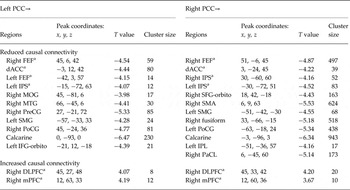
PCC, Posterior cingulate cortex; FEF, frontal eye field; dACC, dorsal anterior cingulate cortex; IPS, intra-parietal sulcus; MOG, middle occipital gyrus; SFG-orbito, superior frontal gyrus orbital pars; MTG, middle temporal gyrus; SMA, supplementary motor area; PreCG, precentral gyrus; SMG, supramarginal gyrus; PoCG, post-central gyrus; IFG-orbito, inferior frontal gyrus orbital pars; IPL, inferior parietal gyrus; PaCL, paracentral lobule; DLPFC, dorsal lateral prefrontal cortex; mPFC, medial prefrontal cortex.
a Regions corresponding to Fig. 2 b.
Results
The sample characteristics are presented in Table 1, showing no differences in age, sex and years of education between the two groups. Compared with the HC group, the SCZ group showed reduced 2-back WM accuracy both for targets (p < 0.001) and non-targets (p < 0.05), and longer reaction time to targets (p < 0.05).
Brain activation patterns during the 2-back WM task
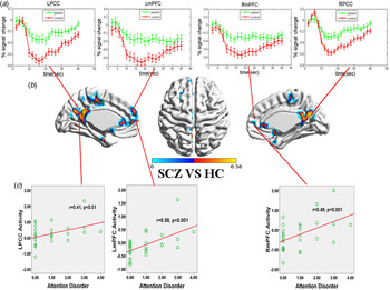
During the 2-back WM task, the DMN regions were deactivated and the EAS regions were activated in both the HC and SCZ groups (online Supplementary Fig. S1). A two-sample t test (FDR correction, p < 0.05) showed that patients had significantly increased brain activity (i.e. ‘non-suppression’) in the DMN regions (Fig. 1 b, and online Supplementary Table S1), including the bilateral mPFC, PCC/PCUN, hippocampus and inferior parietal lobe. Other regions showing increased activity in SCZ included the cuneus, fusiform gyrus and temporal pole (online Supplementary Table S1). We found no areas showing significantly altered activity within the EAS regions in the SCZ patient group relative to the HC group. Averaged across all 2-back blocks, the mean signals detected at the group differential DMN regions are shown in Fig. 1 a.
Group differences in brain activity during the working memory (WM) task between healthy controls (HC) and schizophrenia (SCZ) patients. (a) Comparison of the mean blood oxygen level-dependent (BOLD) signals (percentage of change) averaged across all 2-back blocks in the SCZ group with that in the HC group. The error bars represent the standard error of the mean signal at each time point. LPCC, Left posterior cingulate cortex; LmPFC, left medial prefrontal cortex; RmPFC, right medial prefrontal cortex; RPCC, right posterior cingulate cortex. (b) The brain maps show two-sample t test maps of BOLD differences during the WM task between HC and SCZ (false discovery rate correction, p < 0.05). (c) Significant correlations between attention disorder and brain activity of default mode network regions with group differences during the 2-back task.

Behavioural correlations of brain activities in DMN regions
Activity in the bilateral mPFC (left/right mPFC, r = 0.58/0.49, p < 0.001) and left PCC (LPCC) (r = 0.41, p < 0.01, Fig. 1 c) was positively correlated with attention impairment in the SCZ patients. In addition, the activity of the right PCC (RPCC) showed a trend of significant correlation with attention impairment in the SCZ patients (r = 0.36, p = 0.059).
Consistent with previous studies, in the SCZ group, activity in the left mPFC was significantly and negatively associated with 2-back target accuracy (r = −0.416, p < 0.05); meanwhile, the activities in the right mPFC and LPCC showed a trend of significant correlation with 2-back target accuracy (r = −0.337/−0.312, p = 0.07/0.09). A significant negative correlation between the activity of the left mPFC and 2-back target accuracy (r = −0.424, p < 0.05) was seen in HC.
To examine the between-group difference of correlation between task-induced activation and WM accuracy, we used the Fisher's r-to-z transform and found that the correlation between WM accuracy and task-induced DMN activation in the HC group was not significantly different from that in the SCZ group (LPCC: Fisher's z = 1.78, p = 0.08, RPCC: Fisher's z = 1.33, p = 0.18, left mPFC: Fisher's z = −0.03, p = 0.98, right mPFC: Fisher's z = 0.27, p = 0.79).
Causal interactions between EAS and DMN
In the HC group, the EAS regions, including the bilateral DLPFC, FEF, IPS and dACC, exerted a significant inhibitory influence (negative connectivity) on the four DMN seed regions, while only the bilateral PCC in the DMN exerted an excitatory influence (positive connectivity) on the EAS regions (one-sample t test, family-wise error correction, p < 0.05) (Fig. 2 a and online Fig. S2).
Causal connectivity patterns involving the default mode network and external attention system (EAS) during the working memory (WM) task. (a) One-sample t test of whole-brain voxels-to-seed and seed-to-voxels causal connectivity of the left posterior cingulate cortex (LPCC) in the healthy control (HC) and schizophrenia (SCZ) groups, respectively [family-wise error correction, p < 0.05; see also online Supplementary Fig. S2 for right PCC (RPCC) and left/right medial prefrontal cortex (mPFC)]; the arrows point to the bilateral EAS regions. Colour bars indicate T values. (b) Path diagram of the abnormal causal connectivity from the bilateral PCC to the EAS regions [including the dorsolateral prefrontal cortex (DLPFC), dorsal anterior cingulated cortex (dACC), frontal eye field (FEF) and intra-parietal sulcus (IPS)] and right mPFC (two-sample t test, false discovery rate correction, p < 0.05; see also Table 2). Blue lines represent paths with reduced connectivity and the red lines represent paths with increased connectivity in SCZ patients compared with HC. L, Left; R, right.

Abnormal causal connectivity between EAS and DMN in SCZ patients
Compared with HC, no significant differences in Granger causal connectivity were found from the whole brain, including the EAS regions, to the four seed DMN regions in the SCZ patients (FDR correction, p < 0.05). As for the DMN-to-whole-brain maps, significantly reduced Granger causal connectivity from the DMN nodes (bilateral PCC) to the FEF and IPS in the DAN, and to the dACC in the SN was seen in patients (Table 2). Moreover, we also observed an increase in the Granger causal connectivity from the bilateral PCC to the right DLPFC in the FPN, and to the right mPFC within the DMN network in the SCZ patients (Table 2) (To better visualize the disrupted causal connectivity between the DMN and EAS, see the path diagram; Fig. 2 b.) In addition, Granger causal connectivity from the bilateral PCC to the bilateral visual, supplementary motor and temporal pole regions was also reduced in SCZ patients (Table 2). Of note, except for the increased coupling from the PCC to the DLPFC, the results were very similar with and without motion scrubbing (online Supplementary Fig. S3).
Behavioural correlations with DMN–DAN Granger causal connectivity
In the SCZ group, we only observed significant behavioural correlations in DMN → DAN connectivity (FDR correction, p < 0.05) (Fig. 3 a). The reduced Granger causal influence from the bilateral PCC to anterior DAN nodes (aDAN, bilateral FEF) was significantly related to poorer WM accuracy (left/right FEF: r = 0.546/0.437, p = 0.001/0.022) and higher attentional impairment (left/right FEF: r = −0.498/−0.501, p = 0.003/0.003). Surprisingly, the reduced Granger causal influence from the LPCC to posterior DAN nodes (pDAN, left IPS) was significantly related to better WM accuracy (r = −0.523, p = 0.008) and milder attentional impairment (r = 0.491, p = 0.003) (Fig. 3 a). This similar pattern of behavioural correlations was noted for RPCC Granger causal connectivity maps as well (online Supplementary Fig. S4). In addition, medication doses and illness courses had no significant correlations with this disrupted DMN → EAS causal connectivity.
Behavioural correlations with causal connectivity from the left posterior cingulate cortex (LPCC) to the anterior dorsal attention network (DAN) and posterior DAN. (a) The middle figure shows the t statistics of the between-group differences in the LPCC → whole-brain causal connectivity [blue: schizophrenia (SCZ) < healthy controls (HC); red: SCZ > HC]. Left: correlation statistics of the relationship between 2-back accuracy and the LPCC → whole-brain influence. Right: correlation statistics of the relationship between attention disorder scores and the LPCC → whole-brain influence (red: positive correlation; blue: negative correlation). Anterior DAN (frontal eye field; FEF) and posterior DAN (intra-parietal sulcus; IPS) regions show opposing patterns of correlations. (b) Overlapping areas of between-group difference (blue areas), attention-disorder (red areas) correlation and working memory (WM) accuracy correlation (green areas) of the LPCC → whole-brain effective connectivity. The red rectangles indicate the DAN regions including the FEF and IPS. (c) Behavioural correlation of the LPCC → whole-brain causal connectivity with WM accuracy in the HC group. Anterior DAN (FEF) and posterior DAN (IPS) regions are highlighted within red boxes. L, Left; R, right.

In the HC group, higher WM accuracy was associated with reduced influence from the LPCC to the aDAN (left/right FEF: r = −0.705/−0.698, p = 0.00003/0.00003) and pDAN (left IPS r = −0.604, p = 0.0005) (online Supplementary Fig. 3c). We also found significantly positive correlations between (intact) EAS → DMN negative causal connectivity and WM performance in the HC group (online Supplementary Fig. S5).
We also examined the between-group differences in the relationship between Granger causality connectivity and WM accuracy. The correlation between WM accuracy and PCC → aDAN (FEF) causal connectivity was significantly different between SCZ patients and HC (left FEF: Fisher's z = 5.7, p < 0.001, right FEF: Fisher's z = 5.1, p < 0.001, left IPS: Fisher's z = −0.42, p = 0.67, right IPS: Fisher's z = −0.14, p = 0.89).
Discussion
To our knowledge, this is the first study to decompose the dynamic interactions between large-scale brain networks, particularly the DMN and EAS (including the FPN, DAN and SN), into a pair of directed connections, to investigate a network model of WM deficit in schizophrenia. Beyond the well-known reduced-suppression of the DMN during the WM task in schizophrenia (Garrity et al. Reference Garrity, Pearlson, McKiernan, Lloyd, Kiehl and Calhoun2007; Pomarol-Clotet et al. Reference Pomarol-Clotet, Salvador, Sarro, Gomar, Vila, Martinez, Guerrero, Ortiz-Gil, Sans-Sansa, Capdevila, Cebamanos and McKenna2008; Whitfield-Gabrieli et al. Reference Whitfield-Gabrieli, Thermenos, Milanovic, Tsuang, Faraone, McCarley, Shenton, Green, Nieto-Castanon and LaViolette2009; Fryer et al. Reference Fryer, Woods, Kiehl, Calhoun, Pearlson, Roach, Ford, Srihari, McGlashan and Mathalon2013; Wu et al. Reference Lui, Li, Deng, Jiang, Wu, Tang, Yue, Huang, Chan and Collier2014), a failure of DMN→EAS cooperative interaction was revealed and associated with WM deficit and attention disorder in patients, while the EAS→DMN interaction was relatively unaffected.
Reduced DMN suppression in schizophrenia
Reduced DMN suppression during cognitive tasks has been previously observed in schizophrenia (Garrity et al. Reference Garrity, Pearlson, McKiernan, Lloyd, Kiehl and Calhoun2007; Pomarol-Clotet et al. Reference Pomarol-Clotet, Salvador, Sarro, Gomar, Vila, Martinez, Guerrero, Ortiz-Gil, Sans-Sansa, Capdevila, Cebamanos and McKenna2008; Whitfield-Gabrieli et al. Reference Whitfield-Gabrieli, Thermenos, Milanovic, Tsuang, Faraone, McCarley, Shenton, Green, Nieto-Castanon and LaViolette2009; Fryer et al. Reference Fryer, Woods, Kiehl, Calhoun, Pearlson, Roach, Ford, Srihari, McGlashan and Mathalon2013), even when the samples are performance matched (or adjusted) (Pomarol-Clotet et al. Reference Pomarol-Clotet, Salvador, Sarro, Gomar, Vila, Martinez, Guerrero, Ortiz-Gil, Sans-Sansa, Capdevila, Cebamanos and McKenna2008; Whitfield-Gabrieli et al. Reference Whitfield-Gabrieli, Thermenos, Milanovic, Tsuang, Faraone, McCarley, Shenton, Green, Nieto-Castanon and LaViolette2009; Anticevic et al. Reference Anticevic, Repovs and Barch2013). Efficient DMN suppression has been proposed to be critical when performing attention-demanding tasks, to enable disengaging from distracting stimuli (such as daydreaming) and focusing on goal-related behaviour (Whitfield-Gabrieli et al. Reference Whitfield-Gabrieli, Thermenos, Milanovic, Tsuang, Faraone, McCarley, Shenton, Green, Nieto-Castanon and LaViolette2009; Yaakub et al. Reference Yaakub, Dorairaj, Poh, Asplund, Krishnan, Lee, Keefe, Adcock, Wood and Chee2013). This notion has been supported by the observation that reduced DMN suppression is associated with attentional lapses (Weissman et al. Reference Weissman, Roberts, Visscher and Woldorff2006) and mind wandering (Mason et al. Reference Mason, Norton, Van Horn, Wegner, Grafton and Macrae2007) in healthy controls, and with attention impairments in patients with traumatic brain injury (Bonnelle et al. Reference Bonnelle, Leech, Kinnunen, Ham, Beckmann, De Boissezon, Greenwood and Sharp2011). Our findings offer direct evidence, for the first time, on the involvement of DMN hyperactivity in clinical attentional impairment of schizophrenia.
Competitive and collaborative DMN–EAS interactions and their functional roles in WM processing
In HC, we observed a reciprocal feedback loop, including a negative EAS → DMN influence and a positive DMN → EAS influence (Fig. 2 a and online Supplementary Fig. S2). This excitatory and inhibitory feedback loop may offer the substrate enabling both competitive and cooperative dynamism between the default network and attention network observed in previous studies (Fox et al. Reference Fox, Zhang, Snyder and Raichle2009; Margulies et al. Reference Margulies, Vincent, Kelly, Lohmann, Uddin, Biswal, Villringer, Castellanos, Milham and Petrides2009; Leech et al. Reference Leech, Kamourieh, Beckmann and Sharp2011; Fornito et al. Reference Fornito, Harrison, Zalesky and Simons2012). This lends further support to the emerging notion that the DMN and EAS are not essentially antagonistic systems, but are best viewed as networks that exist in a dynamic stability with reciprocal feedback influence (Cocchi et al. Reference Cocchi, Zalesky, Fornito and Mattingley2013).
The anticorrelation between the DMN and task-positive networks, including the FPN, DAN and SN, has been consistently observed in healthy subjects (Fox et al. Reference Fox, Zhang, Snyder and Raichle2009). A healthy competition between these networks has been suggested to promote adaptive and efficient behaviours (Eichele et al. Reference Eichele, Debener, Calhoun, Specht, Engel, Hugdahl, von Cramon and Ullsperger2008; Kelly et al. Reference Kelly, Uddin, Biswal, Castellanos and Milham2008; Sala-Llonch et al. Reference Sala-Llonch, Pena-Gomez, Arenaza-Urquijo, Vidal-Pineiro, Bargallo, Junque and Bartres-Faz2012). Consistently, the present study also observed that, in the HC, the greater negative EAS → DMN causal-connectivity was related to better WM accuracy (online Supplementary Fig. S5), providing further insight that the inhibitory influence of the EAS over the DMN, along with DMN suppression, may facilitate goal-directed behaviours.
Recent studies using various fMRI paradigms have indicated the possibility of cooperative interactions between the DMN and EAS (Simons et al. Reference Simons, Henson, Gilbert and Fletcher2008; Margulies et al. Reference Margulies, Vincent, Kelly, Lohmann, Uddin, Biswal, Villringer, Castellanos, Milham and Petrides2009; Bluhm et al. Reference Bluhm, Clark, McFarlane, Moores, Shaw and Lanius2011; Leech et al. Reference Leech, Kamourieh, Beckmann and Sharp2011; Fornito et al. Reference Fornito, Harrison, Zalesky and Simons2012). Our results suggest that this cooperative interaction is possibly mediated through excitatory influence from the DMN to the EAS. Of note, the positive influence of the DMN on the EAS was only mediated by the PCC node in our data. Previous structural and functional neuroimaging studies have consistently identified the PCC as a centrality hub in the brain with connectivity to both DMN and EAS subregions (Hagmann et al. Reference Hagmann, Cammoun, Gigandet, Meuli, Honey, Wedeen and Sporns2008; Margulies et al. Reference Margulies, Vincent, Kelly, Lohmann, Uddin, Biswal, Villringer, Castellanos, Milham and Petrides2009; Leech et al. Reference Leech, Braga and Sharp2012), suggesting that the PCC possibly serves as a bridge to support the EAS–DMN dynamic interaction. Consistent with our finding, Uddin et al. (Reference Uddin, Kelly, Biswal, Castellanos and Milham2009) and De Pisapia et al. (Reference De Pisapia, Turatto, Lin, Jovicich and Caramazza2012) also found that the PCC caused the activity of EAS subregions in healthy subjects by using an effective connectivity approach. This observation also concurs with Raichle's hypothesis that the DMN and, in particular, the PCC, acting as an ‘orchestra conductor‘(Raichle, Reference Raichle2010), issues signals to task-relevant regions to coordinate distributed brain systems in preparation for future events. Such cooperative interaction between PCC and EAS subregions has been found to be crucial for various cognitive processes including WM (Leech et al. Reference Leech, Kamourieh, Beckmann and Sharp2011), attention (Wen et al. Reference Wen, Liu, Yao and Ding2013), memory recollection (Fornito et al. Reference Fornito, Harrison, Zalesky and Simons2012) and planning (Spreng et al. Reference Spreng, Stevens, Chamberlain, Gilmore and Schacter2010).
Impaired DMN–EAS collaboration in SCZ patients during WM processing and its implications
Of note, in our study the PCC did not efficiently influence EAS activity (in particular the DAN and SN) in patients, and the weakened DMN (PCC) → aDAN (FEF) excitatory influence was associated with WM/attentional deficits (Fig. 3 and online Supplementary Fig. S4). This finding suggests a critical role of the failed DMN–EAS cooperation in the neuropathology of cognitive impairments in schizophrenia.
It is important to note that the proposed linear correlation between the DMN–EAS interaction and cognitive functioning may be an oversimplified model. Consistent with our observations in the HC group, Prado & Weissman (Reference Prado and Weissman2011) noted that an increased interaction between the DMN and the PFC impeded optimal cognitive performance, suggesting an ‘interference effect’ of the DMN. However, in our study, in addition to reduced PCC to FEF/IPS influence, we noted that an increase of PCC to right FPN (i.e. DLPFC) in patients with schizophrenia, suggesting the possibility of spatially constrained, and possibly ineffective and/or compensatory, cooperative interaction between the DMN and right FPN in patients with prominent WM/attentional deficits. These seemingly conflicting observations that reduced DMN–EAS cooperation and increased DMN–EAS cooperation are both implicated in poor cognitive performance may be attributed to the multiple functional roles underpinned by the DMN, as the DMN (particularly for the PCC) sends not only coordinating signals to independent brain networks as a coordinator (Raichle, Reference Raichle2010; Leech & Sharp, Reference Leech and Sharp2013), but also internal noises as a processor for self-referential information (Buckner et al. Reference Buckner, Andrews-Hanna and Schacter2008). Therefore, too much noise transference from the DMN to the EAS can also lead to poor cognitive performance. As observed in this study, the noise transference from the DMN to the DAN was also implied by the negatively behavioural correlation between WM performance and the DMN (PCC) → pDAN (IPS) causal connectivity in SCZ patients and the DMN → a/pDAN causal connectivity in HC (Fig. 3). However, the increased PCC → DLPFC coupling should be interpreted with caution, as we further showed that this increased coupling may be spurious after ‘scrubbing’ (Power et al. Reference Power, Barnes, Snyder, Schlaggar and Petersen2012) (online Supplementary Fig. S3), which suggests that the subtle head motions may confound the between-group effects on this particular causal connectivity in our data. Nevertheless, future studies with elegant design should lend further insight into the role of over-heightened EAS–DMN cooperation in the cognitive deficits of schizophrenia.
This complicated behavioural correlation pattern also suggests that the altered dynamic interactions of the DMN with aDAN and pDAN may involve distinctly neurological mechanisms with respect to the cognitive deficits in schizophrenia. Recently, convergent evidence from fMRI (Geng & Mangun, Reference Geng and Mangun2009), electroencephalogram (Li et al. Reference Li, Gratton, Yao and Knight2010) and primate single cell studies (Buschman & Miller, Reference Buschman and Miller2007; Premereur et al. Reference Premereur, Vanduffel and Janssen2011) have revealed that the IPS is more probably implicated in bottom-up attention mechanisms and responsible for detecting salient stimuli that are irrelevant to ongoing processing, whereas the FEF may play a more specialized role in top-down attention control (Buschman & Miller, Reference Buschman and Miller2007; Suzuki & Gottlieb, Reference Suzuki and Gottlieb2013). These observations imply that IPS signals bottom-up capture of attention towards saliently goal-irrelevant stimulus, and that FEF activity brings the resolution of subsequent competition between salient distractor and target. Provided the influence from the PCC to FEF is minimal in the SCZ group compared with that in the HC group (as observed in this study), the top-down attention control system anchored on the FEF may not be driven as much by the internal PCC-based noises, thus enabling task performance.
Strengths and limitations
To increase the statistical reliability of the results, we used a seed region to whole-brain scheme to assess the group differences in the Granger causal influences between SCZ patients and HC. In this way, all the group differences identified in this study survived the corrections for multiple comparisons in the whole brain rather than a few selected regions. Apart from the main findings, we also found abnormal interactions between the DMN and distributed brain regions including the sensory and visual cortices in patients. However, these reduced couplings did not show any significant associations with WM accuracy or attentional deficits in SCZ patients. The role of the DMN–sensory/visual cortical interaction in WM deficits in schizophrenia remains to be determined.
To exclude the possible confounding effect of the instant interactions on the causal influences between brain regions, we conducted a FC analysis. By comparing the FC maps between the HC and SCZ groups (please see the online Supplementary Fig. S6 and Supplementary Table S2), we found that the FC between the PCC and both the bilateral FEF and left IPS were significantly reduced in the SCZ group. However, different from the GCA, the FC analysis did not find altered links between the DMN regions to the FPN (i.e. DLPFC) and SN (ACC), but showed reduced connection within the DMN, such as the FC between the PCC and temporal cortex. The above differences suggested that the GCA did bring us new insights beyond the instant interactions between brain regions given by FC analysis. Nevertheless, the partial overlap of findings strengthen the notion that the altered connectivity between the DMN (PCC) and DAN (FEF and IPS) may be seen independent of time-lag patterns.
Our work has highlighted the importance of EAS–DMN interaction in cognitive functioning, and thus points to a potential mechanistic target for developing therapeutic interventions for schizophrenia in the future. Despite this promise, several limitations must be borne in mind while interpreting the results of this study. First, most of our patients were medicated. Although we did not find a significant correlation between the medication doses and the disrupted DMN → EAS causal connectivity in our data, it has been reported that antipsychotics could affect FC among brain regions, at least in the short term (Lui et al. Reference Lui, Li, Deng, Jiang, Wu, Tang, Yue, Huang, Chan and Collier2010). Second, in this study, based on the prior evidence that both the EAS and DMN are implicated in allocation of attentional resources, we hypothesized that disrupted interaction between the two networks might contribute to the schizophrenic attentional deficits. Although we showed robust evidence in SCZ patients that the DMN hyperactivity and failed EAS–DMN cooperation were both correlated with the attentional deficits measured out of the scanner, we did not apply a task to measure attention in this study. Future studies applying an attention task may provide further insight into the role of large-scale networks in the attentional deficits of schizophrenia. Third, due to the low temporal resolution offered by fMRI, the results from the application of GCA to fMRI data must be interpreted with caution. However, despite the criticism based on simulated data, physiologically meaningful differences have been observed when group comparisons in fMRI-based GCA are carried out on real data in disorders wherein the putative primary defect is not of vascular origin (Demirci et al. Reference Demirci, Stevens, Andreasen, Michael, Liu, White, Pearlson, Clark and Calhoun2009; Hamilton et al. Reference Hamilton, Chen, Thomason, Schwartz and Gotlib2011; Schippers et al. Reference Schippers, Renken and Keysers2011; Palaniyappan et al. Reference Palaniyappan, Simmonite, White, Liddle and Liddle2013).
Conclusion
We reported an association between DMN non-suppression during a WM task and clinically measured attention impairment in schizophrenia. At network level, impaired DMN → aDAN influence may contribute to the neuropathology of WM deficits. Our observations underscore the importance of cooperative interaction among large-scale networks in the mechanism of cognitive dysfunction in schizophrenia, and raise the question whether restoring this failed cooperation of large-scale networks should be a treatment target for focused training, neuromodulation or pharmacotherapy approaches that purport to overcome WM deficits – one of the most persistent challenges in treating psychosis.
Supplementary material
For supplementary material accompanying this paper please visit http://dx.doi.org/10.1017/S0033291715002755
Acknowledgements
We thank Zhong He, the Second Xiangya Hospital, Central South University, for his assistance in fMRI data acquisition. This research was supported by grants from the National Natural Science Foundation of China (81401125 to W.P., 81271485 and 81071092 and 81561168021 to Z.L., 11101429 and 11471081 to Q.L., 81171287 to Z.X.), the National 973 Program of China (2011CB707800 to Z.L.), Specialized Research Fund for the Doctoral Program of Higher Education (20110162110017 to Z.L. and 20114307120019 to Q.L.). J.F. is a Royal Society Wolfson Research Merit Award holder. J.F. is also partially supported by the National High Technology Research and Development Program of China (no. 2015AA020507), the key project of the Shanghai Science & Technology Innovation Plan (no. 15JC1400101), and the Shanghai Soft Science Research Program (no. 15692106604). The research was partially supported by the National Centre for Mathematics and Interdisciplinary Sciences (NCMIS) of the Chinese Academy of Sciences and Key Program of the National Natural Science Foundation of China (no. 91230201). L.P. is supported by a Wellcome Trust Fellowship (WT/11/096002).
Declaration of Interest
None.