





Introduction
Lacertidae, commonly known as true or wall lizards, harbour a wide diversity of intestinal parasites, including several helminth groups such as nematodes, cestodes and trematodes (Sharpilo et al., Reference Sharpilo, Biserkov, Kostadinova, Behnke and Kuzmin2001; Bower et al., Reference Bower, Brannelly, McDonald, Webb, Greenspan, Vickers, Gardner and Greenlees2019). The taxonomic composition, diversity and prevalence of these parasites may vary substantially among host populations, influenced by host-specific factors such as body size, genetics and trophic ecology (Filho et al., Reference Filho, Brito, Lima, Pereira, Mesquita, Albuquerque and Almeida2017; Maia-Carneiro et al., Reference Maia-Carneiro, Motta-tavares, Ávila and Rocha2018; Bower et al., Reference Bower, Brannelly, McDonald, Webb, Greenspan, Vickers, Gardner and Greenlees2019; Leung and Koprivnikar, Reference Leung and Koprivnikar2019), as well as by environmental conditions (e.g. habitat structure, climate and degree of habitat fragmentation) (Dobson et al., Reference Dobson, Pacala, Roughgarden, Carper and Harris1992; Ofori et al., Reference Ofori, Ewool, Asamoah, Adarkwah and Oduro2023; Fernandes de Carvalho et al., Reference Fernandes de Carvalho, Brasileiro and Ávila2024) and the presence of intermediate hosts or vectors (Sharpilo et al., Reference Sharpilo, Biserkov, Kostadinova, Behnke and Kuzmin2001; Bower et al., Reference Bower, Brannelly, McDonald, Webb, Greenspan, Vickers, Gardner and Greenlees2019).
Lizards inhabiting Mediterranean ecosystems are typically parasitized by pharyngodonid nematodes (family Pharyngodonidae, superfamily Oxyuroidea) (Roca, Reference Roca1995). This is a highly diverse group of gut pinworms that usually inhabit the posterior region of the digestive tract and are strictly monoxenous, completing their life cycle within a single host species (Anderson, Reference Anderson2000). Their eggs are shed in the feces and subsequently ingested by conspecific hosts by coprophagy or accidental ingestion during licking/feeding (Anderson, Reference Anderson2000). Lack of intermediate hosts can favour persistent host exposure to parasites, leading to host-nematode specificity (Gorelysheva et al., Reference Gorelysheva, Gromov, Lavrenchenko and Spiridonov2021) and relatively low levels of pathogenicity (Falk and Perkins, Reference Falk and Perkins2013).
Lizards confined to insular environments are particularly interesting systems for the study of host-gut parasite diversity and dynamics. Their populations can reach remarkably high densities compared with continental populations due to the absence of predators (Novosolov et al., Reference Novosolov, Raia and Meiri2013; Tenan et al., Reference Tenan, Vallespir, Igual, Moya, Royle and Tavecchia2013; Ruiz De Infante Anton et al., Reference Ruiz De Infante Anton, Rotger, Igual and Tavecchia2014), while their geographical isolation limits the opportunity for parasite dispersal and colonization by new species (Roca, Reference Roca2023). Both aspects can contribute to parasite persistence, genetic structuring and co-diversification with their hosts, favouring the emergence of parasite endemism in island systems (Poulin, Reference Poulin2007).
The Balearic lizard Podarcis lilfordi (Günther, 1874) is endemic to the Gymnesian Islands, part of the Balearic archipelago (Mediterranean, Spain). Although the species was historically present on the main islands of Mallorca and Menorca (both colonized by humans in the third millennium BC (Alcover, Reference Alcover2008), anthropogenic factors (particularly, the introduction of predators) have restricted its current distribution to the surrounding islets (Salvador, Reference Salvador2015). Research on the intestinal helminths of Pod. lilfordi dates mainly from the late 1980s and 1990s (Castaño-Fernandez et al., Reference Castaño-Fernandez, Zapatero-Ramos, Solera Puertas and González-santiago1987; Castaño-Fernández et al., Reference Castaño-Fernández, Solera Puertas and Zapatero-Ramos1988; Roca and Hornero, Reference Roca and Hornero1992, Reference Roca and Hornero1994a,Reference Roca and Hornerob). These early studies, mostly based on examination of dissected digestive tracts from museum and private collections of unclear geographic origin, documented a diverse parasitic fauna, largely dominated by pharyngodonid nematodes, including representatives of the genera Spauligodon Skrjabin, Schikhobalova & Lagodovskaja, 1960, Pharyngodon Diesing, 1861 and Thelandros Wedl, 1862 (Roca and Hornero, Reference Roca and Hornero1994b; Roca, Reference Roca1995). Additional helminth taxa, such as other nematodes, trematodes and acanthocephalans, were sporadically reported, although their occurrence appeared negligible (<1%) (Roca and Hornero, Reference Roca and Hornero1992; Roca, Reference Roca1995). Within the dominant parasite assemblage, two pinworm species were morphologically described as endemic to the Balearic lizard: Parapharyngodon lilfordi and Spauligodon cabrerae (Castaño-Fernandez et al., Reference Castaño-Fernandez, Zapatero-Ramos, Solera Puertas and González-santiago1987; Castaño-Fernández et al., Reference Castaño-Fernández, Solera Puertas and Zapatero-Ramos1988). S. cabrerae was originally described from lizards on Cabrera Island (Castaño-Fernández et al., Reference Castaño-Fernández, Solera Puertas and Zapatero-Ramos1988) and reported on six islets surrounding Menorca with a prevalence of 41.7% (Roca and Hornero, Reference Roca and Hornero1992). Par. lilfordi was originally described in Na Moltona, Na Guardis and Plana, islets surrounding Mallorca and Cabrera archipelago (Castaño-Fernandez et al., Reference Castaño-Fernandez, Zapatero-Ramos, Solera Puertas and González-santiago1987).
Since these foundational studies, no further surveys have been conducted on the diversity, distribution and prevalence of helminths in the Balearic lizard, making current knowledge limited and potentially outdated. Moreover, all previous records relied exclusively on morphological identification with no genetic information on the associated parasite. Importantly, environmental and ecological conditions on these islets have changed since the original studies were conducted. These changes include the eradication of invasive rodents from the islets, which may have directly affected the reptile populations by altering trophic interactions (Nunes et al., Reference Nunes, Mota-ferreira, Sampaio, Andrade, Oliveira, Rebelo and Rocha2022) or vegetation structure that could influence the presence of intermediate hosts (Traveset et al., Reference Traveset, Nogales, Alcover, Delgado, López-darias, Godoy, Igual and Bover2009). Consequently, reassessing helminth communities in Pod. lilfordi is critical to determine whether historically reported parasite species persist, and to explore potential shifts in prevalence and infection patterns due to environmental or ecological changes.
Here we conducted an extensive survey based on nearly 200 fresh fecal samples collected from individuals across eight islets in the Mallorca and Cabrera archipelagos. Using a combination of coprological analyses, microscopy and molecular characterization, this study aimed i) to assess the current diversity of gut helminths infecting Pod. lilfordi; ii) to provide a refined morphological and phylogenetic characterization of the main helminth species; and iii) to quantify current parasite prevalence, coinfection patterns and their association with host intrinsic variables.
Material and methods
Sampling
Sampling encompassed eight insular populations of Pod. lilfordi inhabiting various islets off the coast of Mallorca, namely Dragonera (DR), Na Moltona (NM), Na Guardis (NG), Na Pelada (NP), En Curt (EC), El Toro (TR), Sa Porrassa (PR), and the island of Cabrera (CB) (Figure 1A). Island size ranges from 0.4 to 1150 ha, with average population densities ranging from 30 to 7.800 individuals/ha (Pérez-Mellado et al., Reference Pérez-Mellado, Martín-vallejo, Brown, Picornell, Castro, Ramón, Terrassa, García-díez and Hernández-estévez2008; Ruiz De Infante Anton et al., Reference Ruiz De Infante Anton, Rotger, Igual and Tavecchia2014). Pod. lilfordi is the only lizard species present on these islets (Pérez-Mellado et al., Reference Pérez-Mellado, Martín-vallejo, Brown, Picornell, Castro, Ramón, Terrassa, García-díez and Hernández-estévez2008).
(A) Map of the island of Mallorca (Balearic Islands, Spain), showing location of sampled islets (circled, with names obtained from the ‘Base Topográfica de les Illes Balears (BTIB)’), and (B) prevalence of parasite infection per island according to total fecal samples per island (n).

Figure 1 Long description
A) The map of Mallorca Island highlights the locations of sampled islets: Dragonera, Illot de sa Porrassa, Illot del Toro, Na Guardis, Na Moltona, Na Pelada, Illot d′en Curt and Cabrera. Each islet is circled and labeled. B) Pie charts display the prevalence of parasite infection per islet. The infection status is categorized as co-infection, only Parapharyngodon infection, only Spauligodon infection and uninfected. Cabrera (n=66) shows 9 percent co-infection, 11 percent Parapharyngodon, 9 percent Spauligodon and 71 percent uninfected. Dragonera (n=52) has 10 percent co-infection, 10 percent Parapharyngodon, 10 percent Spauligodon and 70 percent uninfected. En Curt (n=8) shows 25 percent co-infection, 25 percent Parapharyngodon, 0 percent Spauligodon and 50 percent uninfected. Na Guardis (n=12) has 8 percent co-infection, 8 percent Parapharyngodon, 8 percent Spauligodon and 75 percent uninfected. Na Moltona (n=9) shows 11 percent co-infection, 11 percent Parapharyngodon, 0 percent Spauligodon and 78 percent uninfected. Na Pelada (n=9) has 11 percent co-infection, 11 percent Parapharyngodon, 0 percent Spauligodon and 78 percent uninfected. Porrassa (n=19) shows 11 percent co-infection, 11 percent Parapharyngodon, 0 percent Spauligodon and 78 percent uninfected. Toro (n=7) has 14 percent co-infection, 14 percent Parapharyngodon, 0 percent Spauligodon and 71 percent uninfected. The legend explains the infection status categories.
The sampling relied on non-invasive methods due to the conservation status of the Balearic lizard. A total of 201 fresh fecal samples were collected between May and October across three sampling years: 2022 (n = 11), 2023 (n = 21) and 2024 (n = 169) (Supplementary Table S1). Pitfall traps consisted of small plastic containers with tomato juice. Captured lizards were gently handled, and defecation was induced by applying gentle abdominal pressure (occasionally causing direct expulsion of adult nematodes, Figure 2). Faecal material was immediately transferred into sterile 2–mL tubes containing DESS buffer (0.25 M disodium EDTA pH 8.0, 20% DMSO, NaCl saturated) and stored at room temperature for up to 24 h and thereafter at 4 °C. Individuals were photographed, sexed based on the presence of femoral pores (males), weighed and measured for snout–to–vent length (SVL) (Supplementary Table S1). All specimens were released at their exact capture location immediately after sampling. Individual identification was later confirmed through the analysis of ventral scale patterns from digital photographs with the semi-automatic identification software APHIS (Moya et al., Reference Moya, Mansilla, Madrazo, Igual, Rotger, Romano and Tavecchia2015). This procedure allowed confirmation of sex and the exclusion of recaptured individuals from prevalence analyses (none in this study). All fecal samples were initially inspected under a stereomicroscope for the presence of large adult nematodes, identifiable by their white, elongated appearance and movement (Figure 2). Specimens were manually isolated using sterile forceps, transferred into separate tubes containing 95% ethanol, and used for subsequent morphological identification or molecular analysis.
(A) Images of Par. lilfordi specimens protruding from the cloaca of a Pod. lilfordi specimen, and (B) recovered in the feces. Photo credit: Laura Baldo and José de los Ríos-Solera.

Morphological diagnostics of adults
Adult specimens were clarified with lactophenol (25% phenol, 25% lactic acid, 25% glycerin, 25% distilled water) and examined under a Leica DMLB microscope. Images and measurements of representative specimens of each species were taken (when visible, depending on the preservation and quality of adult specimens) with a microscope-mounted Euromex Camera (sCMEX) connected to the optical microscope and scaled using ImageFocus Alpha 1.3.7.27179 program. Confirmation of species and sex was carried out according to previously published diagnostic characters (Castaño-Fernandez et al., Reference Castaño-Fernandez, Zapatero-Ramos, Solera Puertas and González-santiago1987; Castaño-Fernández et al., Reference Castaño-Fernández, Solera Puertas and Zapatero-Ramos1988).
Molecular diagnostics and Phylogenetics
Total genomic DNA was extracted from individual adult nematode specimens using the Extract-N-Amp™ Tissue PCR kit (Merck), following the manufacturer’s guidelines with minor modifications. Each specimen was placed in a 0.2 mL PCR tube containing 1 µL of Proteinase K and an extraction solution adjusted to a 1:4 tissue-to-buffer ratio, with the final volume scaled according to specimen size. Samples were incubated at 55 °C for 1–2 h, depending on specimen size, with occasional vortexing to facilitate digestion. Complete lysis was verified visually under a hand lens. The lysate was subsequently heated at 95 °C for 3 min to inactivate Proteinase K, cooled on ice and an equal volume of neutralization solution was added.
Initially, fragments of the nuclear 18S and 28S rRNA genes were amplified using the primer pairs Nem18SF/R and 28SrD1.2a/28SB, respectively (Whiting, Reference Whiting2002; Floyd et al., Reference Floyd, Rogers, Lambshead and Smith2005). However, inspection of sequencing chromatograms revealed the presence of multiple divergent copies within single specimens, a result that we later corroborated by genomic data (see below). Due to the resulting ambiguity and potential unreliability for phylogenetic inference, these markers were excluded from further analyses. We therefore selected the mitochondrial cytochrome c oxidase subunit I (COI) gene as the primary molecular marker, given its single-copy nature. As no COI sequences were previously available for the target species (except for a partial fragment of S. cabrerae Acc.No. KJ778113), amplification was initially attempted using the universal primer pairs C_NemF1_t1/C_NemR1_t1 (Prosser et al., Reference Prosser, Velarde‐aguilar, León‐règagnon and Hebert2013) and LCO1490_t1/HCO2198_t1 (Folmer et al., Reference Folmer, Black, Hoeh, Lutz and Vrijenhoek1994), but unsuccessfully.
During this process, we generated long-read sequencing data (PacBio) for Par. lilfordi and S. cabrerae from pooled specimens collected from the Dragonera population (unpublished data). Complete COI sequences were retrieved from these reads and used to design species-specific primers based on the following steps. The two COI sequences were BLASTn queried against the NCBI database, and the best hits corresponding to full-length COI sequences were retrieved, including Thelandros galloti (accession number Acc.No. PX608397; later reclassified as Parapharyngodon galloti (Astacio-Arbiza et al. 1989); (De Sousa et al., Reference De Sousa, Jorge, Carretero, Harris, Roca and Perera2019), Spauligodon atlanticus Astasio-Arbiza et al., 1987 (PX608399), Parapharyngodon echinatus (Rudolphi, 1819) (PX608396), Syphacia obvelata (Rudolphi, 1802) (MH427260) and Aspiculuris sp. (ON164650). BLASTn of full-length 18S and 28S rRNA sequences against PacBio reads confirmed the presence of multiple copies of both genes in both Par. lilfordi and S. cabrerae.
COI sequences were aligned using SeaView v 5.0.5 (Gouy et al., Reference Gouy, Guindon and Gascuel2010), and primers were manually designed based on conserved regions across taxa (Table 1). A fragment of approximately 600–700 bp was amplified using two primer pairs: COI_para500/COI_para1100 for Par. lilfordi and COI_spa380/ COI_para1100 for S. cabrerae (Table 1). PCR amplifications were carried out in a total volume of 20 µL containing 1 µL of genomic DNA (either undiluted or diluted 1:10 in molecular-grade water), 10 µL of KAPA HiFi HotStart ReadyMix (2×), 0.5 µL of each primer (10 µM) and 8 µL of molecular-grade water. The PCR conditions consisted of an initial denaturation at 94 °C for 3 min, followed by 35 cycles of 98 °C for 30 sec, annealing at 50 °C for 30 sec and extension at 72 °C for 50 sec, with a final extension at 72 °C for 5 min. PCR products were verified by electrophoresis on a 1.2% agarose gel. Amplicons were purified using ExoSAP–IT PCR Product Cleanup (Thermo Fisher Scientific) following the manufacturer’s instructions and sent to the Genomics Unit at the Centres Científics i Tecnològics (CCiTUB), University of Barcelona, for bidirectional Sanger sequencing.
COI primers designed in this study

Table 1 Long description
Columns list Primer_Name, Sequence 5 prime to 3 prime, Length, F or R, Annealing T in degrees Celsius, and Target_genera. COI_F_para500 has sequence GTTACTRTRTTTAATTTRCGTTC, length 23, F, annealing temperature 50, and targets Parapharyngodon, Syphacia and Aspiculuris. COI_F_spa380 has sequence ATAAGTGCTYTRGTAGATAGAGG, length 23, F, annealing temperature 50, and targets Spauligodon. COI_R_para1100 has sequence GAGCWACHACAAAATAAGTATCAT, length 24, R, annealing temperature 50, and targets Parapharyngodon, Spauligodon, Syphacia, and Aspiculuris.
Chromatograms were manually inspected and edited using 4Peaks v1.8 (https://nucleobytes.com/4peaks/) and consensus sequences were generated using clean forward and reverse sequences. To ensure robust phylogenetic inference, comparative analyses were restricted to closely related taxa with full-length COI sequences and reliable taxonomic identification. All sequences were aligned in SeaView and flanking regions were trimmed to ensure full overlap across all samples, resulting in a final ungapped alignment of 538 bp.
Genetic distances (p-dist) among sequences were calculated with dist.dna function in the R package ape. Phylogenetic relationships were inferred using a Maximum Likelihood (ML) approach in IQ-TREE 3 (Wong et al., Reference Wong, Ly-trong, Ren, Demotte, Baños, Roger, Susko, Bielow, De Maio, Goldman, Hahn, Dos Reis, Vinh, Huttley, Lanfear and Minh2026). Node support was assessed with 1,000 ultrafast bootstrap replicates. The best-fit nucleotide substitution model (TN + F + G4) was selected using ModelFinder based on the Bayesian Information Criterion. The resulting phylogenetic tree was visualized and annotated using iTOL (Letunic and Bork, Reference Letunic and Bork2021).
All newly generated sequences were deposited in GenBank under Acc.Nos. PZ518350- PZ518360 (see also Figure 5).
Coprology
Individual fecal samples were centrifuged, and ethanol was removed. The fecal material was then manually homogenized with 5 mL of Baermann’s solution (93.8% distilled water, 1.13% acetic acid, 5.07% sodium acetate) and 5 mL of ethyl acetate (99.5%) in a 15 mL falcon tube. The mixture was centrifuged at 2,500 rpm (Medifriger BL–S, JP Selecta) for 5 min, after which the supernatant was decanted. The remaining pellet was carefully transferred onto a clean microscope slide, a drop of DESS buffer was added, and the preparation was covered with a coverslip. Slides were examined under an optical microscope (Nikon Eclipse E600).
Eggs were identified based on size and morphology following published descriptions (Castaño-Fernandez et al., Reference Castaño-Fernandez, Zapatero-Ramos, Solera Puertas and González-santiago1987; Castaño-Fernández et al., Reference Castaño-Fernández, Solera Puertas and Zapatero-Ramos1988). Eggs present in the uteri of collected adult female pinworms were used as an internal reference to further confirm species identity. In addition, reference collections stored at the Parasitology Laboratory of the Faculty of Pharmacy, University of Barcelona, were consulted to validate genus–level identifications. The presence or absence of eggs was recorded for each sample, and representative images were taken using a Euromex sCMEX camera mounted on the Nikon Eclipse E600 microscope.
Prevalence and statistical analyses
Host infection status was assessed based on the presence of parasite eggs or adult specimens in fecal samples. Clear morphological differences between the two parasite species at both stages allowed a reliable identification down to species level. Larval stages, although sporadically observed, were excluded from analyses due to their limited diagnostic value for robust taxonomic identification. To reduce false positives, a minimum threshold of two eggs per parasite species was applied to confirm infection status. The prevalence per island was calculated as the proportion of infected individuals among all sampled lizards within each population. Similarly, prevalence per sex was calculated as the proportion of infected males or females among all sampled males or females in a population. Generalised linear models with a binomial error distribution and a logit link function were used to assess the influence of host traits on infection status (infected vs. non-infected). Infection status was treated as the dependent variable, while SVL, body condition, sex and island were included as explanatory variables. Body condition index (BCI) was calculated as the residuals from linear regressions of log-transformed body mass against log-transformed SVL (Schulte-Hostedde et al., Reference Schulte-Hostedde, Zinner, Millar and Hickling2005), fitted separately for each sex due to significant differences in allometric scaling. Island was included as a fixed factor to account for spatial variation in infection probability, allowing the evaluation of the overall effects of SVL, BCI, and sex while controlling for differences between islands. Statistical significance of predictors was assessed using Wald tests.
All analyses were conducted in R v.4.4.2, with a significance threshold of α = 0.05. Bar plots were generated using the ggplot2 package in R.
Results
Diversity of gut helminths
Coprological analyses, combined with detailed morphological examination of adults and eggs, revealed the presence of only two helminth species infecting Pod. lilfordi: S. cabrerae and Par. lilfordi. No additional helminth taxa were detected across the samples. Molecular data obtained from adult specimens further supported the taxonomic identification of both species (see below for phylogenetic analyses).
Metrical data
Complete or partial morphometric data were obtained from 13 adult nematodes (n = 5 for S. cabrerae, n = 8 for Par. lilfordi) collected across four islands (Table 2). Metric characters were measured when visualization was possible, and depending on the quality of the limited number of adults obtained from feces. The two species exhibited marked differences in both morphology and size (Figures 3–4). Females can be easily distinguished based on their cuticle and oesophagus structure, as well as the position of the vulva (Figures 3–4A). Females of Par. lilfordi (4.84–7.90 mm) are substantially larger than females of S. cabrerae (3.58–3.76 mm) , in line with size ranges reported in the original species descriptions (Par. lilfordi: 4.64–5.26 mm; S. cabrerae: 4.657 ± 1.290 mm) (Table 2).
Morphological features of S. cabrerae adult female (A–F) and male (G–L): (A) cephalic region, showing the oesophageal bulb (b); (B) details of the cephalic region with vulva opening (v) and the excretory pore (e); (C) caudal region; (D) detail of tail with spine (s); (E) eggs inside the vulva; (F) single embryonated egg collected in the feces; (G) adult male; (H) lateral view of caudal region; (I) details of the caudal region showing pair of genital papillae (gp1–3) and (L) long caudal tail.

Morphological features of Par. lilfordi adult female (A–F) and male (G): (A) cephalic region, showing the oesophageal bulb (b); (B) details of the mouth; (C) caudal region, with spiculeand eggs inside the body; (D) detail of the oesophagus bulb (b) and the excretory pore (e); (E) eggs inside the vulva; (F) single embryonated egg collected in the feces; and (G) adult male showing short caudal tail.

Morphological measurements of 13 adult nematode specimens (M: male; F: female)

Table 2 Long description
Measurements are listed for 13 adult specimens across two species groups, Spauligodon cabrerae and Parapharyngodon lilfordi, with specimen columns labeled Adult 1 through Adult 13 and sex marked F or M. For Spauligodon cabrerae from Dragonera and Cabrera, length values are 3.580, 1.787, missing for Adult 3, 3.756, and 1.683 micrometers. For Parapharyngodon lilfordi from Dragonera, Cabrera, Na Guardis, and Na Moltona, length values are 6.312, 1.298, 1.651, 1.438, 4.841, 7.897, 4.934, and 7.283 micrometers. Body bulb level values range from 0.199 to 0.365 millimeters in Spauligodon cabrerae where present, and from 0.244 to 0.796 millimeters in Parapharyngodon lilfordi. Length oesophagus values are 0.392 and 0.419 millimeters for Spauligodon cabrerae where present, and 0.4, 0.404, 0.41, 0.951, 1,132, 1,135, 1,184, and 1,255 millimeters for Parapharyngodon lilfordi. Tail length values are 0.273 to 0.769 millimeters for Spauligodon cabrerae where present, and 0.085 to 0.321 millimeters for Parapharyngodon lilfordi. Sex specific rows include female measures such as vulva cephalic distance and egg length and width, and male measures such as number genital papillae and distances for first, second, and third pairs, with several cells left blank, marked missing, or marked not applicable.
NA, not available; , not applicable (i.e. sex-specific trait).
In contrast, the key diagnostic characters in males are the number and size of papillae in the caudal region (Figure 3–4B). Both species exhibit strong sexual dimorphism, with males being considerably smaller than females: males of S. cabrerae (1.30–1.65 mm; 1.47 mm ± 0.312 in the original description) share similar sizes with males of Par. lilfordi (1.68–1.79 mm; 1.78–2.13 mm on average for different islands in the original description) (Table 2).
Morphology of Spauligodon cabrerae
The female of S. cabrerae displays a thin cuticle with visible transverse striations. The mouth has three lips and opens directly into the oesophagus (Figure 3A). Both the vulva and the excretory pore are located in the anterior region, at the level of the oesophageal bulb (Figure 3B). The tail is long and bears spines (Figure 3C–D). The eggs are elongated with rounded ends (Figure 3E–F), matching the original description from Castaño-Fernández et al. (Reference Castaño-Fernández, Solera Puertas and Zapatero-Ramos1988). The male is much smaller (Figure 3G). As in females, the mouth has three lips and opens directly into the oesophagus. The lateral alae begin in the cephalic region and extend along the body until they reach the cloaca (Figure 3G). The caudal alae emerge from the cloaca, which is shorter and terminates near the base of the tail (Figure 3H–L). Males possess three pairs of genital papillae and lack spicules (Figure 3H-I). All these features match the original description of the species (Castaño-Fernández et al., Reference Castaño-Fernández, Solera Puertas and Zapatero-Ramos1988).
Morphology of Parapharyngodon lilfordi
The female of Par. lilfordi has the cuticle with more pronounced transverse striations (Figure 4A–B). The mouth has six lips and opens directly into the oesophagus (Figure 4B). The vulva is situated in the middle of the body, and the excretory pore is in a post-bulbar position (Figure 4D). The tail is short, without spines (Figure 4C). The eggs are asymmetrical: flat on one side and convex on the other, more elongated than wide and possess polar caps at both ends (Figure 4E–F). The male has a straight body with the posterior end curved towards the ventral side (Figure 4G). It lacks caudal alae. At the posterior end, there is a spicule ending in a fine point (Figure 4G). The caudal end has seven papillae. All these features match the original description of the species (Castaño-Fernandez et al., Reference Castaño-Fernandez, Zapatero-Ramos, Solera Puertas and González-santiago1987).
Molecular classification and Phylogenetics
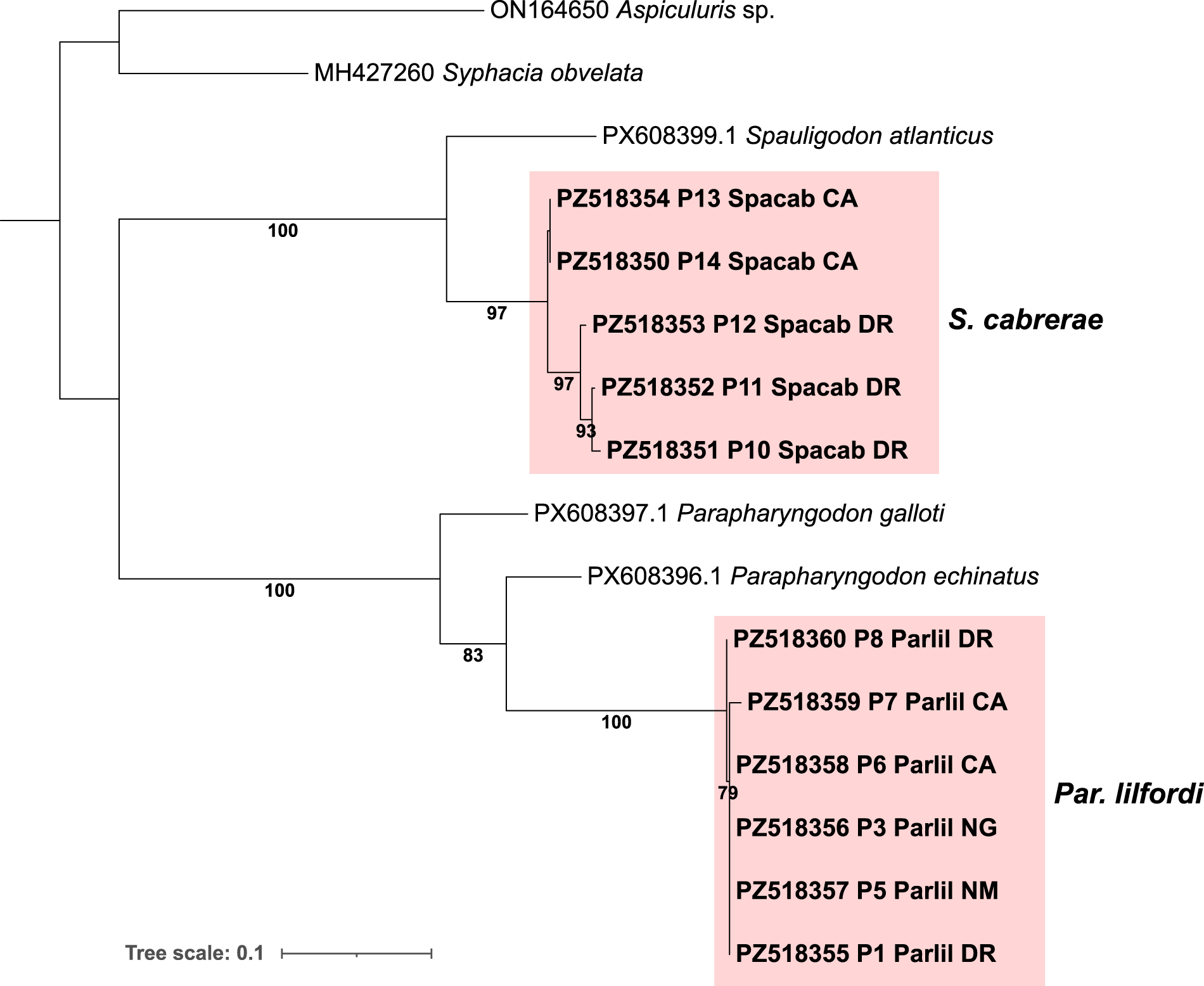
A total of 11 new COI sequences were obtained from Par. lilfordi and S. cabrerae adult specimens collected from four islands (NG, NM, DR and CA), with multiple representative specimens of both species from CA and DR. The ML tree based on partial COI sequences (538 bp alignment) supported the species taxonomic assignments in accordance with morphological data (Figure 5). The two species Par. lilfordi and S. cabrerae were highly divergent (p-dist = 0.23–0.25). All Par. lilfordi sequences clustered into a single, well-supported clade (100% BS), with P. echinatus as the closest relative (p-dist = 0.12). Similarly, all S. cabrerae sequences formed a strong monophyletic clade (100% BS), with S. atlanticus as the closest relative (p-dist = 0.12). The previously reported COI sequence from S. cabrerae (Acc.No. KJ778113) had only 169 bp overlap with the fragment amplified and showed near identity to isolate P12 (two-bp differences at 3’).
Unrooted maximum likelihood (ML) tree based on a 538 bp COI alignment (1000 bootstrap). Specimens sequenced in this study are labelled in bold, with Acc.Nos. followed by isolate name, species abbreviation and island of origin. Only bootstrap support (BS) >60 is shown. CA, Cabrera; DR, Dragonera; NM, Na Moltona; NG, Na Guardis.

Figure 5 Long description
Unrooted Maximum Likelihood tree illustrating relationships among different species based on a 538 base pair COI alignment. The tree is structured horizontally, with branches extending from left to right. Bootstrap support values greater than 60 are shown next to the branches. The top left branch leads to ON164650 Aspiculuris sp., followed by MH427260 Syphacia obvelata. Below, PX608399.1 Spauligodon atlanticus branches into a cluster labeled S. cabrerrae, containing PZ518354 P13 Spacab CA, PZ518350 P14 Spacab CA, PZ518353 P12 Spacab DR, PZ518352 P11 Spacab DR and PZ518351 P10 Spacab DR, with bootstrap values of 97 and 93. Further down, PX608397.1 Parapharyngodon galloti and PX608396.1 Parapharyngodon echinatus are shown. The bottom right cluster labeled Par. lilfordi includes PZ518360 P8 Parlil DR, PZ518359 P7 Parlil CA, PZ518358 P6 Parlil CA, PZ518356 P3 Parlil NG, PZ518357 P5 Parlil NM and PZ518355 P1 Parlil DR, with a bootstrap value of 79. The tree scale is indicated as 0.1.
Within the Par. lilfordi clade, both intra- and interpopulation nucleotide variation were low (maximum p-dist = 0.009), with no amino acid differences. Identical haplotypes were detected across all four islands, with only a few synonymous substitutions across specimens (e.g. P7 from CA and P8 and DR). In contrast, the S. cabrerae clade showed a stronger genetic structure. Specimens from CA and DR formed two distinct, well-supported clades (100% BS). Intrapopulation variation was detected only in DR (maximum p-distance = 0.03), including two nonsynonymous substitutions among isolates P10, P11 and P12.
Prevalence, coinfection and host/environmental variables
Faecal screening of 201 samples collected across eight islands revealed a large number of parasite eggs that could be reliably assigned to one of the two species (N = 708 for Par. lilfordi; N = 104 for S. cabrerae), along with a smaller number of adult specimens (N = 34 for Par. lilfordi, N = 36 for S. cabrerae). No additional helminth taxa were detected. Overall, 52% of the samples tested positive for at least one of the two parasite species. Infection status was primarily determined by the presence of eggs, which accounted for 72% of positive detections, whereas adult worms were observed in 28% of positive samples.
Par. lilfordi was detected in all islets under study, with prevalence ranging from 21% (Porrassa) to 75% (En Curt) (Figure 1B). In contrast, S. cabrerae was restricted to two islands, Dragonera (27% prevalence) and Cabrera (13%). Coinfection by both parasite species occurred on both islands, although with generally low frequency (16.6% of positive samples in Dragonera and 10% in Cabrera) (Figure 1B). None of the predictors (BCI, sex, SVL and island) showed a significant effect on infection probability (all P > 0.05), indicating no detectable influence of host traits or island of origin.
Discussion
In this study, we have confirmed the presence of both taxa historically described in the late 1980s (Castaño-Fernandez et al., Reference Castaño-Fernandez, Zapatero-Ramos, Solera Puertas and González-santiago1987; Castaño-Fernández et al., Reference Castaño-Fernández, Solera Puertas and Zapatero-Ramos1988), Par. lilfordi and S. cabrerae. Both species were originally described on islands sampled in this study (Na Moltona and Na Guardis for Par. lilfordi and the main island of Cabrera for S. cabrerae).
Morphological observations of both species were largely consistent with their original descriptions, including key diagnostic traits distinguishing females (e.g. cuticle structure, oesophagus morphology, vulvar position) from males (caudal papillae). No major morphological differences were detected, except for some variation in body size, which could simply reflect specimen variance. Molecular characterization based on COI sequences aligned with morphological data and supported the classification of S. cabrerae and Par. lilfordi within their respective genera. Genetic distances between the two species and their closest congeners (S. atlanticus and P. echinatus, respectively) were approximately 12%, clearly supporting their recognition as distinct species (Gonçalves et al., Reference Gonçalves, Bianchi, Deprá and Calegaro-marques2021).
The restricted diversity of helminths observed in Pod. lilfordi contrasts with earlier studies reporting a more diverse parasite assemblage (Roca, Reference Roca1995). This discrepancy might be due to methodological differences, as previous studies relied on gut dissection, whereas our coprological approach may underestimate overall diversity, particularly for rare taxa or those that did not shed eggs in the collected fecal samples. Moreover, for parasite taxa using Pod. lilfordi as intermediate host (larval stages), detection by coprology is not adequate. In addition, taxonomic revisions complicate direct historical comparisons. Current taxonomic assignment within this large group remains unstable, as seen for Thelandros species now reclassified to Parapharyngodon (De Sousa et al., Reference De Sousa, Jorge, Carretero, Harris, Roca and Perera2019), despite recent efforts to clarify the systematics (Tomé et al., Reference Tomé, Harris, Jorge, Sousa, Archer, Muñoz-merida, Rato, Perera and Mulder2026). Two species within the Parapharyngodon genus not endemic to the Balearic Islands were previously documented in the Balearic lizard, P. micipsae and P. bulbosus, although no morphological data or island of origin were provided, impeding clear species delineation (Roca, Reference Roca1995). Moreover, the lack or paucity of molecular data in earlier works further introduces uncertainty in species identities, as molecular approaches have increasingly recognized the possibility of the existence of cryptic species across parasitic helminths (Jorge et al., Reference Jorge, Perera, Roca, Carretero, Harris and Poulin2014; Pérez-Ponce de León and Poulin, Reference Pérez-ponce de León and Poulin2018). Importantly, the only previous studies on Parapharyngodon and Spauligodon sp. genetic diversity in Pod. lilfordi relied on 18S and 28S rRNA genes for taxonomic classification (Jorge et al., Reference Jorge, Perera, Roca, Carretero, Harris and Poulin2014; De Sousa et al., Reference De Sousa, Jorge, Carretero, Harris, Roca and Perera2019). However, the occurrence of multiple divergent copies of both genes in our studied species, as assessed by chromatogram analysis and long-reads screening, is consistent with the reported evidence of intragenomic rRNA copy number variation in nematodes (Bik et al., Reference Bik, Fournier, Sung, Bergeron and Thomas2013) and suggests that the use of ribosomal markers in pharyngodonid systematics should be approached with caution or avoided. In this context, the newly designed COI primers, although not broadly tested outside the two target species, constitute a valuable tool for resolving taxonomic uncertainties within this helminth family. Taken together, these issues suggest that historical records on helminth fauna diversity in Pod. lilfordi should be interpreted with caution.
The sampling encompassed islands that varied widely in size, geological age and ecology. Despite these environmental differences, Par. lilfordi was consistently detected across all surveyed insular populations, expanding the record of this species’ presence to five new islands. The occurrence of this species on small islands, such as Na Pelada and En Curt (both less than 1 hectare), suggests that it can persist even in small host populations. Interestingly, identical COI haplotypes were observed across all studied islands, which could indicate a recent demographic expansion, although a recent genomic study on Pod. lilfordi found no evidence of host translocations or recent contact among these insular populations (Otalora et al., Reference Otalora, Riera, Tavecchia, Rotger, Igual, Trotta and Baldo2024), which excludes recent parasite gene flow mediated by lizards. In contrast, S. cabrerae was restricted to the large, old islands of Dragonera and Cabrera, where it co-occurs with Par. lilfordi. Phylogenetic analyses indicated the existence of two separate clades of S. cabrerae between Dragonera and Cabrera, with further genetic subsclustering within each island, both suggesting a spatial segregation restricting gene flow. Moreover, compared to Par. lilfordi, the limited distribution of S. cabrerae may also reflect differences in historical colonization patterns or higher local extinction rates due to specific population and ecological requirements missing in smaller islets. The species was originally described from the island of Cabrera, while also later detected in Menorca (Roca and Hornero, Reference Roca and Hornero1992), suggesting a more widespread distribution. Interestingly, both parasite species were found co-occurring within the same host specimen, although with low frequency, indicating that both nematodes can complete their reproductive cycles within a shared host. Coexistence may be facilitated by differences in microhabitat use along the host gastrointestinal tract (i.e. niche partitioning), or it may simply represent a transient, stochastic colonization pattern in a competing scenario of the two parasites. Overall, broader sampling across the entire host range is necessary to confirm these patterns. Moreover, future population-level genetic studies are required to explore genetic variation and species-specific responses to insularity.
The overall prevalence of nematodes observed in this study was high (52% of all samples carrying at least one species), a likely underestimate when considering the stochastic nature of egg shedding and adult expulsion (Figure 2). Notably, infection status was not associated with host sex, body size, or body condition, suggesting that infection is largely independent of major host intrinsic traits. This pattern is consistent with the lizard being frequently exposed to infective stages (equally for males and females) in high-density populations, facilitated by tongue-flicking and substrate licking. No clear effect on host body condition was observed, indicating generally low virulence or a high degree of host tolerance, as observed, for instance, in Anolis Daudin, 1802 lizards (Falk and Perkins, Reference Falk and Perkins2013). This is in line with the detection of both species across multiple years and seasons (spring and autumn), which supports a temporal stability of this infection. However, targeted seasonal sampling is required to assess potential temporal fluctuations in parasite load. Lizards in the Mediterranean islands do not completely hibernate due to mild winters, a behaviour that may facilitate continuous contact between host individuals and parasite eggs dispersed in the environment.
In conclusion, this study demonstrates the persistence of both endemic helminth species nearly 40 years after their original taxonomic description, expanding their known range of distribution to additional islands. Their widespread occurrence across the islets off Mallorca and the Cabrera archipelago, coupled with high population prevalence, suggests a tight and potentially co-dependent evolutionary relationship with the host Pod. lilfordi, which requires further investigation. Ongoing genomic analyses of both nematode species will provide critical insights to assess the level of virulence and functional relevance of these endemic species, facilitating further study of parasite epidemiology within closed insular environments.
Supplementary material
The supplementary material for this article can be found at https://doi.org/10.1017/S003118202610290X.
Data availability statement
All newly generated COI sequences were deposited in GenBank under Acc.Nos. PZ518350-PZ51836.
Acknowledgements
We thank the Govern de les Illes Balears for granting the sampling permits and the staff of Natural Parc of Cabrera and Dragonera for their logistical support.
Author contributions
Conceptualization: L.B., A. Ribas, J.A.d.L.R.-s., G.T., C.E.; Methodology: L.B., A. Ribas, A. Rotger, J.A.d.L.R.-s., G.T.; Formal analysis and investigation: L.B., A. Ribas, J.A.d.L.R.-s., I.D.; Writing original draft: J.A.d.L.R.-s., L.B., A. Ribas; Manuscript review and editing: all authors; Resources: L.B., A. Ribas, A. Rotger, G.T.
Financial support
This work was supported by the MICIU/AEI/10.13039/501100011033 (L.B. and G.T., grant number PID2022-141578NB-C21 and C22); the Catalan Biogenome Project, Institut d’Estudis Catalans (IEC) (L.B., grant number IEC-BG4-2024-7).
Competing interests
The authors declare no competing interests.
Ethical standards
Permits for specimen manipulation and collection were obtained from the Conselleria del medi ambient i territori, Govern de les Illes Balears (Spain).




 Open access

