Introduction
Parkinson's disease (PD) is a neurodegenerative disease that classically presents with resting tremors, bradykinesia, postural instability, and rigidity (Parkinson, Reference Parkinson2002). However, the non-motor symptoms of PD have attracted a recent, growing interest in the scientific and clinical fields (Schrag, Reference Schrag2004; Martinez-Martin et al., Reference Li, Liu, Skidmore, Liu, Tian and Li2007). One of these non-motor symptoms, depression, affects up to 68.1% of patients with PD and directly influences the quality of life of the afflicted patients (Nakabayashi et al., Reference Mayeux, Stern, Cote and Williams2008; Chagas et al., Reference Chagas, Sanches, Tumas, Marchi and Tirapelli2011).
Etiologically, the onset of depression in PD may be related to multiple causes. Although the degeneration of dopaminergic neurons is considered the central determinant of PD, other neurotransmitter systems are also known to be affected in this disease. The non-motor symptoms observed with PD are most likely precipitated by the involvement of the serotonergic, noradrenergic, and cholinergic systems (Mayeux, Reference Mayberg1990; Mayberg and Solomon, Reference Matsui1995; Hesse et al., Reference Hesse2009).
It has been shown that the depressive symptoms associated with PD may emerge even before the onset of the motor symptoms (Schuurman et al., Reference Schuurman2002; Leentjens et al., Reference Imamura, Okayasu and Nagatsu2003). For this reason, a large number of studies have explored the prodromal or preclinical signs and symptoms of PD, increasing the possibility of early diagnosis and the development of neuroprotective therapeutics. These studies have used different neuroimaging techniques to evaluate the impact of these symptoms on the neurodegenerative process. The purpose of this paper is to review the main neuroimaging studies assessing the comorbidity between depression and PD.
Methods
A review of the Medline and Lilacs databases was conducted using the following key words: magnetic resonance imaging, neuroimaging, functional magnetic resonance, magnetic resonance spectroscopy, SPECT (single-photon emission computed tomography), PET (position emission tomography), DTI (diffusion tensor imaging), tractography, and transcranial US (ultrasound). Further database searches combined these key words with one of the following key words: Parkinson's disease, depression, or depressive disorder.
In total, 296 studies were identified and 20 of these studies were selected because their primary objective was to study the comorbidity between PD and depression. Other studies that evaluated this relationship as a secondary objective were excluded from the analysis. The included papers were full papers written in English, ranging across all available publication dates.
Results
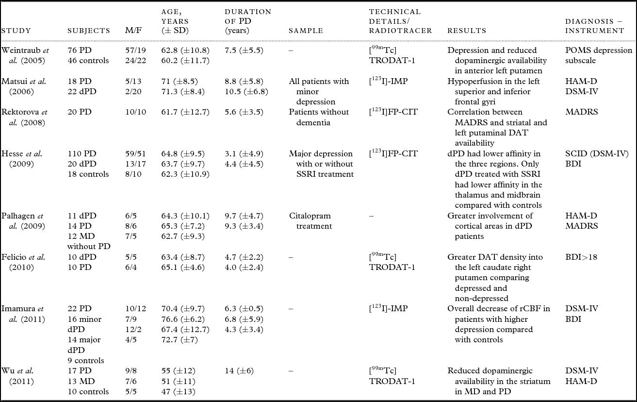
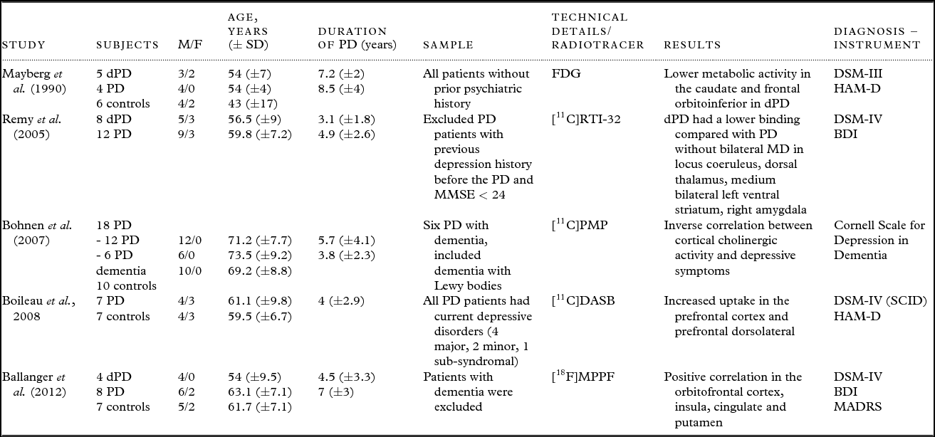
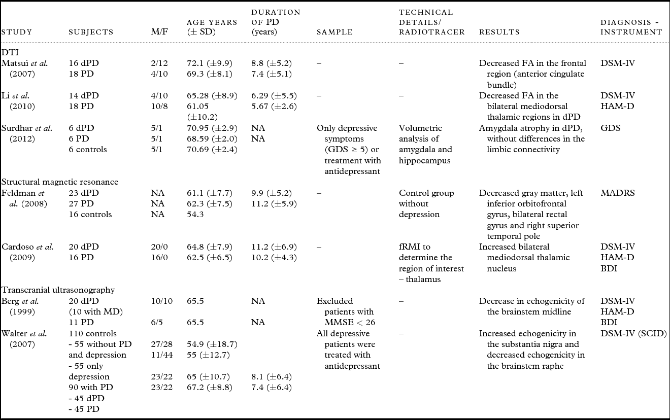
Of the 20 papers included in this review, eight used SPECT (Table 1), five used PET (Table 2), three used DTI, two used structural magnetic resonance imaging (one of these also used functional magnetic resonance), and two used transcranial US (Table 3). None of the studies selected used spectroscopy magnetic resonance imaging.
SPECT studies in depression in PD

PD = Parkinson's disease; dPD = Parkinson's disease with depression; MD = major depression; POMS = profile of mood states; MADRS = Montgomery–Åsberg Depression Rating Scale; SCID = Structured Clinical Interview for DSM disorders; DSM = Diagnostic and Statistical Manual of Mental Disorders; BDI = Beck depression inventory; HAM-D = the Hamilton rating scale for depression; SSRI = selective serotonin reuptake inhibitor; NA = not available; DAT = dopamine transporter.
PET studies in depression in PD
PD = Parkinson's disease; dPD = Parkinson's disease with depression; MD = Major depression; MMSE = Mini-Mental State Examination; FDG = 2-fluoro-2-deoxy-D-glucose, Profile of Mood states; MADRS = Montgomery–Åsberg Depression Rating Scale; SCID = Structured Clinical Interview for DSM disorders; DSM = Diagnostic and Statistical Manual of Mental Disorders; BDI = Beck depression inventory; HAM-D = the Hamilton rating scale for depression.
DTI, structural magnetic resonance, and transcranial ultrasonography studies in depression in PD
DTI = diffusion tensor imaging; PD = Parkinson's disease; dPD = Parkinson's disease with depression; MD = Major depression; MADRS = Montgomery-Åsberg Depression Rating Scale; SCID = Structured Clinical Interview for DSM disorders; DSM = Diagnostic and Statistical Manual of Mental Disorders; MMSE = Mini-Mental State Examination; GDS = Geriatric Depression Scale; BDI = Beck depression inventory; HAM-D = The Hamilton rating scale for depression; fRMI: functional resonance magnetic imaging; FA: fractional anisotropy.
SPECT
Several studies of PD using SPECT were performed; however, only seven studies aimed to evaluate the depressive symptoms in PD. Two of the radiotracers employed, [123I]FP-CIT and [99mTc]TRODAT-1, have high affinities for the dopamine transporter. In addition, [123I]FP-CIT has a high affinity for the serotonin transporter as well. A third radiotracer, [123I]-IMP, was used in some studies to determine the cerebral blood flow (CBF).
Of the studies using [99mTc]TRODAT-1 as a radiotracer to specifically evaluate the dopaminergic system, Weintraub et al. (Reference Weintraub2005) found a negative correlation between depressive symptoms and the availability of the dopamine transporter in the left anterior putamen (r = −0.24, p = 0.05). In addition, Felicio et al. (Reference Felicio2010) compared depressed and non-depressed patients with PD and found differences in the uptake of the radioligand in the left caudate (0.87 ± 0.19 for depressed patients vs. 0.69 ± 0.18 for non-depressed patients; p = 0.02) and in the right putamen (0.37 ± 0.07 for depressed patients vs. 0.28 ± 0.13 for non-depressed patients; p = 0.03). More recently, Wu et al. (Reference Wu, Lou, Huang and Shi2011) reported that dopamine availability is reduced in the striatum in patients with major depression without PD, supporting the hypothesis of dopaminergic hypofunction in depression.
Of the selected studies, two studies used [123I]FP-CIT as a marker, enabling the evaluation of the dopaminergic and serotonergic systems. Rektorova et al. (Reference Rektorova, Srovnalova, Kubikova and Prasek2008) studied the correlation between depressive symptoms and the availability of the dopamine transporter in the striatum region in PD patients without major depression or dementia. The study alluded to a relationship between mild depressive symptoms and decreased dopamine availability in the striatal and left putamen. Hesse et al. (Reference Hesse2009) evaluated patients with major depression and PD and reported low neurotransmitter uptake in regions of the striatum, the thalamus, and the midbrain and brainstem in depressed patients with PD compared with controls without depression. When the same analysis was performed only on patients whose depression was being treated with selective serotonin reuptake inhibitors, the authors found a trend toward lower neurotransmitter uptake in the thalamus (p = 0.075) but no statistically significant differences in other regions.
Of the studies that evaluated CBF, only Matsui et al. (Reference Martinez-Martin2006) evaluated patients with minor depression and PD: they found hypoperfusion in the upper and lower left frontal gyri in patients with depression compared with those without this condition. As a follow-up study, Imamura et al. (Reference Imamura, Okayasu and Nagatsu2011) found an overall reduction in the CBF of patients with PD and major depression compared with healthy controls. Likewise, patients with minor depression and PD showed a decreased uptake compared than healthy controls, in the following brain regions: the posterior cingulate, the hippocampus, the superior parietal lobule, the cuneus, the primary visual areas, the left medial frontal lobe, the parahippocampus, the insula, the thalamus, the putamen, the inferior parietal lobe, the temporal lobe, and the premotor area. There were no differences in CBF between PD patients without depression and healthy controls.
In addition, Palhagen et al. (Reference Palhagen, Ekberg, Walinder, Granerus and Granerus2009) evaluated CBF after treatment with citalopram, a serotonin reuptake inhibitor, in depressed patients with or without PD; in this study, each group was compared with a group of PD patients without depression before the beginning of the clinical trial. PD patients with depression had increased CBF in the left and right frontoparietal areas, the right dorsolateral frontal area, and the left frontal area. After the clinical trial with citalopram, patients with PD and depression exhibited increased CBF in the left dorsolateral frontal area.
PET
Mayberg et al. (Reference Mayberg and Solomon1990) performed the first study aimed at evaluating the impact of depressive symptoms in PD using PET. This study used 2-fluoro-2-deoxy-D-glucose as a marker to determine metabolic activity and compared three groups: PD patients with depression, PD patients without depression, and healthy controls. The metabolic activity in the caudate nucleus and the orbitofrontal region was significantly lower in patients with depression and PD than in PD patients without depression and healthy controls. In addition, there was an inverse correlation between the level of depressive symptoms and the metabolic activity in the frontal lobe and the inferior orbital area.
Remy et al. (Reference Remy, Doder, Lees, Turjanski and Brooks2005) conducted a study aimed at evaluating PD patients with or without histories of depression throughout their lives. This study used [11C]RTI-32, a noradrenalin transporter and dopamine transporter ligand, as a marker. The group of patients with a history of depression and PD had decreased uptake of the marker in the locus coeruleus, the anterior cingulate, the thalamus, the amygdala, and the ventral striatum compared with the group of patients without a history of depression.
Serotonin, another neurotransmitter known to be involved in depression, was investigated in a study using [11C]DASB, a serotonin transporter radioligand. In this study, the authors observed increased uptake in the regions outside the striatum, particularly the prefrontal cortex (68%) and the dorsolateral region (37%). The severity of depressive symptoms was positively correlated with an increase in the uptake of the radioligand (Boileau et al., Reference Boileau2008). More recently, Ballanger et al. (Reference Ballanger2012) used the radiotracer [18F]MPPF, a selective serotonin 1A receptor antagonist, and found a reduction in the tracer uptake in depressed patients compared with patients without depression in the following regions: the left hippocampus, the right insula, the left superior temporal cortex, and the orbitofrontal cortex.
Bohnen et al. (Reference Bohnen, Kaufer, Hendrickson, Constantine, Mathis and Moore2007) used the radioligand [11C]PMP to evaluate the relationship between cholinergic activity and depression. They found an inverse relationship between cholinergic activity and depressive symptoms (ρ = 0.5; p = 0.007).
Diffusion tension imaging
Three studies evaluated the comorbidity between depression and PD using the DTI technique. Matsui et al. (Reference Matsui, Nishinaka, Oda, Komatsu, Kubori and Udaka2007) found that fractional anisotropy values in the left and right frontal cortices were decreased in patients with depression compared with patients without depression. Li et al. (Reference Leentjens, Van den Akker, Metsemakers, Lousberg and Verhey2010) studied changes in the thalamus and found white-matter microstructural differences in the bilateral mediodorsal thalamic regions with decreased fractional anisotropy in PD patients with depression compared with PD patients without depression. Another study with a smaller sample than the previous two found no differences in limbic connectivity (Surdhar et al., Reference Surdhar, Gee, Bouchard, Coupland, Malykhin and Camicioli2012). In this study, smaller amygdala volumes were found in PD patients with depression than in healthy controls. These findings are similar to those found in depression without PD.
Structural magnetic resonance
To date, few volumetric studies have been conducted that evaluate depression in PD. Only two studies were found that had this objective. Feldman et al. (Reference Feldmann2008) compared three groups of subjects: healthy subjects, PD patients with depression, and PD patients without depression. There were no differences in the density of gray matter between the healthy controls and the PD patients. However, when the PD patients were divided into groups with or without depression, there was a decrease in gray matter density in the orbitofrontal regions, the left and right gyrus rectus, and the right superior temporal lobe in PD patients with depression. In addition, there was an inverse correlation between depressive symptoms and changes in the right medial temporal gyrus, the medial and anterior cingulate gyri, and the parahippocampal gyrus.
Another structure that has been linked to depression in PD is the thalamus. One volumetric study demonstrated a bilateral increase in the medial-dorsal thalamic nucleus of patients with depression and PD compared with patients with PD alone (Cardoso et al., Reference Cardoso2009). Furthermore, a functional neuroimaging study was realized using an event-related parametric emotional perception paradigm and showed decreased activation in the left mediodorsal thalamus and medial prefrontal cortex in PD patients with depression compared with those without depression.
Transcranial ultrasound
In studies that used transcranial US, principal changes were observed in the midline of the brain stem, including the raphe nucleus and the pathways related to the limbic system. Berg et al. (Reference Berg1999) found decreased echogenicity in the midline of the brainstem in patients with PD and depression (current or lifetime) compared with PD patients without a history of depression (p = 0.01).
As a follow-up study, Walter et al. (Reference Walter, Hoeppner, Prudente-Morrissey, Horowski, Herpertz and Benecke2007) studied 200 subjects divided into four groups: 55 healthy controls, 55 depression patients without PD, 45 PD patients without depression, and 45 PD patients with depression. The authors found hyperechogenicity of the substantia nigra in 13% of the controls, 40% of the patients with depression only, 69% of the patients with only PD, and in 87% of the patients with PD and depression. The hypoechogenicity of the brainstem was more frequent in the patients with depression than in individuals without depression. Furthermore, the combination of hypoechogenicity of the substantia nigra and hypoechogenicity of the brainstem was associated with a history of depression before the onset of PD.
Discussion
Several neuroimaging techniques have been used to seek a better understanding of the neural substrates involved in the comorbidity between depression and PD. Most of the studies used SPECT and PET. The criteria used to evaluate depression also differed between studies; either psychiatric scales or directed clinical interviews were used to diagnose depression. In addition, some studies did not use major depression as the only possible diagnosis of depressive disorders; dysthymia and minor depression were among the other disorders that were also included. This aspect can be a limitation to more specific conclusions. Furthermore, it is worth mentioning that most of the studies did not compare groups in respect of the use of antidepressant or dopaminergic drugs, which is another potential confounder.
The dopaminergic and serotoninergic systems are cited among the neurotransmitter systems involved in the genesis of depression in PD. It is believed that the reduction in striatal dopamine availability leads to a compensatory reduction of serotonin levels (Mayeux et al., Reference Mayeux1984). Evidence supporting this hypothesis has been observed in studies that have found decreased levels of 5-hydroxyindoleacetic acid, a metabolite of serotonin, in the cerebrospinal fluid of PD patients with depression compared with those without depression; this result indicates the involvement of serotonin levels (Mayeux et al., Reference Mayeux1984; Paulus and Jellinger, Reference Paulus and Jellinger1991). Likewise, neuroimaging studies support the idea of a dysfunctional serotoninergic system (Remy et al., Reference Remy, Doder, Lees, Turjanski and Brooks2005; Boileau et al., Reference Boileau2008; Rektorova et al., Reference Rektorova, Srovnalova, Kubikova and Prasek2008).
Studies have found differences in a dopaminergic system between PD patients with or without depression. The SPECT studies that used TRODAT-1 as a radioligand show conflicting results: two studies (Weintraub et al., Reference Weintraub2005; Wu et al., Reference Wu, Lou, Huang and Shi2011) showed a low uptake of the radioligand in the basal ganglia of PD patients with depression, whereas Felicio et al. (Reference Felicio2010) found increased uptake in this region in PD patients with depression. Studies in non-PD patients also found different results. One possibility is that there is an initial upregulation of the dopamine transporter, primarily in the acute phase of depression; in subjects with more severe PD, this upregulation can no longer balance dopaminergic hypofunction and the neurodegenerative process. In support of this hypothesis, other studies that have used neuroimaging techniques have also seemed to find a greater involvement of the regions related to the dopaminergic system in PD patients with depression compared with non-depressed patients.
Other systems have also been associated with depression in patients with PD. Remy et al. (Reference Remy, Doder, Lees, Turjanski and Brooks2005) suggested the possible involvement of the noradrenergic system based on a finding of lower uptake in the locus coeruleus in patients with depression compared with those without depression. A change in the function of the cholinergic system also seems to be correlated with depressive symptoms.
Changes in regions commonly tied to the neurocircuitry involved in depression in patients without PD, such as the thalamus and the frontal and temporal lobes, are also frequently found in patients with PD and depression. In PD, the degeneration of the substantia nigra leads to losses in the dopaminergic, mesolimbic, and mesocortical pathways and, consequently, the brain's reward system (Fibiger, Reference Fibiger1984). The dysfunction of the dopaminergic efferent pathways to the orbitofrontal and prefrontal cortices could explain the neuroimaging findings in these regions. Furthermore, a dysfunction of the efferent pathways of the orbitofrontal cortex that run to the dorsal raphe nucleus could cause dysregulation in the serotoninergic system (Mayeux, Reference Mayberg1990; Mayberg and Solomon, Reference Matsui1995). It is also worth noting that depressive symptoms in PD may emerge many years before the onset of the typical motor signs of the disease (Schuurman et al., Reference Schuurman2002; Leentjens et al., Reference Imamura, Okayasu and Nagatsu2003). According to Braak et al. (Reference Braak, Tredici, Rub, de Vos, Jansen Steur and Braak2003), the neurodegenerative process involves multiple regions and pursues an ascending course, which could explain, at least in part, the diversity of non-motor prodromal symptoms of PD, including depressive symptoms.
Conclusion
Among the neuroimaging studies that have evaluated depression in PD, the majority have used SPECT or PET techniques. Depression in PD appears to be associated not only with dopaminergic dysfunction but also with the dysregulation of other neurotransmitter systems. However, the impact of current or previous depression on the neurodegenerative process of PD is still unclear. Accordingly, further neuroimaging studies would be valuable and timely.
Conflict of interest
None.
Description of authors’ roles
Marcos Hortes N. Chagas wrote the first manuscript and reviewed the papers. Ila M. P. Linares assisted with writing the manuscript and reviewed the papers. José Alexandre S. Crippa, Jaime Eduardo C. Hallak, Giovana J. Garcia, and Vitor Tumas made contributions to the written manuscript. All authors read and approved the final manuscript.