Introduction
A foreign body is a rare cause of obstructive sialadenitis.Reference Cembranos, Búa, Amezaga and Zuazua 1 Penetration is different in the case of the submandibular and parotid glands: foreign bodies usually migrate into a submandibular gland intra-orally through Wharton's duct,Reference Su, Lao, Zheng, Liang, Huang and Liao 2 whereas most foreign bodies penetrating a parotid gland do so extra-orally through the skin, and are secondary to a traumatic event (there are published cases of a bullet, a pencil tip and a sliver of wood),Reference Levine, Clark and Mandel 3 although a few cases of grass seeds, hair, feathers and fish bones entering the gland through the main parotid duct have also been reported.Reference Yamano, Uzawa, Ito and Tanzawa 4
The global spread of semi-rigid sialendoscopes and miniaturised instruments has allowed surgeons to adopt a gland-sparing approach, rather than traditional sialadenectomy.Reference Gillespie, Koch, Iro and Zenk 5 The recent introduction of minimally invasive techniques for treating obstructive sialadenitis has made the avoidance of surgical complications such as facial nerve injury possible.Reference Overton, Combes and McGurk 6 However, their use has given rise to other risks.Reference Walvekar, Razfar, Carrau and Schaitkin 7 This includes the possible intraductal rupture of a micro-instrument (which may occur during attempts to remove a salivary stone with a wire basket, for instance). The risk of rupture can be increased by: certain anatomical conditions (e.g. kinking and stenosis), the diameter and shape of the stone, or manufacturing defects. The rupture of basket wires during interventional sialendoscopy is a possible event that does not lead to the presence of a foreign body in the ductal system. However, the rupture of the guide wire after the entrapment of a stone is extremely rare. In such cases, surgeons need to be able to resolve the situation in a as minimally invasive a manner as possible.
An intraductal foreign body may be difficult to remove by means of traditional open surgery alone because, regardless of the functional or aesthetic issues, it may be impossible to identify its location precisely. In such cases, it can be useful to adopt a combined external and endoscopic approach,Reference Capaccio, Gaffuri and Pignataro 8 , Reference Koch, Iro and Zenk 9 in order to locate the exact position of the foreign body inside the ductal system and allow its removal with minimal invasiveness.
This paper aimed to describe our experience of removing a broken wire basket containing a 4 mm salivary stone embedded in a secondary branch of Stensen's duct by means of sialendoscopy-assisted transfacial surgery.
Case report
A 37-years-old male patient with recurrent obstructive left parotid gland disease lasting 8 months attended our out-patient ENT department to undergo ultrasonography using a 7.5 MHz Hitachi H21 scanner (Hitachi High Technology, Tokyo, Japan). This revealed a 4 mm stone located in a secondary parenchymal branch of Stensen's duct that was dilated proximally to the stone. He subsequently underwent operative sialendoscopy in November 2013.
The flexible, semi-rigid sialendoscope (Karl Storz, Tuttlingen, Germany) used for the video-endoscopic assessment of the parotid ductal system had a diameter of 0.8 mm and a port for an irrigation pump connected to the system. Sialendoscopy was performed under local anaesthesia, induced by means of 2 per cent lidocaine hydrochloride infiltrated directly into the duct after insertion of the endoscopic device and sedation. The duct was adequately dilated via the insertion of Karl Storz salivary duct probes of increasing diameter (from size 0000 to 6). The sialendoscope was inserted, and its passage through the ductal system was facilitated by means of continuous irrigation with isotonic saline solution through the irrigation port. The procedure allowed the surgeon to visualise the ductal system as far as the stone. A Dormia Stone wire basket (2.5 Fr; Coloplast, Peterborough, UK) was then introduced into Stensen's duct, parallel to the sialendoscope.
The stone was easily engaged, but it became stuck in the ductal branch during withdrawal, and subsequent attempts to retrieve the basket with the stone inside (including the use of miniaturised forceps) failed. The patient was therefore asked to undergo sialendoscopy-assisted transfacial surgery on the following day in order to retrieve the foreign body under general anaesthesia.
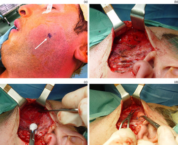
In accordance with McGurk and colleagues' technique,Reference McGurk, MacBean, Fan, Sproat and Darwish 10 a Redon pre-auricular incision was made (Figure 1a, violet line) and the subcutaneous layers were gently dissected (Figure 1b). The skin flap was then raised and the dissection continued, to involve the parotid fascia (Figure 1c), in order to expose the parotid gland and the main duct (Figure 1d).
Stone localisation on the skin surface (arrow) (a). After gently dissecting the subcutaneous layers (b), a skin flap was raised (c), and the dissection was continued through the parotid fascia in order to expose the parotid gland and the proximal tract of Stensen's duct (d).

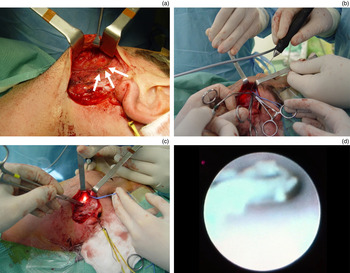
During the blunt dissection, the buccal branch of the facial nerve close to the duct was identified and marked (Figure 2a). The preparation of the best operative field, with complete exposure of the site in which the stone and basket were trapped, required the use of a neurostimulator (Neuro-Pulse; Bovie Medical, Clearwater, Florida, USA), in order to check the correct functioning of the buccal and other possible branches of the VIIth cranial nerve encountered during dissection (Figure 2b). The light at the tip of the endoscope revealed the exact position of the foreign body, in a secondary parenchymal branch (Figures 2c and 2d).
Buccal branch of the facial nerve (arrows) close to the duct (a). The position of the foreign body was confirmed by means of sialendoscopy (b) using the high-power light at the endoscope tip (c). In this case, it was located intra-parenchymally in the proximal tract of Stensen's duct (d).

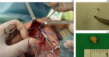
The duct was incised over the foreign body and parallel to its direction using a size 11 scalpel. After gentle dissection using dedicated instruments, the stone engaged in the wire basket was grasped with pincers and removed (Figures 3a–c).
After incising the duct and making a gentle dissection, the foreign body was held by pincers and removed (a). It consisted of a 4 mm stone (c), and the broken wire basket (b) previously used in an attempt to remove the stone from the ductal system.

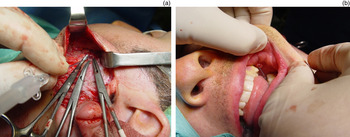
The duct was then irrigated with saline and an endoscopic search was made for any residual stones or debris. Subsequently, a Seldinger arterial catheter (5 Fr Seldicath; Promed, Le Plessis-Bouchard, France) was placed as a stent through the ductal opening (Figure 4a), and secured to the buccal mucosa by means of a 2-0 silk stitch (Figure 4b). The duct wall was then sutured using 6-0 polyglactin sutures (Vicryl; Johnson and Johnson International, Brussels, Belgium), and the parotid fascia was closed. A Redon drainage system was placed for 48 hours, and peri-operative antibiotic prophylaxis was administered.
A Seldinger catheter was placed in Stensen's duct (a) in order to prevent stenosis, and secured to the buccal mucosa (b).

The post-operative course was complicated by a sialocele that developed 3 days after surgery. This was successfully treated by aspirating its contents and administering one injection of botulinum toxin type A (Botox, 60 mouse units; Allergan, Irvine, California, USA) under ultrasonographic guidance (using the 7.5 MHz Hitachi H21 scanner).
The patient was discharged after 7 days. Multiple aspirations were performed and pressure dressings applied in the out-patient department every day for one week after the toxin injection, until the cheek area normalised two weeks after surgery. No other major complications (no facial or auricular nerve damage) occurred, and the patient was satisfied with his facial scar.
After discharge, the patient was closely followed for 12 months in order to check for recurrent symptoms and to evaluate parotid salivary flow. The follow-up clinical examinations showed that the affected parotid gland was normal in size and consistency, and normal clear saliva flow could be induced by massage. No relapses or post-operative complications occurred during the follow-up period.
Discussion
Technical advances in miniaturisation and endoscopy over the last 20 years have favoured the development of minimally invasive and conservative procedures for the major salivary glands, thus avoiding the major complications associated with traditional surgery.Reference Overton, Combes and McGurk 6 A number of studies have validated the safety and effectiveness of diagnostic sialendoscopy. Koch et al. showed that it provides excellent and direct information on duct pathology in cases of salivary duct obstruction secondary to stones, strictures or foreign bodies.Reference Koch, Zenk, Bozzato, Bumm and Iro 11 Furthermore, interventional sialendoscopy can be performed during the same procedure, and is currently considered to be a successful means of removing mobile stones and stones of less than 5 mm in diameter.Reference Gillespie, Koch, Iro and Zenk 5 , Reference Koch, Iro and Zenk 9 , Reference Capaccio, Torretta, Ottavian, Sambataro and Pignataro 12 , Reference Witt, Iro, Koch, McGurk, Nahlieli and Zenk 13
We used interventional sialendoscopy to retrieve a 4 mm parotid duct stone, but encountered an unexpected complication. To the best of our knowledge, there are no published cases of miniaturised instruments breaking inside the ductal system, and ours is the first involving a stone engaged in a broken wire basket offering no further possibility of sialendoscopic retrieval. Sialendoscopy certainly plays a fundamental role in conservative treatment. However, despite its apparent simplicity, sialendoscopy is still technically challenging and requires sequential learning, as indicated by the fact that success rates seem to be proportional to the level of experience.Reference Marchal and Dulguerov 14 In addition, the technique is not devoid of risks, and it is likely that complication rates will increase with its worldwide use.Reference Capaccio, Torretta, Osio, Minorati, Ottaviani and Sambataro 15
Walvekar et al. reported that the major complication in their case series was the avulsion of Stensen's duct during an attempt to retrieve an impacted sialolith.Reference Walvekar, Razfar, Carrau and Schaitkin 7 In such cases, surgeons should avoid excessive traction when the stone is engaged in a wire basket; the bigger the stone, the more difficult the sialendoscopic retrieval, especially if it is located in a narrow secondary branch of the ductal system as it was in our case. On the basis of this experience, the removal of stones of more than 3 mm cannot always be guaranteed by interventional sialendoscopy, especially in the absence of a stone breaker like laser device.Reference Capaccio, Torretta, Ottavian, Sambataro and Pignataro 12
The availability of minimally invasive and conservative techniques allowed us to treat our patient without the need for traditional invasive surgery: the entrapped stone and basket were removed by means of sialendoscopy-assisted transfacial surgery.Reference Capaccio, Gaffuri and Pignataro 8 Furthermore, normal salivary function was preserved (something not possible in the case of an invasive parotidectomy), although the post-operative course was complicated by a sialocele despite the positioning of a salivary duct stent (a 5 Fr Seldinger arterial catheter). However, this problem was resolved by the use of botulinum toxin type A, along with pressure dressings applied for one week. Together with conservative procedures, or the surgical closure of the ductal system or sialoadenectomy, botulinum toxin therapy is currently considered a valid treatment option for salivary disorders such as sialoceles and salivary fistulas.Reference Capaccio, Torretta, Osio, Minorati, Ottaviani and Sambataro 15 The low rate of side effects during the post-surgical management of salivary disordersReference Capaccio, Torretta, Osio, Minorati, Ottaviani and Sambataro 15 – Reference Kruegel, Winterhoff, Koehler, Matthes and Laskawi 17 justifies its use in patients refractory to a new surgical procedure after a failure.
-
• A foreign body is a rare cause of parotid gland obstructive sialadenitis; intra-oral penetration via Stensen's duct is unusual
-
• The use of miniaturised endoscopes and instruments during interventional sialendoscopy can cause intraductal rupture resulting in an iatrogenic foreign body
-
• Sialendoscopy-assisted transfacial surgery is an effective and safe method of retrieving a foreign body stuck in Stensen's duct
Finally, scarring was minimal, and the patient was satisfied with both the functional and aesthetic results.
Conclusion
Parotid duct foreign bodies penetrating Stensen's duct are extremely rare, but the advent of minimally invasive interventional sialendoscopy has added possible new causes of iatrogenic duct obstruction. In the case of instrumental breakage inside the ductal system, when the foreign body is stuck and cannot be retrieved endoscopically, sialendoscopy-assisted transfacial surgery is a safe, effective and well-tolerated means of resolving the situation in a minimally invasive manner. This novel approach, which has also been successfully used to treat obstructive sialadenitis secondary to large impacted parotid stones (of more than 7 mm in diameter), favours gland preservation and symptom control.Reference Capaccio, Gaffuri and Pignataro 8 In contrast, the only alternative option is traditional parotidectomy, as recently demonstrated in a case of obstructive sialadenitis.Reference Gillespie, Koch, Iro and Zenk 5 Based on our experience, patients should be properly informed about the possibility of a foreign body being left in the ductal system during sialendoscopic procedures; this event will require a second informed consent to plan subsequent sialendoscopy-assisted transfacial surgery for the removal of the foreign body and the stone.