Implications
Matured mammalian oocytes can be used for many biotechnologies, for example, fertilization, cloning and transgenesis. Under in vitro conditions, these oocytes deteriorate very quickly due to processes called oocyte aging. The development of embryos produced from aged oocytes is rather poor. In addition, knowledge about the process of aging is limited. In this study, we observed that one of the significant features of oocyte aging – fragmentation of oocyte – can be completely suppressed by treatment with substances activating protein kinases C. The oocytes undergoing such treatment maintain their developmental competence for a longer time, and there is hope that they can be used for various biotechnologies.
Introduction
Advanced reproductive biotechnologies depend on a sufficient source of oocytes. Oocytes of domestic animals are used for in vitro fertilization, for cloning using the transfer of nuclei from somatic cells and for transgenesis (Kikuchi et al., Reference Kikuchi, Kashiwazaki, Nagai, Nakai, Somfai, Noguchi and Kaneko2008). Owing to the difficulties encountered in production of in vivo matured oocytes, in vitro matured oocytes are widely used. Unfortunately, the necessary quality is not always met in these oocytes, and their developmental competence is lower when compared to oocytes matured in vivo. The causes of the reduced developmental competence are multifactorial. In addition to genetic variations, environmental effects on oocyte donors or the in vitro environment, the culture conditions remained one of the important factors. Therefore, the search for better culture systems continues.
During in vitro maturation, an oocyte undergoes germinal vesicle breakdown and proceeds through further stages of meiosis up to the stage of metaphase II, when meiosis is spontaneously blocked (Wassarman, Reference Wassarman1988). The quality of oocytes can be improved by prolonged culture, that is, by culturing an oocyte for some time after it has reached the stage of metaphase II. Cattle oocytes cultured for an additional 16 h were used for in vitro fertilization (Pavlok et al., Reference Pavlok, Kalab and Bobak1997), human oocytes cultured for an additional 48 h were used for cloning (Hall et al., Reference Hall, Compton, Stojkovic, Nesbitt, Herbert, Murdoch and Stojkovic2007) or pig oocytes cultured for an additional 12 h were used for the production of parthenogenetic embryos (Jolliff and Prather, Reference Jolliff and Prather1997). However, a culture prolonged beyond a certain critical period threatens oocytes with the complex process designated as ageing. The developmental capability of aged oocytes could be significantly reduced (Nagano et al., Reference Nagano, Katagiri and Takahashi2006), and to this end treatments reducing or delaying ageing have been investigated.
Many aged oocytes do not remain at the stage of metaphase II. Many processes occur within an ageing oocyte that complicate or exclude the use of these oocytes, for example, they undergo spontaneous parthenogenetic activation, fragmentation or lysis (Petrová et al., Reference Petrová, Sedmíková, Chmelikova, Svestkova and Rajmon2004). Fragmentation is a manifestation of apoptosis (Tarin et al., Reference Tarin, Perez-Albala and Cano2001), which is an integral part of oocyte ageing (Yuce and Sadler, Reference Yuce and Sadler2001). The activation of many specific molecules, for example, caspases, proteases that are responsible for dismantling of the cell, and many pro-apoptotic factors of the Bcl-2 gene family are necessary for the triggering of apoptosis (Adams and Cory, Reference Adams and Cory1998).
Oocyte ageing could be regarded as an alternative process to oocyte activation occurring after fertilization. Many processes that are involved in the activation of a fertilized oocyte act against oocyte ageing and protect the oocyte from apoptosis (Sadler et al., Reference Sadler, Yuce, Hamaratoglu, Verge, Peaucellier and Picard2004). Activation of mammalian oocytes is triggered by the signaling of free calcium ions, which is followed by the mobilization of downstream calcium-dependent signaling pathways. These pathways also include protein kinases C (PKCs). PKCs are known as transducers of signaling of calcium ions and play the role of calcium transducers in mammalian oocytes as well (Fan et al., Reference Fan, Tong, Chen and Sun2003). When compared to the role of PKCs during oocyte activation, knowledge about the role of PKC in oocyte ageing is much more limited.
PKCs belong to the serine/threonine kinases and have many isotypes that can be divided into three classes. Conventional PKCs (calcium-dependent PKC isotypes (cPKCs)) are represented by isotypes PKC-α, -βI, -βII and -γ. These PKCs are activated by free calcium ions or diacylglycerols (DAGs). The so-called novel PKCs (nPKCs) involve isotypes PKC-δ, -ε, -η, -μ and -θ. These PKCs are independent of calcium ions, but can be activated by DAGs. Atypical PKCs (aPKCs) include isotypes PKC-ζ, -λ and -τ. These PKCs are independent of both calcium ions and DAGs (Liu and Heckman, Reference Liu and Heckman1998).
The expression of individual PKC isotypes depends on the cell type and its developmental stage (Aderem, Reference Aderem1995). Various authors have detected calcium-dependent isotypes PKC-α, -βI, -βII, -γ; calcium-independent isotypes PKC-δ, -ε, -μ, or atypical isotypes PKC -λ and -ζ in matured rodent oocytes (Gangeswaran and Jones, Reference Gangeswaran and Jones1997; Raz et al., Reference Raz, Eliyahu, Yesodi and Shalgi1998; Luria et al., Reference Luria, Tennenbaum, Sun, Rubinstein and Breitbart2000; Pauken and Capco, Reference Pauken and Capco2000; Downs et al., Reference Downs, Cottom and Hunzicker-Dunn2001; Eliyahu and Shalgi, Reference Eliyahu and Shalgi2002; Viveiros et al., Reference Viveiros, O’Brien, Wigglesworth and Eppig2003). However, the data concerning the spectra of PKC isotypes are contradictory. In matured pig oocytes, calcium-dependent isotypes PKC-α, -βI, -γ (Fan et al., Reference Fan, Li, Tong, Chen, Xia, Song, Schatten and Sun2002a and Reference Fan, Tong, Li, Lian, Chen, Schatten and Sun2002b) and calcium-independent PKC-δ (Sedmíková et al., Reference Sedmíková, Rajmon, Petr, Svestkova, Chmelikova, Bantirgu, Rozinek and Jílek2006) were detected. Very limited data are available concerning the role of different classes of PKCs during the ageing of pig oocytes.
The broader objectives of this study were to discover the factors that can suppress the fragmentation of ageing oocytes. We can suggest that PKCs are markedly involved in the regulation of oocyte fragmentation. Our objective was to investigate the effects of different classes of PKCs on the ageing of pig oocytes. Using specific PKC activators, we attempted to suppress fragmentation of ageing oocytes and to prolong the time after which development of preimplantation embryos could be successfully induced using parthenogenetic activation. For nonselective activation of both calcium-dependent and calcium-independent PKCs, we used phorbol-12-myristate-13-acetate (PMA) or 1-stearoyl-2arachidonoyl-sn-glycerol (STEAR). For specific activation of calcium-dependent PKCs, we used 1-oleoyl-2-acetyl-sn-glycerol (OLE). Dipalmitoyl-l-α-phosphatidylinositol-3,4,5-triphosphate heptaammonium salt (DIPALM) was used for specific activation of calcium-independent PKCs.
Material and methods
Isolation and culture of oocytes
Pig ovaries were obtained from a local slaughterhouse from gilts at an unknown stage of the estrous cycle and transported to the laboratory within 1 h in a saline solution (0.9% natrium chloride) at 39°C. Fully grown oocytes were collected from follicles by aspirating follicles that were 2 to 5 mm in diameter with a 20-gauge needle. Only oocytes with compact cumuli were selected for further study.
Growing oocytes of different size categories were obtained from thin strips (10 to 15 mm long, 1 to 2 mm wide) dissected from the surface of the ovaries using a scalpel. The strips of ovarian tissue were placed in Petri dishes containing a culture medium. The oocytes were released from their follicles by opening the follicular wall using the tip of a 25-gauge needle. The internal diameter of the oocytes (without zona pellucida) was measured with an ocular micrometer mounted on a microscope. Only oocytes surrounded by several layers of cumulus cells were selected for further experiments.
Before culture, the oocytes were washed three times in a maturation culture medium.
They were cultured in a modified M199 medium (GibcoBRL, Life Technologies, Paisley, Scotland) containing sodium bicarbonate (0.039 ml of a 7.0% solution per milliliter of the medium), calcium lactate (0.6 mg/ml), gentamicin (0.025 mg/ml) HEPES (1.5 mg/ml), 13.5 IU eCG : 6.6 IU hCG/ml (P.G.600 Intervet, Boxmeer, Holland) and 10% fetal calf serum (GibcoBRL, Life Technologies, Karlsruhe, Germany).
The oocytes were cultured for 48 h in 3.5-cm diameter Petri dishes (Nunc, Roskilde, Denmark) containing 3.0 ml of the culture medium at 39°C in a mixture of 5.0% CO2 in the air.
Evaluation of the oocytes
At the end of the culture, the oocytes or embryos were mounted on slides, fixed with acetic alcohol (1 : 3, v/v) for at least 24 h, and stained with 1.0% orcein. The oocytes were examined under a phase contrast microscope. Activation was considered to have occurred if the oocytes were in the pronuclear stage. Oocytes remaining at metaphase II or arrested at anaphase II or at telophase II were not considered as activated. In cleaved parthenogenetic embryos, the number of nuclei was counted at the end of the culture.
Oocyte activation and culture of embryos
Oocytes were activated using the method described by Jílek et al. (Reference Jílek, Huttelova, Petr, Holubova and Rozinek2001). Briefly, oocytes matured in vitro were denuded of their cumulus cells and were subjected to a 5-min treatment with 25 μM calcium ionophore A23185. The calcium ionophore was diluted in a modified M199 medium without fetal calf serum and without bovine serum albumin. After treatment, the oocytes were washed in a M199 medium supplemented with bovine serum albumin and cultured for 2 h in an NCSU23 culture medium (Petters and Wells, Reference Petters and Wells1993) supplemented with 2 mM 6-dimethylaminopurine (DMAP). The oocytes were then carefully washed and cultured in a DMAP-free NCSU23 medium in four-well Petri dishes (Nunc, Roskilde, Denmark), each well containing 1.0 ml of the culture medium. The eggs were cultured at 39°C in a mixture of 5.0% CO2 in the air for 7 days.
Arrangement of experiments
The purpose of experiment 1 was to investigate the fates of pig oocytes aged in vitro. In vitro matured oocytes were denuded of cumulus cells, placed in an M199 culture medium and cultured for another 1, 2, 3, 4 or 5 days.
The purpose of experiment 2 was to investigate the effects of PKC activators on the fragmentation of ageing pig oocytes in vitro. The oocytes matured in vitro were devoid of cumulus cells and cultured for another 3 days with a respective PKC activator. PMA and STEAR were used as activators of both calcium-dependent and calcium-independent PKCs. PMA was used in concentrations of 0, 10, 25, 50 and 100 nM. STEAR was used in concentrations of 0, 25, 50, 100 and 200 μM. The specific activator of calcium-dependent PKCs OLE was used in concentrations of 0, 10, 25, 50 and 100 μM. Specific activator of calcium-independent PKCs DIPALM was used in concentrations of 0, 100, 200, 500 and 1000 nM. The combined effect of activation of both calcium-dependent and calcium-independent PKCs was tested by culture of the oocytes in a medium supplemented with 10 μM OLE and 100 nM DIPALM.
All activators were dissolved in dimethylsulfoxide (DMSO) and then diluted to the respective concentration in the culture medium. The concentration of DMSO in the culture medium did not exceed 0.3%. In control experiments, we verified that DMSO at concentrations below 1% has no effect on the fragmentation of pig oocytes in vitro. In each experiment, we used a control group of oocytes that underwent ageing in a medium without DMSO or in a medium with respective concentrations of DMSO. The results of these control experiments did not differ significantly, and we present only the data from oocytes cultured in a medium without DMSO as ‘control’.
In experiment 3, we investigated the course of ageing of oocytes exposed to the effect of 100 nM PMA. In vitro matured oocytes were devoided of cumulus cells and then cultured in a culture medium supplemented with 100 nM PMA for another 0, 1, 2 and 3 days.
In experiment 4, we investigated the capability of aged oocytes to be parthenogenetically activated by ionophore and the capability of parthenogenetic development of oocytes aged under the influence of 100 nM PMA. In vitro matured oocytes were devoid of cumulus cells and further cultured for 1 day in a medium supplemented with 100 nM PMA. The oocytes were then parthenogenetically activated using calcium ionophore A23187 and 6-DMAP as described above. After this treatment, the oocytes were cultured for another 22 h in a culture medium without DMAP, and the ratio of parthenogenetically activated oocytes was assessed. Parthenogenetic development of oocytes after their parthenogenetic activation was assessed in an experiment in which matured oocytes were devoid of cumulus cells, parthenogenetically activated using calcium ionophore and DMAP, and then cultured in a medium of NCSU23 for another 7 days.
Statistical analysis
The data from all the experiments were subjected to statistical analysis. Each experiment was performed four times. The data were evaluated by ANOVA (Statistica 6.0). A P-value of <0.05 was considered significant.
Results
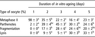
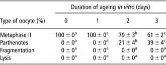
After in vitro culture for 48 h, 98% of the oocytes reached the stage of metaphase II. The remaining oocytes were at the stage of metaphase I, anaphase I or telophase I. The ageing of oocytes during their culture in vitro during experiment 1 is shown in Table 1. After the first day of ageing, almost all the oocytes remained at the stage of metaphase II. After the second day of ageing, there was a significant decrease in oocytes remaining at the stage of metaphase II and a significant increase in oocytes that underwent spontaneous parthenogenetic activation or fragmentation. After 3 days of ageing, there was a further significant decrease in oocytes remaining at the stage of metaphase II and an increase in fragmented oocytes. On the fourth day of ageing, there was an increased ratio of lysed oocytes. There were no significant changes on the fifth day of ageing.
The fate of pig oocytes during their aging in vitro

The oocytes matured in vitro for 48 h and then were further cultured for 1, 2, 3, 4 or 5 days. Each treatment group consisted of 120 oocytes. The data are presented as mean ± s.e.m.
a,b,cStatistically significant differences between oocytes of the same type during different lengths of ageing (i.e. differences within a row) are indicated by different superscripts.
We selected 3-day ageing for further experiments that investigated the effect of PKC activation on oocyte ageing, because at days 4 and 5 a high portion of ageing oocytes undergoes lysis.
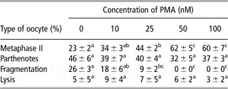
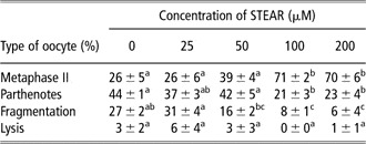
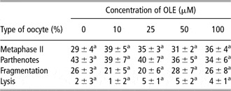
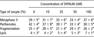
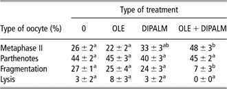
In experiment 2, we observed a statistically significant decrease of fragmented oocytes after 3 days of ageing in the presence of PMA, an activator of both calcium-dependent and calcium-independent PKCs (see Table 2). STEAR, which is similar to PMA, as it acts as an activator of both calcium-dependent and calcium-independent PKCs, had similar effect as PMA (Table 3). However, the investigated concentrations of OLE, an activator of calcium-dependent PKCs, did not suppress the fragmentation of the pig oocytes aged in vitro (Table 4). Similarly, we did not observe a significant effect of the investigated concentrations of DIPALM, a specific activator of calcium-independent PKCs, on the fragmentation of the oocytes aged in vitro (Table 5). The positive effect of activation by both calcium-dependent and calcium-independent PKCs was supported by the results of an experiment in which combined treatment with OLE and DIPALM significantly reduced the ratio of fragmented oocytes (Table 6).
Effect of PMA on pig oocytes’ ageing in vitro

PMA = phorbol-12-myristate-13-acetate.
The oocytes were cultured in vitro for 48 h and then further cultured for 3 days in a medium supplemented with PMA. Each treatment group consisted of 120 oocytes. The data are presented as mean ± s.e.m.
a,b,cStatistically significant differences in the type of oocytes between different treatments (i.e. differences within a row) are indicated by different superscripts.
Effect of STEAR on in vitro ageing of pig oocytes

STEAR = 1-stearoyl-2 arachidonoyl-sn-glycerolu.
The oocytes were cultured in vitro for 48 h and then further cultured for 3 days in a medium supplemented with STEAR. Each treatment group consisted of 120 oocytes. The data are presented as mean ± s.e.m.
a,b,cStatistically significant differences in the type of oocytes between different treatments (i.e. differences within a row) are indicated by different superscripts.
Effects of OLE on in vitro ageing of pig oocytes

OLE = 1-oleoyl-2-acetyl-sn-glycerol.
The oocytes were cultured in vitro for 48 h and then further cultured for 3 days in a medium supplemented with OLE. Each treatment group consisted of 120 oocytes. The data are presented as mean ± s.e.m.
aDifferences in the type of oocytes between different treatments (i.e. differences within a row) are statistically insignificant.
Effect of DIPALM on in vitro ageing of pig oocytes

DIPALM = dipalmitoyl-l-alpha-phosphatidylinositol-3,4,5-triphosphate heptaammonium salt.
The oocytes were cultured in vitro for 48 h and then further cultured for 3 days in a medium supplemented with DIPALM. Each treatment group consisted of 120 oocytes. The data are presented as mean ± s.e.m.
aDifferences in the type of oocytes between different treatments (i.e. differences within a row) are statistically insignificant.
Effect of DIPALM and OLE on in vitro ageing of pig oocytes

DIPALM = dipalmitoyl-l-alpha-phosphatidylinositol-3,4,5-triphosphate heptaammonium salt; OLE = 1-oleoyl-2-acetyl-sn-glycerol.
The oocytes were cultured in vitro for 48 h and then further cultured for 3 days in a medium without any supplement (0), a medium supplemented with 10 μM OLE (OLE), a medium supplemented with 100 nM DIPALM (DIPALM) or a medium supplemented with 10 μM OLE and 100 nM DIPALM (OLE + DIPALM). Each treatment group consisted of 120 oocytes. The data are presented as mean ± s.e.m.
a,bStatistically significant differences in the type of oocytes between different treatments (i.e. differences within a row) are indicated by different superscripts.
We used PMA, the PKC activator of both calcium-dependent and calcium-independent PKCs, in our further experiments.
Experiment 3 showed the course of ageing in oocytes exposed to the effects of PMA. The oocytes aged for 3 days in vitro in a culture medium supplemented with 100 nM PMA had completely suppressed fragmentation. However, a significant proportion (39%) of these oocytes underwent spontaneous parthenogenetic activation (Table 7). This is approximately the same ratio of spontaneous parthenogenetic activation as in oocytes aged for the same time without PMA (33% of oocytes with pronuclei). After 2 days of culture with PMA, the ratio of oocytes with spontaneous parthenogenetic activation was significantly lower than in oocytes aged without PMA (pronuclei in 21% v. 41% oocytes). After 1 day of ageing with PMA, all the oocytes were at the stage of metaphase II. This was not significantly different from oocytes aged for 1 day in a PMA-free culture medium.
Progression of ageing in pig oocytes cultured with PMA

PMA = phorbol-12-myristate-13-acetate.
The oocytes were cultured in vitro for 48 h and then further cultured for 1, 2 or 3 days in a medium supplemented with 100 nM PMA. Each treatment group consisted of 120 oocytes. The data are presented as mean ± s.e.m.
a,b,cStatistically significant differences in the type of oocytes between different treatments (i.e. differences within a row) are indicated by different superscripts.
In experiment 4, we investigated artificial parthenogenetic activation of pig oocytes aged under the effects of PMA and their subsequent parthenogenetic development. After the treatment with calcium ionophore and DMAP, the activation rate was similar in oocytes aged 1 day in a medium with 100 nM PMA (86% of activated oocytes), oocytes aged 1 day without PMA (93% of activated oocytes) or oocytes treated under the same activation protocol immediately at the end of maturation in vitro (87% of activated oocytes).
Cleavage rate in oocytes artificially activated after 1-day ageing in PMA was significantly higher than the activation rate in oocytes aged in a medium without PMA and was similar to the activation rate observed in oocytes artificially activated immediately at the end of in vitro maturation. Concerning the ratio of oocytes developing to the morula stage (10%) or blastocyst stage (12%) after ageing for 1 day with PMA, the subsequent artificial parthenogenetic activation was the same as in oocytes aged without PMA. The ratio of oocytes developing to the blastocyst stage was lower than in oocytes that did not age and which were artificially activated immediately at the end of maturation (Table 8).
Parthenogenetic development in oocytes activated by artificial stimulus after 24 h aging

PMA = phorbol-12-myristate-13-acetate; DMAP = supplemented with 2 mM 6-dimethylaminopurine.
The oocytes matured in vitro were further cultured for 24 h in a medium supplemented with 0 or 100 nM PMA. These oocytes were then parthenogenetically activated using an artificial stimulus (5 min in 25 μM calcium ionophore and then 2 h in 2 mM DMAP) and subsequently cultured in the medium NCSU23 for 7 days. Fresh oocytes that did not age in vitro were activated using the same protocol at the end of their maturation and were used as a control. Each treatment group consisted of 120 oocytes. The data are presented as mean ± s.e.m. a,bStatistically significant differences between oocytes at the same stage of parthenogenetic development after ageing under different treatments (i.e. differences within columns) are indicated by different superscripts.
Discussion
In this study, we demonstrated that the activation of PKCs during the culture well beyond the time necessary for reaching the stage of metaphase II reduces fragmentation of pig oocytes and supports metaphase II arrest. This effect resulted only when we used the activators PMA or STEAR, which activate both calcium-dependent and calcium-independent PKCs. A similar effect was also achieved after combined treatment of ageing oocytes with the activator of calcium-dependent PKCs OLE and the activator of calcium-independent PKCs DIPALM. We can propose that positive effect on fragmentation of ageing pig oocytes is due to simultaneous activation of both classes of PKCs, since OLE or DIPALM alone was ineffective.
Postovulatory ageing of mammalian oocytes matured to the stage of metaphase II is a very complex process, and it represents an alternative pathway to fertilization and embryonic development (Fissore et al., Reference Fissore, Kurokawa, Knott, Zhang and Smyth2002). Aged oocytes have damaged mitochondria (Wilding et al., Reference Wilding, Dale, Marino, di Matteo, Alviggi, Pisaturo, Lombardi and De Placido2001), a changed balance of pro-apoptotic and anti-apoptotic factors, for example, of proteins of the Bcl-2 gene family (Perez et al., Reference Perez, Tao and Tilly1999), damaged cytoskeleton (Webb et al., Reference Webb, Howlett and Maro1986), and reduced activity of maturation promoting factor (MPF; Kikuchi et al., Reference Kikuchi, Naito, Noguchi, Shimada, Kaneko, Yamashita, Aoki, Tojo and Toyoda2000). In addition to other changes, aged oocytes also exhibited altered signaling of calcium ions (Takahashi et al., Reference Takahashi, Saito, Hiroi, Doi and Takahashi2000).
PKCs are involved in calcium-signaling in oocytes (Tatone et al., Reference Tatone, Delle Monache, Francione, Gioia, Barboni and Colonna2003). However, PKCs are also significantly involved in regulation of the mitochondria function (Baines et al., Reference Baines, Song, Zheng, Wang, Zhang, Wang, Guo, Bolli, Cardwell and Ping2003), regulation of the cytoskeletal function (Ma et al., Reference Ma, Koch and Viveiros2008) and also influence the MPF in mammalian oocytes (Yu et al., Reference Yu, Zheng, Yu, Shi, Liu, Wu, Fu and Yang2004). The effect of PKC activators observed in our study can be explained by several mechanisms due to the many targets of PKC. Our experimental design did not permit us to answer this question. Moreover, we cannot exclude that nonspecific effects of PKC activators also play important role. PMA and other PKC activators promote the activation of other kinases in addition to PKCs, particularly in higher concentrations.
Under our conditions, 24-h ageing had no effect on the capability of oocytes to be parthenogenetically activated using the artificial stimulus of calcium ionophore and DMAP. This capability was not significantly changed even when the oocytes were aged for 24 h in the presence of PKC activators. Parthenogenetic activation by standard activation protocol based on the combined effect of calcium ionophore and DMAP (Jílek et al., Reference Jílek, Huttelova, Petr, Holubova and Rozinek2001) was similarly effective in oocytes aged under the effect of PMA and in oocytes aged in a PMA-free culture medium. The ratio of parthenogenetically activated oocytes was similar in oocytes aged 1 day with PMA and ‘fresh’ oocytes exposed to parthenogenetic activation immediately after finishing their maturation.
Ageing can be viewed as an alternative pathway to fertilization and subsequent embryonic development. However, the role of particular PKC isotypes is not fully understood even in the regulation of oocyte activation during fertilization or parthenogenesis. Some authors suggest that cPKCs are involved in oocyte activation. Luria et al. (Reference Luria, Tennenbaum, Sun, Rubinstein and Breitbart2000) demonstrated the activation of PKC-α aPKC-β1 in fertilized mouse oocytes. Raz et al. (Reference Raz, Eliyahu, Yesodi and Shalgi1998) observed relocalization of cPKC isotypes PKC-α and PKC-β during activation of rat oocytes. Fan et al. (Reference Fan, Tong, Li, Lian, Chen, Schatten and Sun2002b) observed PKC-α, β1 and γ in matured pig oocytes and described the relocalization of these PKCs after oocyte fertilization or oocyte parthenogenetic activation using calcium ionophore A23187.
Other authors suggest that calcium-independent PKC isotypes (nPKCs) play a key role during fertilization and parthenogenetic activation. Gangeswaran and Jones (Reference Gangeswaran and Jones1997) described nPKC isotype PKC-δ and aPKC isotype PKC-λ as the dominant isotypes of PKC in mouse oocytes. According to Viveiros et al. (Reference Viveiros, O’Brien, Wigglesworth and Eppig2003) and Tatone et al. (Reference Tatone, Delle Monache, Francione, Gioia, Barboni and Colonna2003), PKC-δ is a key isotype for activation of mouse oocytes after fertilization or parthenogenetic activation. The important role of PKC-δ in parthenogenetic activation of pig oocytes was demonstrated by Sedmíková et al. (Reference Sedmíková, Rajmon, Petr, Svestkova, Chmelikova, Bantirgu, Rozinek and Jílek2006). The significant role of nPKC for exit from meiotic metaphase is also supported by the observation that in mouse oocytes PKC-δ regulates the exit from metaphase I and progression of meiosis to the stage of metaphase II (Viveiros et al., Reference Viveiros, Hirao and Eppig2001).
The roles of different PKC isotypes are indispensable during the full activation of mammalian oocytes, but there are differences between species. In matured mouse oocytes, parthenogenesis can be induced simply by an activator of calcium-independent PKCs (Tatone et al., Reference Tatone, Delle Monache, Francione, Gioia, Barboni and Colonna2003). However, pig oocytes cannot be activated by stimulation of PKCs (Fan et al., Reference Fan, Li, Tong, Chen, Xia, Song, Schatten and Sun2002a). This is in agreement with our results, since the treatment of ageing oocytes with PMA, which is an activator of both calcium-dependent and calcium-independent PKCs, did not induce parthenogenetic activation during the first day or even the second day of ageing, and therefore spontaneous activation was significantly lower than in oocytes aged in a PMA-free medium. Only on the third day of ageing was the ratio of spontaneously activated oocytes comparable between oocytes aged with PMA and control oocytes aged for 3 days in a PMA-free medium. Oocyte activation, chromatin decondensation and formation of pronuclei in aged oocytes depended on the decrease of the activity of MPF and the decrease of the activity of mitogen-activated protein kinase (MAP) (Kikuchi et al., Reference Kikuchi, Naito, Noguchi, Shimada, Kaneko, Yamashita, Aoki, Tojo and Toyoda2000). Therefore, we can speculate that activation of PKCs using PMA prevents at least temporarily (by two days of aging) a decrease of the activity of the MPF and MAP. There is also an interesting possibility that oocytes that remained arrested at the stage of metaphase II (with high MPF activity) may be less likely to undergo fragmentation/lysis.
In our experiments, we confirmed that aged oocytes have lower developmental competence when compared with fresh oocytes that just finished their meiotic maturation. The activation of PKCs during oocyte ageing cannot prevent this decrease in developmental competence. The ability of oocytes to undergo parthenogenetic activation in response to an artificial stimulus was not affected even when the oocytes were aged for 24 h in the presence of PMA. This indicates that PMA did not affect the viability of oocytes, their ability to exit from metaphase II and to start cleavage. This opinion is further supported by the fact that the ratio of cleaved oocytes after an artificial stimulus was similar in oocytes aged with PMA and in fresh oocytes exposed to an artificial stimulus immediately after completion of their maturation. The ratio of cleavage in oocytes exposed to an artificial stimulus after 1 day of ageing in a PMA-free medium was significantly decreased.
In mouse oocytes, the activation of PKC diminished the cleavage of early embryos (Pauken and Capco, Reference Pauken and Capco2000). In our experiments, the ageing of pig oocytes in PMA impaired development in the more advanced stages of preimplantation development triggered by artificial stimulus. It is known that PKCs are involved not only in the exit of oocytes from metaphase II after fertilization, but also in morula compaction (Winkel et al., Reference Winkel, Ferguson, Takeichi and Nuccitelli1990). However, it is not clear whether the lower rate of blastocyst count in oocytes aged with PMA was due to the activation of PKC during ageing. With regard to the fact that the same ratio of blastocysts was observed in oocytes aged for 1 day without PMA, we can suggest that this suppression of development to the blastocyst stage is not a specific effect of the PKC activator. On the basis of our data, we can conclude that activation of PKCs suppressed some aspects of ageing in pig oocytes, especially their fragmentation. This effect appears only after simultaneous activation of calcium-dependent and calcium-independent PKCs. The activation of PKCs allowed oocytes aged for 24 h to retain their capability of cleavage after parthenogenetic activation by an artificial stimulus. However, it did not prevent a decrease in the capability of developing to the blastocyst stage.
Acknowledgements
This study was supported by grants from MZe ČR grant numbers MZE 0002701401 and QG50052 and MSM 6046070901. The authors thank s Lois Russell for her editorial assistance with this manuscript.