Introduction
The chemical composition of marbling fat is important from both consumer acceptance and human health perspectives. Fatty acid synthesis in ruminant adipose tissue has been studied for decades and the composition, particularly the nutraceutical component of ruminant fat, may be enhanced through dietary manipulation (Scollan et al., Reference Scollan, Hocquette, Nuernberg, Dannenberger, Richardson and Moloney2006). The strategic supplementation of cattle diets can generate beef with increased conjugated linoleic acid (CLA) and n-3 polyunsaturated fatty acid (PUFA) concentrations, a lower quantity of fat and a fatty acid profile more compatible with current human dietary recommendations (Moloney et al., Reference Moloney, Mooney, Kerry and Troy2001). More recently, however, it has been shown that the simultaneous accretion of tissue concentrations of n-3 PUFA and CLA appears to be hampered by the apparent negative effects of n-3 PUFA on tissue CLA synthesis (Waters et al., Reference Waters, Kelly, O’Boyle, Moloney and Kenny2009). Our group has shown that increases in dietary n-3 PUFA supplementation decreased mRNA levels for Δ-9 desaturase in muscle (Waters et al., Reference Waters, Kelly, O’Boyle, Moloney and Kenny2009). Furthermore, tissue concentration of the n-3 PUFA, eicosapentaenoic acid (EPA), was found to exhibit a significant negative relationship with Δ-9 desaturase gene expression. There is also evidence that the transcription factor – sterol regulatory element-binding protein (SREBP)-1c – controls the activation and expression of Δ-9 desaturase in humans (Renaville et al., Reference Renaville, Mullen, Moloney, Larondelle, Schneider and Roche2006). To date, there is no information on the effect EPA specifically has on the transcriptional regulation of Δ-9 desaturase in the bovine. This requires further investigation and understanding of the molecular mechanisms involved, particularly at a cellular level.
Adipogenesis is the process of adipocyte cell development whereby differentiation results in its transformation from a fibroblast-like cell to a lipid-filled cell with the expression of transcription factors and molecular markers for the mature fat cell phenotype and capable of metabolizing lipid (Fernyhough et al., Reference Fernyhough, Vierck, Hausman, Mir, Okine and Dodson2005). There appears to be differences in adipocyte development and its regulation in subcutaneous and intramuscular depots (Hausman et al., Reference Hausman, Poulos, Pringle and Azain2008). The development and maintenance of primary adipocytes in culture and conducting reproducible experiments have been difficult (Viravaidya and Shuler, Reference Viravaidya and Shuler2002). It is acknowledged that there is limited information available on optimal culture conditions for bovine adipocytes, particularly from different adipose depots and for their differentiation (Grant et al., Reference Grant, Ortiz-Colòn, Doumit and Buskirk2008).
In non-ruminants, several preadipocyte cell lines such as 3T3-L1 and Ob 17 have proven to be useful models for the study of adipocyte differentiation (Forest et al., Reference Forest, Grimaldi, Czerucka, Negrel and Alihaud1983), growth and development (Fernyhough et al., Reference Fernyhough, Vierck, Hausman, Mir, Okine and Dodson2005), function (Morganstein et al., Reference Morganstein, Christian, Turner, Parker and White2008) and diseases characterized by increased fat depots (Natal et al., Reference Natal, Fortuño, Restituto, Bazán, Colina, Díez and Varo2008). Importantly, in vitro differentiation of adipocytes has been shown to possess many characteristics of adipose cells in vivo (Gregoire et al., Reference Gregoire, Smas and Sul1998). In ruminants, most studies investigating fatty acid synthesis or accretion in vitro have employed tissue explants, slices or fat cells in short-term primary culture (Vernon and Taylor, Reference Vernon and Taylor1988; Faulconnier et al., Reference Faulconnier, Thévenet, Fléchet and Chilliard1994). While no mature intramuscular adipocyte cell line has been developed for the bovine species, Aso et al. (Reference Aso, Abe, Nakajima, Ozutsumi, Yamaguchi, Takamori, Kodama, Hoshino and Takano1995) established the first clonal bovine intramuscular preadipocyte line from white adipose tissue of the Musculus longissimus thoracis of Japanese black cattle. Unfortunately the preadipocyte cell line has since been lost. There is no report of the existence of a mature validated intramuscular adipocyte cell line for the bovine species. Therefore, the objective of the current study was to develop and validate a mature bovine intramuscular adipocyte cell line and to employ this resource to examine the effect of level of EPA supplementation on the mRNA expression of Δ-9 desaturase and SREBP-1c genes. An in vitro approach was applied to avoid possible confounding effects of inter-animal variance in feed intake and adiposity.
Material and methods
Adipocyte cell isolation
Intramuscular adipose tissue was obtained from the M. longissimus thoracis of an Angus × Charolais beef heifer in a licensed abattoir (Jennings Meats, Ballinrobe, Co. Mayo, Ireland) within 20 min of slaughter. The heifer was fed a diet of grass silage supplemented with 3 kg of a cereal-based concentrate, 6 weeks prior to slaughter. Adipose tissue was stored in sterile phosphate-buffered saline containing Penicillin (5000 IU/ml) (Sigma-Aldrich Ireland Ltd, Dublin, Ireland) and Streptomycin (5 mg/ml) (Sigma-Aldrich Ireland Ltd) (PBS-PS) on ice during the 45 min transport to the laboratory. Visible connective and muscle tissue were carefully dissected away from the intramuscular adipose tissue. Adipocytes were isolated using an adaptation of the methods described by Fernyhough et al. (Reference Fernyhough, Vierck, Hausman, Mir, Okine and Dodson2005). Briefly, adipose tissue was washed four times in sterile PBS-PS and was cut into approximately 1 cm3 pieces. Tissue was subsequently treated with 25 ml of PBS containing glucose (1.5%) (Sigma-Aldrich Ireland Ltd), bovine serum albumin (BSA) (0.1%) (Invitrogen, Carlsbad, CA, USA) and collagenase (1.5%) (Sigma-Aldrich Ireland Ltd) for 1 h at 37°C, with agitation. Following enzymatic digestion, the collagenase was removed by centrifugation for 5 min at 1500 r.p.m., and the tissue was then washed (3×) in sterile PBS. The upper adipose layer was removed and transferred to 25 cm3 culture flasks (Sarstedt Ltd, Drinagh, Wexford, Ireland).
Culture of adipocytes
Mature adipocytes were cultured using an inverted ceiling culture method to facilitate adherence of cells to the bottom of culture flasks (Fernyhough et al., Reference Fernyhough, Vierck, Hausman, Mir, Okine and Dodson2005). The upper adipose layer was transferred to culture flasks (25 cm3) and filled with 24.5 ml Dulbecco’s modified Eagle’s medium/nutrient F-12 (DMEM/F12) culture medium (Sigma-Aldrich Ireland Ltd) supplemented with 25 mmol NaHCO3 (VWR International Ltd, Lutterworth, Leicestershire, UK), 10% foetal calf serum (FCS; Invitrogen), 0.03% l-glutamine (Sigma-Aldrich Ireland Ltd), 0.009% Penicillin G (Sigma-Aldrich Ireland Ltd), 0.0145% Streptomycin sulphate (Sigma-Aldrich Ireland Ltd), 1% fungizone (Invitrogen) and inverted in an CB210 CO2 incubator (Binder GmbH, Tuttlingen, Germany) set at 37°C and 5% CO2, with the lid loosened to allow passage of CO2 into the culture. Following 5 days, unattached cells were removed and the flasks were placed in an upright position. Fresh culture medium, which was equilibrated to 37°C to avoid heat shocking of adipocytes, was added to the flasks every 2 to 3 days and attachment to the bottom of the flasks was monitored daily using a Nikon oxivert inverted light microscope (Nikon Precision Europe GmbH, Livingston, Scotland).
Trypsinization and passaging of adipocytes
Cells were routinely passaged at 70% confluency. Culture media was removed and cells were incubated in 2 ml 0.05% trypsin-ethylenediaminetetraacetic acid (EDTA) (Invitrogen) solution for 5 min at 37°C to enzymatically detach cells from the wall of the flask. Efficiency of cell detachment was monitored under a microscope as prolonged incubation in trypsin can damage cells. Once detachment was complete, 2 ml of FCS was added to the flask to neutralize the trypsin. The cell suspension was then removed and centrifuged for 5 min at 1500 r.p.m. The supernatant was discarded and the pellet re-suspended in culture medium. Cells were cultured for 30 passages. Adipocytes from all passages were stored in liquid nitrogen.
Storage and thawing of adipocytes
Cells were trypsinized, centrifuged for 5 min at 1500 r.p.m. and excess medium was aspirated and discarded. Cells from one flask (25 cm3) were frozen down to a 1 ml volume for long-term storage. Freezing medium (1 ml; 10% dimethyl sulfoxide (DMSO), 90% FCS) (Sigma-Aldrich Ireland Ltd) was added to the cell pellet in a cryovial (Invitrogen). As freezing medium contains DMSO, which is a cryoprotectant and is toxic to cells at temperatures greater than 4°C, this process was carried out as quickly as possible. Cryovials were stored overnight at −80°C followed by transfer into liquid nitrogen (−180°C).
To thaw cells, a cryovial was carefully removed from liquid nitrogen and placed in a beaker of pre-warmed water (37°C). When thawed (approximately 2 min), contents were transferred to a centrifugation tube (25 ml) (Sarstedt Ltd). Culture medium (9 ml), pre-warmed to 37°C, was added to the centrifuge tube and the suspension was centrifuged at 1500 r.p.m. for 5 min. The supernatant was discarded, the pellet was re-suspended in 1 ml of culture medium and the sample was repeat-pipetted to ensure homogeneity. A total of 1 ml of the cell suspension was added into a culture flask and 9 ml of pre-warmed culture medium was added to the flask and cells were cultured until approximately 70% confluency.
Evaluation of lipid accumulation of adipocytes
Both Sudan IV and Oil red O were initially assessed as staining methods to establish accumulation of intracellular lipid in adipocytes. Sudan IV penetrated cells to a greater extent and was therefore used throughout this study to determine the ability of adipocytes to accumulate lipid at passage 10, 20 and 30. Briefly, culture medium was removed from the flask and cells were washed with PBS and then with 70% ethanol. Cells were fixed on a glass slide by the addition of 10% formalin and incubated for 10 min. Sudan IV stain (5 mmol) was added to the slide and cells were incubated for 5 min. Cells were washed with PBS and basophilic structures such as nuclei were counterstained by the addition of 2 ml haematoxylin for 2 to 3 min. Cells were finally washed with PBS and examined using a Nikon oxivert inverted light microscope.
Fatty acid analysis
Analysis of fatty acids was conducted in spent media and adipocytes at passage 30 in the development of the cell line using gas chromatography. Fatty acid methyl esters (FAME) were also measured in spent media and adipocytes following treatment of cells with EPA. Cells were trypsinized, centrifuged for 5 min at 1500 r.p.m. and excess medium was aspirated and analysed separately. Total lipids were extracted from a pellet of adipocytes as well as 1 ml of spent media using chloroform : methanol (2:1 v/v) as described by Folch et al. (Reference Folch, Lees and Sloane Stanley1957). Methylation was carried out for both adipocytes and spent media using the method described by Park and Goins (Reference Park and Goins1994). The FAME were separated on a CP Sil 88 column (100 m × 0.25 mm i.d., 0.20 μm film thickness; Chrompack, Middleburg, The Netherlands) using a gas liquid chromatograph (3400; Varian, Harbor City, CA, USA) fitted with a flame ionization detector. Helium (37 psi) was used as the carrier gas. The injector temperature was held isothermally at 225°C for 10 min and the detector temperature was 250°C. The column oven was held at an initial temperature of 140°C for 8 min and then programmed to increase at a rate of 8.5°C/min to a final temperature of 200°C, which was held for 41 min. Data were recorded and analysed on a Minichrom PC system (VG Data System, Manchester, UK). Prior to fatty acid analysis, a hemocytometer was used to determine cell density in flasks. A 10 μl sample was introduced into one of the V-shaped wells of the hemocytometer, and using a microscope adipocytes were counted. These data were then used to normalize fatty acid concentrations determined in adipocytes.
RNA isolation
To validate the transcriptional activity of the adipocytes, the expression of markers of fat metabolism was identified by reverse transcription-PCR (RT-PCR). Adipocytes were typsinized and cells collected by microcentrifugation at 12 000 r.p.m. for 5 min at 4°C in sterile 1.5 ml eppendorfs. The supernatant was removed and the pellet was immersed and stored in RNA later (Ambion, Applera Ireland, Dublin, Ireland) at −20°C. Total RNA was isolated from adipocytes using TRIzol reagent and chloroform and subsequently precipitated using isopropanol. RNA quantity was determined by absorbance at 260 nm using the Nanodrop spectrophotometer (Labtech International Ltd, East Sussex, UK). RNA quality was assessed using 28S/18S ratio and RNA integrity number (RIN) on the Agilent Bioanalyzer 2100 with the RNA 6000 nano labchip kit (Agilent Technologies Ireland, Dublin, Ireland).
Reverse transcription-PCR and sequence analysis
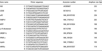
RT-PCR was performed to determine the expression of markers of fat metabolism in the cell line (Table 1). RNA was DNase-treated with RQ1 RNase-free DNase (Promega UK Ltd, Southhampton, UK) and purified using the RNeasy mini kit (Qiagen Ltd, Crawley, UK). RNA (1 μg) was DNase-treated and reverse transcribed to generate cDNA, using the Reverse Transcription system according to manufacturer’s instructions (Promega UK Ltd). cDNA was then quantified using the Nanodrop spectrophotometer (Labtech International Ltd), diluted to 50 ng/l working stocks and stored at −20°C.
Bovine specific oligonucleotide primers used in this study

FAS = fatty acid synthase; aP2 = adipocyte lipid-binding protein; FABP-4 = fatty acid-binding protein-4; MTP = microsomal triglyceride transfer protein; SREBP-1c = sterol regulatory element-binding protein-1c; PPAR-γ = peroxisome proliferator-activated receptor-gamma; HNF4α = hepatocyte nuclear factor four alpha.
Details of primer sets used in this study are listed in Table 1. Primers for RT-PCR were commercially synthesized (Sigma-Aldrich Ireland Ltd). Primers were designed to amplify specific fragments (approximately 200 bp) of the following genes, which are adipocyte markers, that play important roles in adipocyte metabolism and function, including peroxisome proliferator-activated receptor (PPAR)-γ, fatty acid synthase (FAS), fatty acid-binding protein (FABP)-4, adipocyte long chain fatty acid-binding protein (aP2), CD36, Δ-9 desaturase, the transcription factor SREBP-1c, leptin and microsomal triglyceride transfer protein (MTP) (Table 1). The liver-specific hepatocyte nuclear factor-4 alpha (HNF4α) was applied as the negative control (Sladek, Reference Sladek1993). Cycling parameters applied in PCR were as follows: 95°C for 2 min and 30 cycles of 95°C for 30 s, 60°C for 30 s, 72°C for 30 s (Eppendorf Master Cycler; Unitech, Dublin, Ireland), followed by 72°C for 5 min and PCR products were stored at 4°C. Analysis of amplified PCR products (10 μl) was carried out on 1% agarose gels using 1× Tris acetate EDTA (TAE) running buffer, pH 8.3 in a model EC-360-M Maxicell gel system (EC Apparatus Corporation, St. Petersburg, USA). Agarose gels were stained using 5 μg/ml of ethidium bromide. DNA was observed using a UV transilluminator (254 nm) and the image was photographed using a Bio ID Gel Documentation system (Vilber Lourmat, Cedex, France). All amplified PCR products generated were purified using the PCR purification kit (Roche, Basel, Switzerland), and sequenced (Macrogen; Nucleics Pty Ltd, Bendigo, Australia) and analysed using the Basic Local Alignment Search Tool (BLAST) on the National Centre for Biotechnology Information (NCBI) web page, to verify their identity.
Eicosapentaenoic acid supplementation of adipocyte culture media
The novel adipocyte cell line was subsequently used to determine the effects of the n-3 PUFA, EPA, on the transcriptional regulation of Δ-9 desaturase in an in vitro bovine intramuscular adipocyte cell culture model. EPA stock solution (50 mg/500 μl; Cayman Chemical, Ann Arbor, MI, USA) was diluted to 1 mmol in pre-heated sterile DMEM/F12 medium containing 33 mg/ml fatty acid-free BSA. The remaining ethanol was evaporated under a gentle stream of nitrogen and the EPA was stored at −20°C. The solution was incubated for 2 h at 37°C to ensure binding of EPA to BSA. This 1 mmol stock solution was then further diluted to 100 and 50 μM in their respective culture media. Ethanol was used as a vehicle to incorporate EPA into the culture medium and was added at the same concentration to the control (0 μM EPA).
Mature adipocytes (passage 30) were removed from storage and grown for 21 days to maximum confluency, n = 60 flasks (75 ml). Prior to treatment, cells were serum starved for a 24 h period and were subsequently incubated in a serum-free medium containing 1% insulin, selenium and transferrin as substitutes for FCS. Following 24 h, the medium was replaced with serum-free medium containing one of three concentrations of EPA, viz. 0 (control), 50 or 100 μM, n = 16 (8 for fatty acid analysis and 8 for gene expression analysis) for each treatment. The EPA concentrations selected were based on in vivo EPA concentrations recorded in bovine muscle tissue as part of an in vivo experiment at our laboratory (Waters et al., Reference Waters, Kelly, O’Boyle, Moloney and Kenny2009). Following the 24 h incubation, cells and media were harvested. Cells were collected using trypsin–EDTA and spun at 1500 r.p.m. for 5 min. Supernatant was collected and stored at −20°C for fatty acid analysis. One of the duplicate cell pellets were immersed and stored in 1 ml RNA later at −20°C while the other duplicate was transferred to a sterile cryovial, snap frozen in liquid N2 and stored at −80°C for fatty acid analysis. Analysis of adipocyte FAME was carried out using gas chromatography as described above.
Gene expression
RNA was isolated, its quality analysed and cDNA synthesized as described above. Primers were designed to amplify specific fragments of Δ-9 desaturase and SREBP-1c (Table 1). β-actin was used as a single standard reference gene for this experiment (Bahar, Reference Bahar2006). Primers were first tested using end point PCR to optimize amplification conditions. All amplified PCR products from this study were also sequenced to verify their identity.
Each real-time PCR reaction was carried out in a total volume of 20 μl with 1 μl cDNA (50 ng/μl), 10 μl SYBR Green I master mix (Langanbach Services Ltd, Bray, Co. Wicklow, Ireland), 1 μl forward and reverse primers (20 ng each) and 8 μl nuclease-free H2O. Dissociation curves were examined for the presence of a single PCR product. Real-time RT-PCR was performed using a Corbett Rotor-Gene™ 3000 quantitative PCR system (Corbett Life Sciences, Sydney, Australia) with the following cycling parameters: 95°C for 15 min followed by 40 cycles of 95°C for 30 s, 60°C for 30 s, 72°C for 30 s, followed by amplicon dissociation (95°C for 1 min, 50°C for 45 s, increasing 0.5°/cycle until 95°C was reached). Gene expression results were calculated using the 2−ΔΔCT method (Livak and Schmittgen, Reference Livak and Schmittgen2001).
Statistical analysis
Data were analysed using the Statistical Analysis Systems software package version 9.1 (SAS Institute, Cary, NC, USA). Data were examined for adherence to normality using PROC UNIVARIATE (SAS, 2001). Data were then analysed to determine the effect of EPA treatment on fatty acids in spent media and adipocytes, and Δ-9 desaturase and SREBP-1c expression levels using mixed models ANOVA (PROC MIXED, SAS, 2001). The Tukey critical difference test was used to determine statistical difference between treatment means. Regression analyses were carried out to establish relationships between supplemental EPA concentrations and relative gene expression of Δ-9 desaturase and SREBP-1c using PROC REG (SAS, 2001).
Results
Staining and microscopy
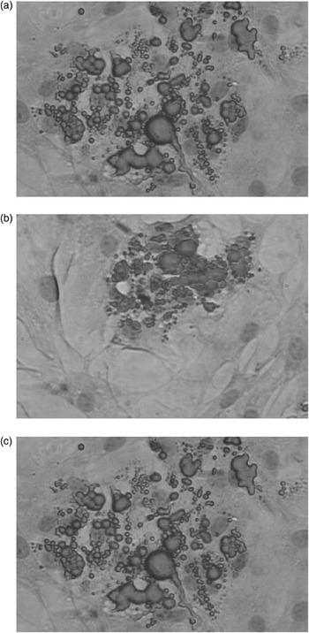
Staining and microscopy analysis showed that isolated cells accumulated lipid over time. Figure 1 (a) to (c) clearly demonstrated that fat droplets increased in number and size over time with large fat-filled vesicles observed at 30 passages. Cell nuclei were stained blue with haematoxylin, identifying basophilic structures such as ribosomes, nucleus and the cytoplasmatic regions rich in RNA, thus demonstrating the functionality of the cell line. Adipocyte cell doubling time was recorded as 2 to 3 days.
Analysis of lipid accumulation in primary intramuscular adipocyte cell line at passages (a) 10, (b) 20 and (c) 30 using the Sudan staining and microscopy. The lipid droplets are stained red with Sudan IV and nuclei counterstained with haematoxylin.

Fatty acids
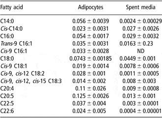
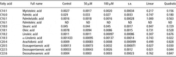
Concentration of fatty acids in spent media and adipocytes is presented in Table 2. Thirteen fatty acids ranging from saturated fatty acid, tetradecanoic acid (C14:0), to the long chain PUFA, docosahexaenoic acid (DHA) (C22:6), were detected and quantified.
Concentrations of fatty acids detected in unsupplemented adipocytes and spent media

ND = non-detectable.
Fatty acid results are presented as mg of fatty acid/ml of media and mg 10−7 cells. These are mean (±s.e.) for eight replicates.
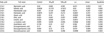
The effect of level of EPA supplementation on the concentration of FAME in spent media and adipocytes is presented in Table 3. Results show that treatment of adipocytes with EPA resulted in a linear increase in the concentration of stearic (P = 0.04), α-linolenic (P = 0.004), EPA (P = 0.001), docosapentaenoic acid (P = 0.004) and DHA (P = 0.004) with increasing concentrations of supplemental EPA. The effect of EPA supplementation on the concentration of myristoleic (P = 0.01), palmitic (P = 0.003) and palmitelaidic acids (P = 0.001) was predominantly quadratic in nature (Table 3a).
Effect of dietary eicosapentaenoic acid supplementation on adipocyte fatty acid concentration (mg 10−7cells)

Effect of dietary eicosapentaenoic acid supplementation on media fatty acid concentration (mg/ml)

ND = non-detectable.
Fatty acid results are presented as mg of fatty acid/ml of media and mg 10−7 cells.
In the media, there was a linear increase in EPA concentration with increasing supplemental EPA (P = 0.027) and docosapentaenoic acid (P = 0.021). There was no effect of supplementation on the concentration of any other fatty acids measured (P > 0.05). There was a quadratic increase in α-linoleic acid with increasing supplemental EPA (P = 0.022) (Table 3b).
Reverse transcriptase-PCR and sequence analysis
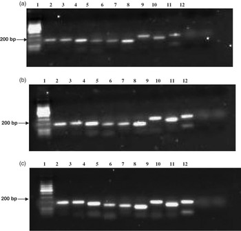
High-quality total RNA was successfully isolated from adipocytes. Results of 28S/18S ratios for all samples were between 1.8 and 2 and RIN of 9 to 10. In addition, yields of RNA recovered from 25 ml flasks were in the region of 12 μg per flask. Markers of fat metabolism PPAR-γ, FAS, FABP-4, aP2, CD36, Δ-9 desaturase, leptin, SREBP-1c and MTP were all shown to be expressed at passages 10, 20 and 30 using RT-PCR (Figure 2). No expression of the negative control HNF4α was observed. PCR products were sequenced and subjected to BLAST analysis to establish their identity. In the case of all PCR products sequenced, DNA sequences were 100% identical to the published sequences.
Reverse transcription-PCR analysis of marker genes involved in fatty acid metabolism in bovine intramuscular adipocyte cells line. Lane 1, 100 bp ladder; Lane 2, Δ-9 desaturase; Lane 3, SREBP-1c; Lane 4, CD36; Lane 5, PPAR-γ; Lane 6, MTP; Lane 7, leptin; Lane 8, aP2; Lane 9, FABP-4; Lane 10, FAS; Lane 11, HNF4α and Lane 12, water control: (a) passage 10, (b) passage 20 and (c) passage 30. aP2, adipocyte lipid-binding protein; FABP-4, fatty acid-binding protein-4; FAS, fatty acid synthase; MTP, microsomal triglyceride transfer protein; PPAR-γ, peroxisome proliferator-activated receptor-gamma; SREBP, sterol regulatory element-binding protein.

EPA supplementation and adipocyte gene expression
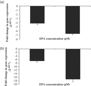
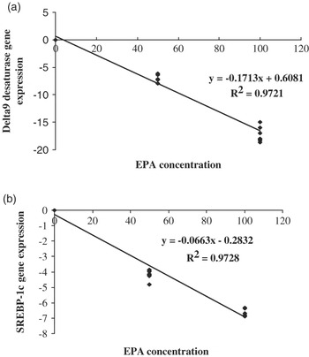
Compared with the control, supplementation of the culture media with either 50 or 100 μM EPA led to respective decreases in Δ-9 desaturase expression of five- and seven-fold, respectively. Similarly, mRNA transcripts for SREBP-1c were decreased 6- and 18-fold following the addition of 50 and 100 μM EPA, respectively (Figure 3). There was a strong linear negative relationship between the gene expression of both Δ-9 desaturase and SREBP-1c and EPA concentrations (R 2 = 0.97 and 0.98, respectively, P < 0.0001; Figure 4). Additionally, a positive relationship was observed between SREBP-1c and Δ-9 desaturase gene expressions (R 2 = 0.90; P < 0.0001).
Effect of concentration of EPA in culture media on the gene expression of (a) Δ-9 desaturase and (b) SREBP-1c in an in vitro intramuscular adipocyte cell culture model. Gene expression results expressed as mean fold change ± s.e. in comparison to control.

Regression analysis of EPA concentration v. gene expression of (a) SREBP-1c and (b) Δ-9-desaturase.

Discussion
In this study, we developed and validated a biologically functional bovine intramuscular primary adipocyte cell line. The cells displayed in vivo adipocyte functionality, determined by both histological and molecular methods. Furthermore, the results of this study are consistent with other recent in vivo findings from our laboratory (Waters et al., Reference Waters, Kelly, O’Boyle, Moloney and Kenny2009) showing that EPA treatment decreased gene expression of both Δ-9 desaturase and SREBP-1c.
A mature bovine intramuscular adipocyte cell line resource is of huge importance in implementing hypothesis-driven research and in the investigation of the biochemical and molecular mechanisms of tissue fatty acid synthesis, metabolism and accretion in beef cattle. The increasing cost of large animal experiments, increased legislative requirements on the ethical use of animals (Radzikowski, Reference Radzikowski2006), together with considerable inter-animal variation in feed intake (Nkrumah et al., Reference Nkrumah, Sherman, Li, Marques, Crews, Bartusiak, Murdoch, Wang, Basarab and Moore2007), hepatic function (Dorroch et al., Reference Dorroch, Goldammer, Brunner, Kata, Kühn, Womack and Schwerin2001), resistance to disease (Morris, Reference Morris2007) and inherent fatty acid concentrations in cattle even within breeds fed the same diets (Pitchford et al., Reference Pitchford, Deland, Siebert, Malau-Aduli and Bottema2002) results in marked difficulties in investigating dietary effects on tissue fatty acid accretion. While there is notable genetic variation associated with beef fatty acid composition (Zhang et al., Reference Zhang, Knight, Reecy and Beitz2008), there is also variation in the expression of a number of genes controlling fatty acid constituents of beef, such as SREBP-1c (Hoashi et al., Reference Hoashi, Ashida, Ohsaki, Utsugi, Sasazaki, Taniguchi, Oyama, Mukai and Mannen2007) and Δ-9 desaturase (Lehnert et al., Reference Lehnert, Byrne, Reverter, Nattrass, Greenwood, Wang, Hudson and Harper2006). Consequently, the use of more controlled environments to test specific biological hypotheses is desirable.
Microbial contamination constitutes a frequent and important problem encountered when culturing cells, especially as it has been shown to impede cell cycle, thus triggering cell death under various conditions (Darin et al., Reference Darin, Kadhom, Brière, Chretien, Bébéar, Rötig, Munnich and Rustin2003). Fortunately, no contamination was encountered during the course of this study. It was also noted that the novel cell line developed in this study remained a primary culture and did not spontaneously immortalize. Results clearly show that the isolated mature intramuscular bovine adipocytes retained their ability to accumulate lipid in culture. The cells underwent normal differentiation characterized by vesicular accumulation of lipid. Two histological staining techniques were initially carried out to evaluate cellular composition with Sudan IV stain, which was found to be a superior method for staining lipid compared with Oil Red O, targeting triglycerides present in the cells and confirming the presence of lipid within the intracellular vesicles. Comparative studies by Dux et al. (Reference Dux, Mesaros, Rohan, Gajdos, Jakab and Guba1981), evaluating simple stains for typing skeletal muscle fibres, also showed that the Sudan stain was superior to Oil Red O. Optimum staining was obtained using both fixed and unfixed sections using this method.
In the current study, high-quality total RNA was successfully isolated from adipocytes with 28S/18S ratios of 1.8 to 2 and a RIN number of 9 to 10, indicating that the cell line was transcriptionally active. Yields of RNA recovered from a 25 cm3 flask were in the region of 12 μg per flask, which is sufficient for real-time PCR studies and microarray analysis and would therefore not require amplification, which potentially introduces some degree of bias in gene expression analyses (Li et al., Reference Li, Adams, Schwartz and Bumgarner2003).
PPAR-γ (Kershaw et al., Reference Kershaw, Schupp, Guan, Gardner, Lazar and Flier2007), FAS (Kuhajda et al., Reference Kuhajda, Jenner, Wood, Hennigar, Jacobs, Dick and Pasternack1994), FABP-4 (Wolnicka-Glubisz et al., Reference Wolnicka-Glubisz, King and Noonan2005), aP2 (Cabrero et al., Reference Cabrero, Alegret, Sánchez, Adzet, Laguna and Vázquez2001), CD36 (Sun and Yang, Reference Sun and Yang2003), Δ-9 desaturase (Su et al., Reference Su, Han, Yang, Mancuso, Chen, Bickel and Gross2004), leptin (Zhou et al., Reference Zhou, Jiang, Holik, Chawla, Hagan, Leszyk and Czech2008), SREBP-1c (Seo et al., Reference Seo, Moon, Noh, Lee, Jeong, Yoo, Kim, Park, Youn, Kim, Park and Kim2004) and MTP (Stalenhoef et al., Reference Stalenhoef, Armstrong, Steinmetz, Taskinen and Zechner1995) were selected as adipocyte markers due to their roles in fatty acid metabolism and synthesis, lipid transport, adipogenesis and differentiation. All were shown to be expressed using RT-PCR. HNF4α, a liver-enriched transcription factor, which plays a critical role in transcriptional regulation of many liver-specific gene promoters (De Simone and Cortese, Reference De Simone and Cortese1992; Sladek, Reference Sladek1993), was applied as a negative control in the current study and was not detected. Thus, mRNA expression of adipocyte markers and the failure to amplify the negative control in combination with the ability of the cells to accumulate lipid over time confirm that the primary adipocyte cell line was mature and functional at passage 30. It therefore seems possible that mature adipocytes may not be a terminally differentiated cell form, as previously thought. Instead, these adipocytes may be capable of re-entering the cell cycle and forming proliferative-competent precursor cells like preadipocytes, adipofibroblasts or even other forms of cells, similar to the finding of Dodson et al. (Reference Dodson, Fernyhough, Vierck and Hausman2005).
Gas chromatography analysis showed that an array of both saturated and PUFA could be readily separated and quantified in both adipocytes and spent media. The ability to measure these fatty acids in vitro is of huge benefit in lipid research using cell lines, particularly in examining lipid uptake and metabolism as well as assessing strategies to improve the fatty acid quality of beef. Treatment of adipocytes with EPA resulted in significant accumulation in concentration of the fatty acids: myristoleic acid, palmitic acid, stearic acid, α-linoleic acid, EPA, docosapentaenoic acid and DHA. Cellular accumulation of EPA methyl esters validated its uptake into cells. These results indicate that following EPA supplementation, cells underwent an altered state of fatty acid metabolism. As the concentration of docosapentaenoic and DHA was increased, it appears that a process of fatty acid elongation may have occurred following EPA supplementation. EPA was found to have increased in the media following treatment, indicating that EPA was present in excess concentrations and not all was incorporated into adipocytes. In addition, there was an increase in the concentration of α-linolenic acid following EPA supplementation. This may be due to the breakdown of excess EPA in the media. This study demonstrates that the lipid content of cultured cells can be modified by supplementing the culture media with specific lipids. Supplementation of smooth muscle cells with EPA altered the phospholipid and intracellular fraction in a time-dependent manner and altered the membrane potential (Asano et al., Reference Asano, Nakajima, Hazama, Iwasawa, Tomaru, Omata, Soma, Asakura, Mizutani, Suzuki, Yamashita and Okuda1998). More recently, Bate et al. (Reference Bate, Tayebi, Diomede, Salmona and Williams2008) demonstrated that EPA and DHA altered the membrane composition of neurons and certain fatty acids are known to modify the kinetics of cellular metabolism (Szekely et al., Reference Szekely, Kitajka, Panyi, Marian, Gaspar and Krasznai2007).
This is the first report demonstrating that EPA treatment of bovine intramuscular adipocytes decreased gene expression of both Δ-9 desaturase and SREBP-1c in vitro. Even the short-term exposure of cells to EPA decreased the expression of both Δ-9 desaturase and SREBP-1c genes in a dose-dependent manner, also seen in the human model study by Renaville et al. (Reference Renaville, Mullen, Moloney, Larondelle, Schneider and Roche2006) investigating the effects of EPA on Δ-9 desaturase and SREBP-1c in intestinal cell in vitro. Acute (24 h) treatment with EPA reduction in mRNA Δ-9 desaturase expression was possibly mediated through a decrease in the gene expression of SREBP-1c. These results are consistent with recent in vivo findings from our laboratory (Waters et al., Reference Waters, Kelly, O’Boyle, Moloney and Kenny2009), thus validating the functionality of the in vitro model. These data have significant implications for both ruminant and human nutrition. The resource provides an important tool for the future study of lipid metabolism in cattle as well as the elucidation of limitations associated with simultaneous augmentation of tissue n-3 PUFA and CLA (Waters et al., Reference Waters, Kelly, O’Boyle, Moloney and Kenny2009) and the molecular mechanisms involved. It will also permit future investigations on the cellular mechanisms involved, controlling differences in fat depots and important traits like marbling in beef. Moreover, this resource can be used in RNA-interference-mediated research to gain a greater understanding of the role of various genes and the molecular pathways and mechanisms they control. For example, similar technology has recently been applied in the 3T3-L1 adipocytes in mice (Zhou et al., Reference Zhou, Jiang, Holik, Chawla, Hagan, Leszyk and Czech2008).
In vitro cell line-based studies have been instrumental in advances in research. They act as a rapid, cost-effective method of carrying out various hypothesis-driven research studies prior to investing in large animal experiments. In terms of marbling, differences in the development of preadipocytes among adipose depots in cattle are poorly understood. Grant et al. (Reference Grant, Ortiz-Colòn, Doumit and Buskirk2008) developed an in vitro cell culture system to examine differences in differentiation between intramuscular and subcutaneous bovine preadipocytes. Many in vitro cell lines have produced vital results in investigating factors that effect de novo tissue synthesis of CLA. De La Torre et al. (Reference De La Torre, Gruffat, Chardigny, Sebedio, Durand, Loreau and Bauchart2005) found that vaccenic acid was highly metabolized by bovine hepatocytes followed by its conversion to CLA.
With mandatory legislation controlling the use of animal models for experimentation, research is becoming restrictive with governments only granting licences to those who demonstrate the need to conduct the research. There is increasing pressure on scientists to use non-animal techniques, such as cell culture methodologies, to perform preliminary research (Matfield, Reference Matfield1996). This novel cell line resource will facilitate a more economical, rapid screening of research hypotheses circumventing experimental animals.