Pulmonary stenosis is a common abnormality in congenital heart disease. This may be isolated valvar stenosis or in combination with other defects such as tetralogy of Fallot. There are also a large number of patients with acquired pulmonary valve dysfunction after surgical reconstruction necessitating a right ventricle to pulmonary artery conduit including but not limited to those with common arterial trunk, pulmonary atresia, complex transposition, and complex left ventricular outflow tract obstruction necessitating a Ross autograft. It is because of this that the replacement of the pulmonary valve, with or without a conduit, is such a relatively common congenital heart operation.Reference Jacobs, Jacobs, Mavroudis, Lacour-Gayet and Tchervenkov 1 For patients with pulmonary stenosis necessitating treatment – surgical or percutaneous – most are left with some degree of right ventricular outflow tract dysfunction following intervention – residual pulmonary stenosis, pulmonary regurgitation, or both. For some patients, this is modest dysfunction and well tolerated for years. For many others, late right ventricular outflow tract dysfunction often leads to right ventricular dilation, hypertention, or dysfunction, which necessitates repeat surgical intervention usually involving surgical pulmonary valve replacement.Reference Davlouros, Karatza, Gatzoulis and Shore 2 – Reference Frigiola, Tsang and Bull 5 For patients with an early need for an right ventricle to pulmonary artery conduit, the need for repeat surgical intervention years down the road is a given, and deliberately oversizing conduits does not add to their clinical longevity.Reference Karamlou, Ungerleider and Alsoufi 6
It is for this combination of reasons that congenital interventionalists sought methods to extend conduit life and thereby widen the interval between surgeries. This has included first the use of conduit balloon angioplastyReference Zeevi, Keane, Perry and Lock 7 – Reference Lloyd, Marvin, Mahoney and Lauer 9 and later balloon-expandable stenting.Reference Hosking, Benson, Nakanishi, Burrows, Williams and Freedom 10 – Reference Powell, Lock, Keane and Perry 12 Stenting proved to allow more consistent extension of conduit life, but safety issues such as coronary compression and stent embolisation made this a more challenging intervention. Even with these new challenges, several groups have reported the ability to extend conduit life by addressing conduit stenosis with bare metal stenting.Reference Hosking, Benson, Nakanishi, Burrows, Williams and Freedom 10 – Reference Powell, Lock, Keane and Perry 12 Stenting allowed for significant pressure gradient reduction, but did nothing to address pulmonary regurgitation, often making it worse. Despite this, in one of the largest and longest series to date, stenting has allowed patients >5 years of age with stenotic conduits a median freedom from re-intervention of almost 4 years.Reference Peng, McElhinney and Nugent 13
It was the pioneering work of Bonhoeffer et alReference Bonhoeffer, Boudjemline and Saliba 14 that led to the first human transcatheter pulmonary valve implantation over 10 years ago. That valve was a modified bovine jugular vein valved conduit sutured into an expanded balloon-expandable platinum iridium stent. This valved conduit/stent combination was crimped over a large-diameter balloon delivery system. After several years of development and clinical trials, the valve now known as the Melody® (Medtronic, Minneapolis, Minnesota, United States of America) was approved for general use in Europe and Canada in 2006, and in the United States in 2010.Reference Zahn, Hellenbrand, Lock and McElhinney 15 – Reference McElhinney, Hellenbrand and Zahn 16 The Melody® has been implanted in over 3000 patients worldwide to date. Being able to address both pulmonary stenosis and insufficiency, the Melody® valve can extend conduit life in a way not possible with bare metal stenting alone and allows maximal improvement and correction of abnormal right ventricular outflow tract haemodynamics without the inherent risks of repeat sternotomy and cardiopulmonary bypass. The potential benefits of the Melody® over surgery are legion and include but are not limited to avoiding the inherent risks of repeated surgery: cardiac injury risk of re-entry sternotomy, risk of myocardial and neurologic injury from cardiopulmonary bypass, risk of air embolus – especially in the presence of residual shunts – risk of bleeding, and risk of infection. The other obvious diminished morbidity advantages of the Melody include the markedly shorter length of stay – 24–48 hours for most patients – and the ability to return to work or school within days not weeks.
When to implant a transcatheter pulmonary valve
As is true for a patient who is a candidate for percutaneous intervention or surgery, one starts the assessment with a complete review of all prior patient encounter data including clinic notes, echocardiography, cardiac catheterisation reports, prior angiograms, and operative summaries. For most patients, a magnetic resonance imaging study or computerised axial tomography is necessary to review right and left ventricular size and function parameters, conduit anatomy, and branch pulmonary artery disease. Occasionally, especially in younger patients with a good echocardiographic window, clear marked conduit stenosis alone, and normal right ventricular size and function, one can selectively go to the cardiac catheterisation laboratory solely on the basis of an echocardiography. The indications for transcatheter pulmonary valve implantation at this point need to mirror those for surgical pulmonary valve replacement. They should be divided into indications for an asymptomatic patient and those for a symptomatic patient.
For an asymptomatic patient with the physiologically predominant problem being pulmonary insufficiency, we continue to follow the guidelines put forth by Geva.Reference Geva 17 These guidelines include the patient having at least moderate pulmonary regurgitation, with moderate equating to a magnetic resonance imaging-derived regurgitant fraction of ≥25% in the presence of significant right ventricular dilation and or dysfunction.
For asymptomatic patients in whom the physiologically predominant issue is right ventricular outflow tract obstruction, the indication for intervention is an echocardiographic mean gradient >35 mmHg, a right ventricular pressure greater than or equal to two-thirds systemic, or diminished right ventricular systolic function in the presence of a slightly lower ouflow tract gradient.
For a symptomatic patient, a less conservative approach is utilised,Reference McElhinney, Hellenbrand and Zahn 16 but efforts should be made to appropriately document congestive heart failure symptoms, fatigue, and dypsnoea. Exercise testing is often helpful in sorting out the degree of dypsnoea with diminished oxygen consumption lending support to the need for transcatheter pulmonary valve implantation. Often, patients self-adjust to pulmonary insufficiency by developing a sedentary lifestyle and have few complaints in the presence of marked activity intolerance and poor right ventricular haemodynamics.
The other important issue in patient selection is coronary safety evaluation. This is something that needs to be considered at all stages of evaluation and when reviewing prior patient data. The only method for confirming coronary safety is balloon-dilating a patient's conduit to the expected final size at which transcatheter pulmonary valve implantation will occur, and during balloon inflation performing an aortic angiogram. Sometimes, selective coronary angiography is necessary during balloon inflation. Operators cannot be content with a lack of acute electrocardiographic changes during balloon inflation as a method for ensuring coronary safety during right ventricle to pulmonary artery conduit stenting.
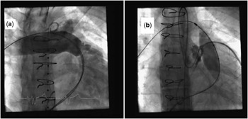
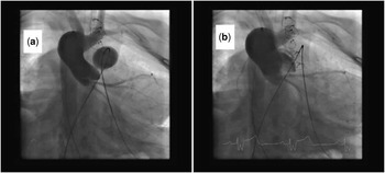
We have been surprised to find that patients in whom a coronary issue was known have still been found to be transcatheter pulmonary valve implantation candidates (Fig 1). The patient in Figure 1 had an initial diagnosis of tetralogy of Fallot and had undergone initial repair followed years later by placement of a surgical right ventricle to pulmonary artery conduit. The patient was known to have the left anterior descending artery coming off the right coronary artery coursing across the right ventricular outflow tract. At transcatheter pulmonary valve implantation, the left anterior descending indeed did course adjacent to the right ventricle to pulmonary artery conduit but was far enough away that no compression was noted and pre-stenting and valve implant were successful. We have also been just as surprised to find patients confirmed with conduit angioplasty not to be candidates (Fig 2). The patient in Figure 2 had an initial diagnosis of tetralogy of Fallot with pulmonary atresia, and on prior angiograms his right coronary artery origin was clearly high, leftward, and posterior, as is often noted to some degree in all patients with tetralogy of Fallot. Just how close the right coronary artery was to the right ventricle to pulmonary artery conduit was not appreciated until balloon dilation to 18 mm showed complete but transient right coronary artery occlusion. This was a diameter at which we hoped to deliver the Melody® valve. This patient was clearly not a candidate for transcatheter pulmonary valve implantation.
Panel ( a ) shows initial conduit angiogram and ( b ) selective right coronary artery angiogram during dilation of a 22-mm balloon in the conduit showing unimpeded flow in the left anterior descending.

Panel ( a ) shows complete right coronary artery occlusion with expansion of the stenotic conduit to 18 mm and ( b ) balloon deflation shows that the right coronary artery is widely patent and directly medial to the conduit.

How to implant a transcatheter pulmonary valve
Similarly to all complex interventions, procedural planning occurs well before the patient enters the cardiac catheterisation laboratory. Laboratory inventory needs to reflect the common need for multiple large-diameter balloons; a large number of the strongest available radial strength stent for extensive conduit preparation; the sheaths necessary to allow for stent delivery; the availability of covered stents or the ability to make covered stents; Ensemble delivery systems; Melody® valves; and the wires necessary to allow the delivery of the above. It is also very important to plan your likely valve delivery route before the procedure, and if a patient has a suspected access issue this should always be investigated at least by ultrasound before the procedure.
Transcatheter pulmonary valve implantation is a prolonged and complex interventional catheterisation and is usually performed under general anaesthesia. We always have two units of blood available in the cardiac catheterisation laboratory and two large bore peripheral intravenous access lines. All transcatheter pulmonary valve implantations are coordinated with the cardiac surgical schedule. We try to ensure that this is a light of day procedure and never schedule more than two procedures on a given day.
It all starts with access. For groin access procedures, we perform a venogram to ensure that the accessed femoral and iliac vein are normal in appearance, and if so the vein is often “pre-closed” with two Perclose® sutures (Abbott, Abbott Park, Illinois, United States of America) placed at a 90° angle from one another. The venous access is upsized to a large-diameter entry sheath (16 or 18 French, 30-cm long; Cook, Bloomington, Indiana, United States of America). For jugular access patients, we will begin with placement of a shorter large-diameter sheath (also from Cook). The activated clotting time is measured and heparin is given. Goal activated clotting time level at valve implant is 250 seconds. A right heart catheterisation is performed with a balloon tipped end-hole catheter, and a retrograde left heart catheterisation is performed in the routine manner. The venous end-hole catheter is advanced to whichever branch pulmonary artery seems easiest to get to and allows the straightest course. A MULTI-TRACK™ catheter (NuMED, Hopkington, New York, United States of America) is advanced to the main pulmonary artery and right ventricle for angiograms, and the conduit is measured. An ascending aortogram is performed.
If the conduit is noted to be rather stenotic, we proceed with a graduated dilation, often using several increasing diameter balloons to sort out how compliant the stenotic conduit segments really are, working towards a final diameter judged adequate for final valve implantation. Once the right ventricle to pulmonary artery conduit is dilated to its anticipated final size, ascending aortic angiograms are performed during balloon inflation in a steep caudal and lateral projection in order to ensure that the coronaries are not impinged upon. Sometimes, selective coronary angiograms are necessary. If a coronary appears impinged, the procedure is ended. After each dilation, a main pulmonary artery angiogram should be performed to ensure no right ventricle to pulmonary artery conduit disruption or dissection.
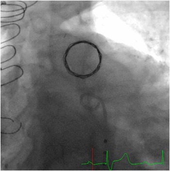
It is at this point that one should proceed with right ventricle to pulmonary artery conduit pre-stenting, and not before. The most important and controllable safety issue is sorting out the coronaries and coronary safety needs to be completely confirmed before implanting any rigid hardware. In our experience, all patients should receive at least one conduit stent before transcatheter pulmonary valve implantation to ensure an adequate landing zone and to minimise the risk of early and medium-term stent fracture and recurrent conduit stenosis. It is at this point that the wire used for ballooning is usually exchanged for a very stiff Lunderquist exchange length wire with a hand-moulded distal curve (Cook) to facilitate stent delivery. The vast majority of patients receive a 3- or 4-cm-long XL series stent (Cordis, Miami, Florida, United States of America), delivered on an 18-, 20-, or 22-mm outer diameter BIB® balloon (NuMED). This stent should always be delivered via a long sheath. The stent is often post-dilated with an Atlas® balloon (Bard, Tempe, Arizona, United States of America) or Mullins™ balloon (NuMED) to ensure no residual waist following initial implantation, and at this point the delivered stent is closely examined for evidence of stent recoil. If recoil of >1–2 mm is noted with non-compliant balloon deflation, a second stent should be implanted at the same diameter. Stent recoil should be reassessed, and occasionally several stents are implanted at this point. Again angiograms should be performed after each step to ensure good stent position and no conduit disruption or dissection. A well-prepared pre-stented right ventricle to pulmonary artery conduit demonstrates a very circular appearance in a steep caudal view (Fig 3).
The barrel/steep caudal view showing a circular appearance of an extensively pre-stented conduit before transcatheter pulmonary valve implantation.

Once pre-stenting is completed, often the most straightforward manoeuvre is valve implantation. The large-diameter sheath is exchanged for a 22-French dilator (Cook). The appropriate sized Ensemble® (Medtronic, Minneapolis, Minnesota, United States of America) delivery system is flushed and inner and outer balloons are negatively prepped. We do not use a pressure inflation manometer on the inner balloon, but always do on the outer. The valve undergoes two sequential 1-minute saline baths to wash off gluteraldehyde and its serial number is confirmed. The valve is inspected to ensure appropriate leaflet function and coaptation direction. The expanded Melody® valve is sequentially hand-crimped, first over a 3-mm syringe and then over the prepped Ensemble® balloons. A time-out is taken to ensure that the upstream and downstream parts of the valve are appropriately mounted on the Ensemble – blue to blue and white to white colour codes are checked. The covering sleeve is flushed, and very slowly and gently advanced over the loaded valve. The loaded Melody® is brought to the catheterisation laboratory table, the 22-French dilator is removed, and the loaded Melody® is advanced over the wire into the venous system. Often, a gentle back and forth twisting motion of the loaded valve facilitates traversing the subcutaneous tissue. The Ensemble's® haemostasis adapter is advanced into the entry site. The loaded valve is advanced over the wire and brought to straddle the just placed pre-stent and often just a bit past it. The valve covering sleeve on the Ensemble® is withdrawn, often leading to the valve rocking forward several millimetres. The final positioning to overlap the pre-stent is finalised, and the inner and outer balloons are sequentially inflated deploying the valve. The outer balloon is often used to post-dilate. The Ensemble® is removed, the large-diameter venous sheath is re-advanced into the vein, and haemodynamics and angiograms are repeated. If the transvalvar gradient is >10 mmHg, the valve is often further post-dilated with the same non-compliant balloon used to post-dilate the pre-stent, and measurements are repeated. The procedure is ended, the “pre-closed” vein entry site is closed, and the patient is awakened from anaesthesia. All patients are observed in the intensive care unit overnight and receive 24 hours of intravenous antibiotic coverage. Patients usually go home the next day after chest film, electrocardiogram, and echocardiogram.
Why implant a transcatheter pulmonary valve?
There are several reasons to develop and employ transcatheter pulmonary valve implantation. Clearly, there is a large and divergent group of patients with congenial heart disease who wind up with a dysfunctional pulmonary valve, and with ever-improving survival from early cardiac surgery these numbers will only burgeon further. Without transcatheter pulmonary valve implantation, the only option for these patients is repeated lifetime repeat surgical valve replacement. With transcatheter pulmonary valve implantation, the concept of minimisation of lifetime cumulative morbidity from repeated surgery becomes an achievable goal. The risks of repeated sternotomy and cardiac injury increases, particularly after the second operation.Reference Holst, Dearani and Burkhart 18 The risks of air embolism, myocardial injury, central nervous system injury, and systemic organ injury with surgery remain rare but ever present, and rare risks over time are nothing but additive. It is also clear that, if anything, we may be waiting too long to replace the pulmonary valves for some patients and that a more aggressive approach may aid in maintaining late right ventricular size and function.Reference Frigiola, Tsang and Bull 19 A safer, less-invasive option should eventually lower the clinical threshold for pulmonary valve replacement and therefore ensure that fewer patients develop irreversible adverse right ventricular changes.
It is true that the current widely available transcatheter pulmonary valve implantation has shown occasional early dysfunction due to stent fracture leading to recurrent conduit stenosis, but this can also be often addressed with a second catheterisation, additional conduit stenting, and a second transcatheter pulmonary valve.Reference McElhinney, Cheatham and Jones 20 It is also clear that no current surgical pulmonary valve replacement offers a perfect long-term solution, as a marked number of patients following homograft right ventricle to pulmonary artery conduit and Gore-Tex® (Gore, Flagstaff, Arizona, United States of America) bivalve pulmonary valve replacement have good but far from ideal medium and late-term function.Reference Oosterhof, Meijboom and Vliegen 21 , Reference Quintessenza, Jacobs, Chai, Morell and Lindberg 22
It was into this imperfect clinical world that transcatheter pulmonary valve implantation was introduced, and this is why in lifetime planning for patients requiring pulmonary valve replacement surgery needs to be performed, allowing this technology to become a viable late treatment option.