Implications
In this work, we demonstrate the promoter-specific regulation of acetyl-CoA carboxylase α (ACC-α) in mammary and adipose tissue during milk fat depression (MFD) induced by trans-10, cis-12 CLA. This demonstrates a novel nutrient–gene interaction coordinating metabolism between tissues and provides insight into the physiological consequences of bioactive fatty acids (FAs).
Introduction
Long-chain FAs and their derivatives can act as bioactive molecular regulators affecting cellular activity. An example is trans-10, cis-12 CLA that causes diet-induced MFD. During MFD, mammary lipogenic capacity and expression of key mammary lipogenic genes are coordinately down-regulated (Bauman et al., Reference Baumgard, Corl, Dwyer, Sæbø and Bauman2011). ACC-α (EC 6.4.1.2) and fatty acid synthase (FASN) (EC 2.3.1.85) (Kim, Reference Kim1997) are two essential and regulated enzymes involved in FA synthesis in eukaryote cells and both are down-regulated in mammary tissue during MFD (Baumgard et al., Reference Bauman, Harvatine and Lock2000). During lactation, ACC-α plays a key role to facilitate the partitioning of FA precursors for synthesis of milk fat in the mammary gland. This occurs through repression of ACC-α in adipose tissue and, in particular, ACC-α is induced in the mammary gland, in part, by matching levels of messenger RNA (mRNA) in both tissues (Barber et al., Reference Barber, Price and Travers1997).
Previous studies with rodents, ovine and bovine have shown that ACC-α is transcribed from multiple promoters (P) in a tissue-specific manner with some differences between species (Barber and Travers, Reference Barber, Clegg, Travers and Vernon1995; Barber et al., Reference Barber and Travers2003). Constitutive expression from PII occurs in many tissues, whereas PI expression is limited to adipose tissue and liver under lipogenic conditions (Barber et al., Reference Barber and Travers2003) and the lactating bovine mammary gland (Mao et al., Reference Mao, Marcos, Davis, Burzlaff and Seyfert2001). However, expression from PI is low in the ovine mammary gland and the regulation of the species differences has not been elucidated (Molenaar et al., Reference Molenaar, Mao, Oden and Seyfert2003). A third promoter driving ACC-α expression (PIII) was identified in sheep (Barber and Travers, Reference Barber and Travers1998) and it is highly expressed in lactating ovine and bovine mammary tissue with ~15-fold increase during lactation (Mao et al., Reference Mao, Marcos, Davis, Burzlaff and Seyfert2001). In addition, Barber et al. (Reference Barber, Vallance, Kennedy and Travers2005) showed the existence of a fourth promoter expressed predominantly in the brain of ruminants showing the complexity of the regulation of ACC-α. The mechanisms controlling ACC-α expression in different tissues and stages of lactation is not well understood, but a role for the sterol regulatory element-binding protein (SREBP1) family of transcription factors was proposed based on their function as global regulators of expression for many genes involved in lipid synthesis (Harvatine and Bauman, Reference Harvatine and Bauman2006). The scenario becomes even more obscure during altered physiological conditions such as MFD caused by bioactive isomers of CLA (i.e. trans-10, cis-12 CLA isomer).
The objective of this study was to quantify ACC-α gene expression from PI, PII and PIII during MFD induced by supplementation of trans-10, cis-12 CLA at three stages of lactation that represent different physiological states due to changes in milk yield and energy balance.
Material and methods
Animals and treatments
Santa Catarina State University ethical committee approved all animal management and care procedures. In all, 12 cross-bred Lacaune/Texel ewes weighting 62±1.8 kg (mean±SE), mean parity 3.1±0.23 and producing 1.8±0.12 kg milk/day were arranged in a completely randomized design, and fed one of the following treatments: Control (forage+0.9 kg of concentrate on a dry matter (DM) basis) and CLA (control+27 g/day of CLA Luta-60; BASF AG, Sao Paulo, Brazil). The CLA supplement consisted of FA methyl esters with the following composition: 4.1% palmitic acid, 3.6% stearic acid, 27.4% oleic acid, 1.2% linoleic acid, 29.8% cis-9, trans-11 CLA, 29.9% trans-10, cis-12 CLA and 3.0% other FA. This CLA supplement has been reported to induce MFD and change the milk FA profile in sheep by Oliveira et al. (Reference Oliveira, Bauman and Harvatine2012) and Baldin et al. (Reference Baldin, Dresch, Martelo, Souza and Oliveira2013a), and in goats by Baldin et al. (Reference Baldin, Dresch, Souza, Fernandes, Gama, Harvatine and Oliveira2013b and Reference Baldin, Gama, Dresch, Harvatine and Oliveira2014) and Fernandes et al. (Reference Fernandes, Gama, Ribeiro, Lopes and Oliveira2014). In addition, de Veth et al. (Reference de Veth, Griinari, Pfeiffer and Bauman2004) reported that abomasally infused CLA methyl esters had equal potency in inhibition of milk fat synthesis compared with CLA in the free FA form. In the current experiment, CLA was orally dosed once a day with a syringe (30 ml). Our previous work with a similar procedure in ewes resulted in a 3.2±0.6% (0.28 g/day) transfer of trans-10, cis-12 CLA to milk (Oliveira et al., Reference Oliveira, Bauman and Harvatine2012).
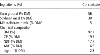
The treatments were applied to the same animals at 15 (early), 70 (mid) and 120 (late) days in milking and each treatment period lasted 14 days with no treatment for 55 and 50 days between the early and mid and mid and late phases, respectively. All ewes grazed six paddocks of festuca (Festuca arundinacea Schreb.) and white clover (Trifolium repens L.) with free access to water and were supplemented with 1.0 kg of corn silage overnight. The concentrate supplement composed of ground corn, soybean meal and a commercial vitamin and mineral mix (Table 1). The diet was balanced to meet or exceed energy, protein, vitamin and minerals requirements according to National Research Council (2007) and predicted DM intakes based on Institut National De La Recherche Agronomique (2007) taking into account live weight, milk yield and milk composition.
Ingredients and chemical composition of the concentrate

DM=dry matter.
* Sheep mineral and vitamin premix (Nutrimasa Agroindustrial Feeds, Chapecó, Brasil). Major minerals (g/kg) were Ca, 190; Na, 35; Mg, 30; S, 18; P, 69. Trace minerals (mg/kg) were Zn, 1440; Fe, 840; Mn, 1550; Co, 3.4; Se, 22. Vitamins (IU/kg) were vitamin A, 125; vitamin D3, 10; vitamin E, 500.
Milk sampling and analysis
On day 14 of each treatment period, milk samples were collected at the a.m. and p.m. milkings. Harvatine et al. (Reference Harvatine, Perfield and Bauman2009) reported that trans-10, cis-12 CLA decreased milk fat and expression of lipogenic enzymes and regulators after 3 days. The 14-day period in the current experiment was selected to allow complete adaptation to diet-induced MFD. Milk samples were stored at 4°C with a preservative (bronopol tablet; D & F Control Systems Inc., San Ramon, CA, USA). All milk samples were proportionally composited according to a.m. and p.m. yields. Milk fat, protein, lactose and total solids were determined by IR analysis (Association of Official Analytical Chemists, 2004).
Tissue biopsies
All mammary biopsies were taken between 1 and 4 h after the a.m. milking on day 14 of each treatment period with an average duration of 15 min/ewe. Lidocaine hydrochloride subdermal block (3 ml/ewe) was administered above the incision site. A 0.5 cm incision was made in the skin at the midpoint of the rear quarter and a biopsy was collected using a Bard Max-Core Disposable Core Biopsy Instrument (Bard Biopsy Systems, Covington, GA, USA). Briefly, a 16-gauge biopsy needle was inserted 5 cm into the mammary gland through the incision. Tissue samples (~10 mg tissue/biopsy) were inspected to verify tissue homogeneity and snap frozen in liquid N2. Samples were placed in cryotubes containing 1 ml of Dulbecco’s phosphate-buffered saline (Gibco Laboratories, Grand Island, NY, USA) and stored in liquid N2 until RNA extraction. Immediately after removal of the biopsy needle, a purse string suture was placed around the incision with number 1 Nylon. The biopsy procedure resulted in minimal bleeding and milk appeared normal in two to four milkings following the biopsy. No intra-mammary infections were observed.
The adipose tissue biopsy was collected from the tail head region immediately cranial and lateral to the last lumbar vertebra (dorsal subcutaneous depot). Before the biopsy, lidocaine hydrochloride subdermal block was administered in a circular pattern surrounding the incision site (3 ml/ewe). Once the block was effective, an incision was made in the skin and adipose tissue was dissected. The adipose tissue sample (~100 to 400 mg) was divided into two samples and snap frozen as described above. The incision was irrigated and closed with number 1 Nylon using a blanket stitch. Flunixin meglumine (1.1 mg/kg of BW) was administered following the biopsies. Biopsy sample size was based on expected RNA yield as mammary tissue has a higher and adipose has a lower RNA yield per milligram of tissue.
Acetyl-CoA carboxylase α transcripts quantification
Total RNA was extracted from mammary and adipose tissue samples with the RNeasy Lipid Tissue Mini Kit (Qiagen Sciences, Germantown, MD, USA) with on-column DNase treatment (RNase-free DNase set; Qiagen Sciences). The RNA concentration and protein contamination was measured using a spectrophotometer (NanoDrop ND-2000; NanoDrop Technologies, Wilmington, DE, USA). Sample 260/280 ratio ranged between 1.8 and 2.0. Agarose gel electrophoresis was used to determine RNA integrity. Briefly, 1 µl of total RNA and 1 µl of GelRed™ Nucleic Acid Gel Stain, 10 000× in water (Biotrend Chemikalien GmbH, Köln, Germany) was resolved on a 1.5% agarose gel at a constant 100 V and after visualized under UV light. Two discrete bands corresponding to the 28S and 18S ribosomal RNA were observed for all samples.
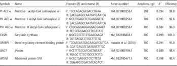
Primers were designed using Primer Express software (PE Applied Biosystems, version 3.0) using gene sequences publically available in GenBank (National Center for Biotechnology Information; http://www.ncbi.nlm.nih.gov/genbank/). Details including primer sequences and performance are listed in Table 2. Briefly, promoter-specific primers to amplify ACC-α PI transcripts included the forward primer in exon 1 and reverse in exon 5, ACC-α PII transcripts included the forward primer at the junction of exon 2 and reverse primer in exon 5, and ACC-α PIII primers were in exon 5A. The expression of SREBP1, FASN and leptin was also measured.
Sequences (5' to 3') and efficiencies of the primers that were used in the reverse-transcription quantitative PCR

The mRNA was transcribed to complementary DNA (cDNA) using the High-Capacity cDNA Reverse Transcription kit (Applied Biosystems, Foster City, CA, USA) with random primers. PCR amplification was performed in triplicate in a 15 µl reaction with 30 ng of cDNA and 7.5 µl SYBR Green Select Master Mix (Applied Biosystems) in a StepOne Real-Time machine (Applied Biosystems). The PCR program was as follows: 95°C for 2 min, 40 cycles of 95°C for 15 s, 62°C for 1 min and 95°C for 15 s. The results were normalized using the threshold cycle (CT) method for the expression of the reference genes, the ribosomal protein S18 (RPS18) gene and β-actin gene (BACT). The data were analysed with StepOne software version 2.1 (Applied Biosystems).
A validation assay was performed to demonstrate formation of a single product and the amplification efficiencies of each primer pair of the target and reference genes (Table 2) using the standard curve method of pooled cDNA from mammary or adipose tissue for each sample where the standard curve is graphically represented as a semi-log regression line plot of CT value v. log of nucleic acid and the slope as the efficiency for the reaction.
Statistical analysis
Data were analysed using the MIXED procedure of SAS Institute (2009) and the model included the fixed effect of animal and treatment. The geometric mean of the housekeeping genes (RPS18 and BACT; Vandesompele et al., Reference Vandesompele, De Preter, Pattyn, Poppe, Van Roy, De Paepe and Speleman2002) was used as a covariate in the model for gene expression data and was removed if not significant. Data points with studentized residuals outside of ±2.5 were considered outliers and excluded from analysis. Milk yield and milk components were also analysed by the MIXED procedure using ewe and treatment and milk yield on the day before each period as a covariate (removed if not significant). LSMEANS were used to compare treatments and significance was declared at P<0.05.
Results
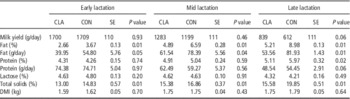
The effect of CLA on performance and milk composition at each stage of lactation are shown in Table 3. Milk yield was unaffected by CLA supplementation, whereas milk fat concentration and yield were reduced by CLA in all three periods (P<0.01; Table 3). Compared with Control, CLA decreased milk fat yield 27%, 21% and 35% and concentration 28%, 26% and 42% in early, mid and late lactation, respectively. Milk protein concentration was not altered by CLA supplementation in early and mid lactation (Table 3), but was decreased 14% by CLA in late lactation (P=0.02).
Performance and milk composition of dairy ewes supplemented with 27 g/day of CLA at different stages during the lactationFootnote *

CON=Control; DMI=dry matter intake.
* n=6 (Control) and 6 (CLA); CLA supplement: 4.1% palmitic acid, 3.6% stearic acid, 27.4% oleic acid, 1.2% linoleic acid, 29.8% cis-9, trans-11 CLA, 29.9% trans-10, cis-12 CLA and 3.0% other fatty acids.
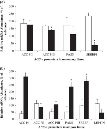
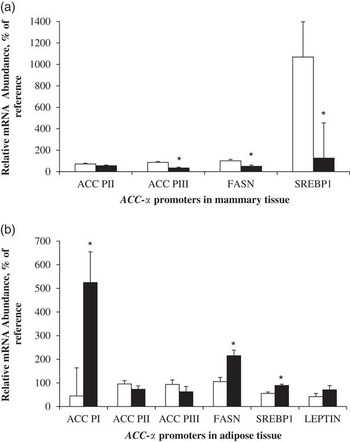
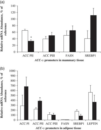
Expression of ACC-α PI was below the level of detection in mammary tissue in agreement with similar observations in the ovine by Molenaar et al. (Reference Molenaar, Mao, Oden and Seyfert2003). In early lactation, CLA reduced mammary PIII ACC-α transcripts by 17.3% (P<0.05; Figure 1), but did not change mammary PII-derived transcripts (P=0.37). In adipose tissue, transcripts from PI increased by 69.6% (P<0.01) when ewes received CLA in early lactation. There was no treatment effect on PII and PIII transcripts in the adipose tissue during early lactation (P>0.05). Mammary ACC-α transcripts from PIII were reduced 59.7% (P=0.04) by CLA treatment during mid lactation (Figure 2), whereas transcripts from PII were not affected by treatment (P=0.21). Similar to early lactation, during mid lactation ACC-α transcripts from PI were up-regulated in adipose tissue by CLA supplementation (P<0.02).
Effects of supplementation with CLA on acetyl-CoA carboxylase α (ACC-α) transcripts from promoters (PI, PII and PIII), fatty acid synthase (FASN) and sterol regulatory element-binding protein (SREBP1) in (a) mammary and (b) adipose tissues from ewes fed a Control (□, n=6) or a rumen-unprotected CLA (■, n=6) supplement in early lactation. Values are mean±SE. *P<0.05 between treatments. mRNA=messenger RNA.

Effects of supplementation with CLA on acetyl-CoA carboxylase α (ACC-α) transcripts from promoters (PI, PII and PIII) , fatty acid synthase (FASN) and sterol regulatory element-binding protein (SREBP1) in (a) mammary and (b) adipose tissues from ewes fed a Control (□, n=6) or a rumen-unprotected CLA (■, n=6) supplement in mid lactation. Values are mean±SE. *P<0.05 between treatments. mRNA=messenger RNA.

Effect of CLA on ACC-α transcripts was different in late lactation (Figure 3), as CLA did not change expression of PIII transcripts in the mammary gland (P=0.83). However, transcripts from PII were reduced 49.4% by CLA in mammary tissue (P<0.01). During late lactation, CLA had no effect on ACC-α transcripts in adipose tissue (Figure 3). To verify regulation of lipogenesis, expression of FASN and SREBP1 were determined in adipose and mammary tissue. In the mammary gland, SREBP1 was reduced in early and mid lactation (P=0.001) and not changed in late lactation, similar to ACC-α PIII transcripts. However, FASN expression in the mammary gland was only changed in mid lactation (P<0.05). In adipose tissue, SREBP1 and FASN increased in CLA treated in early and mid lactation (P=0.01), but were unchanged in late lactation similar to the ACC-α PI transcripts. Interestingly, leptin in adipose tissue was not changed through the lactation cycle (P>0.05).
Effects of supplementation with CLA on acetyl-CoA carboxylase α (ACC-α) transcripts from promoters (PI, PII and PIII), fatty acid synthase (FASN) and sterol regulatory element-binding protein (SREBP1) in (a) mammary and (b) adipose tissues from ewes fed a Control (□, n=6) or a rumen-unprotected CLA (■, n=6) supplement in late lactation. Values are mean±SE. *P<0.05 between treatments. mRNA=messenger RNA.

Discussion
In this study, primers were designed to measure promoter-specific expression of ACC-α in mammary and adipose tissue during CLA treatment at three stages of lactation. This allowed the investigation of the tissue and promoter-specific regulation of ACC-α during MFD induced by trans-10, cis-12 CLA. Barber et al. (Reference Barber, Price and Travers1997) identified PIII as the promoter most important to milk fat synthesis because its expression is dramatically increased in mammary tissue during lactation. In the current experiment, only PIII transcripts were decreased in the mammary gland by CLA treatment in early and mid lactation and PIII appears to be the predominant point of CLA regulation.
Expression of ACC-α from PI is not thought to be crucial for milk fat synthesis in sheep and PI transcripts were not quantifiable in mammary tissue in the current study. Similarly, Travers et al. (Reference Travers, Vallance, Gourlay, Gill, Klein, Bottema and Barber2001) established that the contribution of PI to mammary ACC-α expression is low in sheep because expression is low at all stages of lactation and does not change from pregnancy to lactation. The importance of mammary PI-derived transcripts for synthesis of milk fat has also been questioned in cows, although PI transcripts are highly abundant in adipose tissue (Mao et al., Reference Mao, Marcos, Davis, Burzlaff and Seyfert2001). Transcripts from PI are also the highest in the mammary gland of the cow, but its concentration does not increase during lactation. Considering the high exon 1 sequence conservation between bovine and sheep (92.3%), the species-specific difference is not clear. Although the bovine lactating mammary gland has little adipose tissue, it may contain some adipocytes, which may explain the low level of PI-derived transcripts in lactating and non-lactating mammary tissue (Mao et al., Reference Mao, Marcos, Davis, Burzlaff and Seyfert2001). Future studies should include techniques allowing to isolate the different mammary cell types and study their gene expression.
In late lactation, no changes in expression of PI-derived ACC-α transcripts were observed in adipose tissue. The possible explanations for that could be as follows: first, would be an increase in hepatic lipid synthesis, even though ruminants have low capacity for hepatic lipid synthesis. Second, would be a reduction in feed intake (not significant) during CLA supplementation limiting deposition in adipose tissue or because the adipose tissue was already near a maximal rate of lipogenesis or may be above a metabolic set-point and not responsive to increases in nutrient availability. A third explanation for no change in the gene expression of the measured genes in adipose tissue in late lactation may be due to changes at the enzymatic level or activity. Other studies have noted that there were no changes in ACC-α expression on diets inducing MFD in cows and mouse (Thering et al., Reference Thering, Graugnard, Piantoni and Loor2009; Rudolph et al., Reference Rudolph, Monks, Burns, Phistry, Marians, Foote and Neville2010), whereas the previous study obtained changes at the enzymatic level measured by western blot. Harvatine et al. (Reference Harvatine, Perfield and Bauman2009) and Thering et al. (Reference Thering, Graugnard, Piantoni and Loor2009) reported that expression of lipogenic enzymes and transcription factors were increased in adipose tissue during CLA-induced MFD in the cow. Tissue-specific expression of ACC-α may explain opposite responses in adipose and mammary tissue during CLA treatment and in the current experiment, as promoter-specific regulation of ACC-α was observed between adipose and mammary tissues. It is noteworthy that the promoters down-regulated in mammary gland did not increase in adipose tissue in the same lactation period. In early and mid lactation, the main promoter up-regulated in adipose tissue was PI and the main promoter down-regulated in mammary tissue was PIII. Interestingly, SREBP1, that it is considered a global regulator of lipid synthesis (Eberlé et al., Reference Eberlé, Hegarty, Bossard, Ferré and Foufelle2004), had a similar response to the ACC-α PIII transcripts in the mammary gland, whereas in adipose tissue had a similar response to ACC-α PI transcripts and FASN. SREBP1 regulates enzymes which synthesize FA and cholesterol from precursor molecules (Horton et al., Reference Horton, Shah and Warrington2003; Goldstein et al., Reference Goldstein, DeBose-Boyd and Brown2006) and SREBP1 is a major regulator of mammary de novo FA synthesis and the response to dietary factors that modify milk fat in mouse (Anderson et al., Reference Anderson, Rudolph, McManaman and Neville2007) and cow (Harvatine and Bauman, Reference Harvatine and Bauman2006).
Care should be taken in interpreting studies across lactation as milk production may be affected by different mechanisms in each period (cell differentiation and lactogenesis during start of lactation, galactogenesis during lactation and apoptosis during established lactation; Bauman et al., Reference Baumgard, Corl, Dwyer, Sæbø and Bauman2011). It is well established that CLA inhibits SREBP1 activity in vivo, although the critical points that regulate this inhibition have not been described. Specifically, a direct or indirect link between trans-10, cis-12 CLA and SREBP1 signal has not been elucidated, nor the mechanism for inhibiting SREBP1 by other bioactive FA (Bauman et al., Reference Baumgard, Corl, Dwyer, Sæbø and Bauman2011). Moreover, it is important to make a mention that the observed positive effects of trans-10, cis-12 CLA on SREBP1, ACC-α and FASN in adipose tissue (lipogenesis) probably is not a direct effect because it was shown that this CLA isomer is able to inhibit the lipogenesis in human (Brown and McIntosh, Reference Brown and McIntosh2003) and bovine (Lengi and Corl, Reference Lengi and Corl2010) adipocytes. Despite the positive effect on lipid synthesis genes in adipose tissue, the short time of each experimental period (15 days) may not have been adequate for fat accumulation to stimulate expression of leptin. The difference in response of the ACC-α transcripts in the mammary gland to CLA during the different stages of lactation might be explained by the magnitude of MFD or source of milk FA. Previous work in cows has demonstrated that higher doses of trans-10, cis-12 CLA are required to induce MFD during early lactation compared with mid and late lactation (Castaneda-Gutierrez et al., Reference Castaneda-Gutierrez, Overton, Butler and Bauman2005; Odens et al., Reference Odens, Burgos, Innocenti, VanBaale and Baumgard2007). This may be due to transcript-specific regulation of ACC-α. Milk fat concentration was higher during late lactation in the current experiment and may indicate a different cellular environment regulating milk fat synthesis during this stage. Other possible difference in late lactation could be changes in cell physiology during which in a late stage the remaining mammary epithelial cells may have a reduction in PIII transcripts and remaining PII transcripts as shown by Molenaar et al. (Reference Molenaar, Mao, Oden and Seyfert2003). Oliveira et al. (Reference Oliveira, Gama, Fernandes, Tedeschi and Bauman2014) found that ACC-α transcripts from PII and PIII were down-regulated by CLA treatment that decreased milk fat by 24% in mid lactation dairy cows. In our study, there was no difference in transcripts from PII at mid lactation, but the magnitude of the MFD was greater when the PII-derived transcript was down-regulated during late lactation. Milk FA profile was not measured in the present experiment so a correlation of transcript expression with de novo and preformed FA contribution to milk fat is not possible, but should be investigated in future experiments.
Conclusion
This study demonstrated that ACC-α transcripts are down-regulated in the mammary gland and up-regulated in adipose tissue of lactating ewes during CLA-induced MFD through a promoter and tissue-specific regulation. The promoter-specific regulation of ACC-α in the mammary gland is a breakthrough in the study of MFD and future studies should investigate the differences between PI-derived transcripts and those from PII and PIII of ACC-α.
Acknowledgements
The authors gratefully acknowledge Conselho Nacional de Desenvolvimento Científico e Tecnológico – CNPq (grant no. 474094/2012-0 to D. E. O.) for the financial support. The authors also would like to thank Paulo Gregianin (manager of Pinheiro Seco farm), they also thank Dr Daniel Rico for revising the manuscript. The assistance of Ana Povaluk, Mauricio Camêra and Camila Renneberg was gratefully appreciated.