Introduction
Papillomas are benign epithelial sinonasal tumours which represent 0.5 to 4 per cent of all nasal neoplasms. They are thought to be derived from the Schneiderian mucosa, which is of ectodermal origin. These tumours are notoriously locally aggressive and have the propensity to recur after excision. They can be associated with malignant changes in 2 to 56 per cent of cases.Reference Nachtigal, Yoskovitch, Frenkiel, Braverman and Rochon1, Reference Bhaya and Har-El2
Rarely, more than one type of carcinoma may coexist. Hybrid tumour is defined as a focus of non-verrucous squamous cell carcinoma which arises synchronously with verrucous carcinoma.Reference Batsakis, Suarez and El-Naggar3 Only five cases of hybrid tumour of the paranasal sinuses have been reported in the literature.Reference Orvidas, Lewis, Olsen and Weiner4–Reference Letichevsky, Talmon, Samet and Cohen8 We present a rare case of sinonasal hybrid tumour within an inverted papilloma, with an iatrogenic cerebrospinal fluid (CSF) leak which was repaired successfully on revision surgery.
Case report
A 60-year-old Chinese man, a non-smoker, had presented to a private ENT centre with a history of left nasal obstruction for a few years, which had worsened over the last five months. There had been associated anosmia. However, he had not complained of epistaxis, persistent rhinorrhoea, diplopia or visual blurring.
This patient had been treated in a private specialist centre (prior to presentation to our institution) and had undergone surgical endoscopic removal of an inverted papilloma. The procedure had been complicated by a CSF leak. Intra-operatively, the tumour had been found to be attached to the middle turbinate, extending to the posterior choana and involving the anterior and posterior ethmoids. Superiorly, it had been found to have caused pressure necrosis of the anterior skull base and to be adherent to the dura. During endoscopic sinus surgery, the dura had been inadvertently opened, causing the CSF leak.
On referral, we admitted the patient to our ward and inserted a lumbar drain. Intravenous antibiotics (ceftriaxone and metronidazole) were commenced.
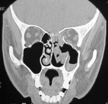
A computed tomography (CT) scan (Figure 1) showed a bony defect of the floor of the anterior cranial fossa, measuring 12×6 mm, with evidence of pneumocranium.
Coronal computed tomography scan taken after the patients' initial endoscopic sinus surgery, showing bony defect at the anterior skull base and residual papilloma within the posterior ethmoids.

Endoscopic repair of the CSF leak was performed, which revealed a dural tear measuring 15×5 mm in the region of the ethmoid and sphenoid junction, along with remnants of the papilloma. The left lamina papyracea was absent. Intrathecal sodium fluorescein was used intra-operatively in order to assist visualisation of the defect. The defect was repaired with an underlay fat plug and an onlay mucoperichondrial flap with fibrin glue application.
The lumbar drain was left in situ for another three days. The nasal pack was removed on the fourth post-operative day. Recovery was uneventful, and the patient was discharged on the fifth post-operative day. Antibiotics were administered for 10 days in total (including in patient treatment).
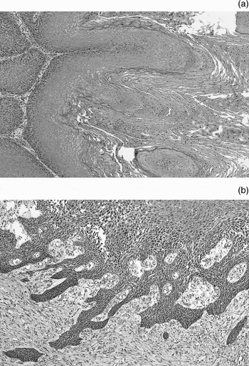
Histopathological examination showed a Schneiderian inverting papilloma with areas of verrucous carcinoma with keratinisation, together with foci of moderately differentiated squamous epithelium with high mitotic activity and invasive edges (Figure 2). Therefore, a histopathological diagnosis of hybrid verrucous squamous cell carcinoma was concluded.
Photomicrographs showing (a) verrucous carcinoma with keratinisation and adjacent papilloma (H&E; ×100), and (b) invasive squamous cell carcinoma (H&E; ×100).

In view of this diagnosis, the patient received adjuvant radiotherapy of 60 Gray given in 30 fractions.
During post-treatment surveillance, a suspicious polypoidal lesion at the left cribiform plate was seen on nasal endoscopy, 19 months after completion of radiotherapy. There was no recurrence of the CSF leak. A biopsy of the mass was performed, which showed Schneiderian papilloma with squamous metaplasia. There was no evidence of abnormal mitosis. A repeat magnetic resonance imaging (MRI) scan showed a soft tissue lesion at the left cribiform plate. The adjacent intracranial changes were most probably due to previous radiotherapy. Further repeated biopsies from the polypoidal growth showed Schneiderian papilloma with no evidence of malignancy.
At the time of writing, after 42 months of follow up, the patient remained clinically well, with no rapid growth of the lesion or presence of cervical metastases.
Discussion
Inverting papilloma usually presents as a unilateral nasal mass with symptoms of progressive nasal obstruction. It is rarely bilateral and multicentric in origin. The aetiology of inverting papilloma is still unknown, but smoking, allergens, chemical pollutants, chronic sinusitis and human papilloma virus have been indicated as possible predisposing factors.Reference Pasquini, Sciarretta, Farneti, Modugno and Ceroni9
Pre-operative radiological investigation, including CT and MRI, is imperative in the treatment plan for patients with inverting papilloma. A CT scan, using both soft tissue and bone settings, allows study of the tumour extent and the presence of surrounding bone remodelling, dehiscence or erosion. Features of erosion would strongly suggest the possibility of carcinoma. In the presence of skull base or orbital involvement, MRI is very helpful to delineate the tumour extent and the presence of intra-orbital or intra-axial extension.
Previously published data indicated that inverting papillomas with maxillary, ethmoid and sphenoid involvement are significantly associated with the presence of carcinoma. These authors also indicate a significant relation between inverting papillomas of multicentric origin and malignancy. Multicentricity is defined as separate lesions, with no mucosal or submucosal continuity between the lesions.Reference Nachtigal, Yoskovitch, Frenkiel, Braverman and Rochon1
Surgery is the mainstream treatment for inverting papilloma. However, the choice of surgery is tailored towards the nature of this tumour, which is characteristically aggressive and locally destructive, and is associated with malignancy; it is also known to recur if not completely excised. Open surgery for inverting papilloma may take the form of lateral rhinotomy, midfacial degloving, or osteoplastic or craniofacial resection. The choice of approach is dependent on the extent of the tumour and the surgical expertise available. Contrary to previous belief, recurrence rates following open and endoscopic approaches are similar, provided that proper pre-operative selection criteria are observed.Reference Pasquini, Sciarretta, Farneti, Modugno and Ceroni9, Reference Kraft, Simmen, Kaufmann and Holzmann10 Lawson et al. quote a recurrence rate of 16 per cent for open technique, compared with 12 per cent for endoscopic technique.Reference Lawson, Kaufman and Biller11
With the advent of endoscopes and powered instrumentation, extensive inverting papilloma (as presented in our patient) may be treated endoscopically. However caution should be taken where the anterior skull base is involved and the expertise for possible complications such cerebrospinal fluid leak should be at hand. In our case, we have illustrated that an endoscopic approach was feasible for excision of inverting papilloma involving the anterior skull base with no evidence of frank intracranial extension.
Our patient was referred for further management of the CSF leak encountered during the first surgery, and for completion of the tumour excision. Computed tomography was repeated in order to assist in detecting the site of the skull base defect. Intrathecal fluorescein was used intra-operatively to help localise the defect. The intra-operative findings correlated well with the pre-operative imaging study, and the defect was closed in a multilayer fashion with autologous grafts and fibrin glue.
Histopathological examination is an important routine procedure in confirming the diagnosis of any nasal tumour. It is essential to alert the histopathologist to possible suspicious features of the tumour which may suggest associated malignancy. Inverting papilloma is usually large at presentation, and therefore the presenting pathological specimen is equally large. The associated carcinoma within the tumour may be present in a small section of the tumour. Therefore, the histopathologist will invariably need to examine the various small sections of the tumour.
Squamous cell carcinoma is seen in 5 to 21 per cent of cases. Initially, there is squamous metaplasia of the natural nasal respiratory epithelium. Once epithelial dysplasia is established, carcinoma in situ and invasive squamous cell carcinoma could follow.Reference Pasquini, Sciarretta, Farneti, Modugno and Ceroni9 There are three situations in which inverting papilloma is associated with squamous cell carcinoma. First, the two may coexist together as a synchronous tumour. Second, the latter may present as a foci of carcinoma within the papilloma. Third, carcinoma may arise metachronously at the site of previously removed papilloma.Reference Pasquini, Sciarretta, Farneti, Modugno and Ceroni9, Reference Lawson, Kaufman and Biller11
Our patient was diagnosed with hybrid verrucous squamous cell carcinoma on histopathological examination. In this tumour, tissue sections showed areas of squamous carcinoma synchronously within the verrucous carcinoma, as defined by Batsakis et al. Reference Batsakis, Suarez and El-Naggar3 This type of hybrid tumour has been described in the oral cavity and larynx. The treatment for hybrid tumour of non-verrucous origin should be as for squamous cell carcinoma, that is, wide tumour resection with adjuvant radiotherapy.Reference Batsakis, Suarez and El-Naggar3, Reference Gomez, Mendenhall, Tannehill, Stringer and Cassisi12 On the other hand, in cases of verrucous carcinoma, radiotherapy is avoided because the tumour is insensitive to radiotherapy; furthermore, such treatment may alter the nature of the tumour to anaplastic carcinoma.Reference Go, Oh and Ko5, Reference Batsakis and Suarez6 Verrucous carcinoma is a highly differentiated form of squamous cell carcinoma. It has the ability to invade surrounding tissues, but metastases are rare. Histologically, it must be distinguished from benign papilloma, verrucous hyperplasia and well differentiated non-verrucous squamous cell carcinoma.
• Only five cases of hybrid tumour have been reported in the literature
• Hybrid tumour associated with inverted papilloma is even rarer
• Hybrid tumour is defined by a focus of non-verrucous carcinoma arising synchronously within a verrucous carcinoma
• Treatment of hybrid tumour should be as for squamous cell carcinoma, i.e. wide surgical resection with adjuvant radiotherapy
Life-long endoscopic follow-up examinations of these patients are required, given the possibility of recurrent papilloma and metachronous malignancy. At follow-up appointments, there should be no hesitation in taking tissue biopsy samples to test for evidence of recurrent polypoidal lesions.
Conclusion
Inverting papilloma is a benign sinonasal tumour of ectodermal origin which commonly arises from the lateral nasal wall. It is known for its aggressive and locally destructive behaviour, its propensity for recurrence if not completely excised, and its association with malignant change. Pre-operative CT imaging is essential for surgical planning. Magnetic resonance imaging may be invaluable in cases with skull base or orbital involvement. Awareness of features suggesting malignancy is important, and thorough histopathological examination is imperative to detect evidence of associated carcinoma. The presented case illustrates the fact that the complete histopathological diagnosis could well have been missed, resulting in the patient not receiving adequate treatment. The treatment of hybrid verrucous squamous cell carcinoma should follow the regime for squamous cell carcinoma. Long term follow up is essential in these patients in order to detect recurrence and metachronous malignancy.