Introduction
An important rationale for integrating MR images into radiotherapy treatment planning is the high-grade soft tissue contrast, resulting in improved tumour visualisation. Prostate gross tumour volume (GTV) delineation shows that the MR-based GTV is smaller ranging from 19 to 32% compared to CT-only-based GTV. Reference Rasch, Barillot, Remeijer, Touw, van Herk and Lebesque1 Also, the inter- and intra-observer variations are reduced when MR is included in CT planning. Reference Debois, Oyen and Maes2 In prostate radiotherapy (PR), magnetic resonance imaging (MRI) has the potential to identify intra-prostatic changes (during the treatment) for dose escalation apart from the prostate delineation itself. Reference Parker, Damyanovich, Haycocks, Haider, Bayley and Catton3,Reference Ménard, Paulson and Nyholm4 To date, in regard to image fusion, most publications have considered the actual irradiated volume in PR. GTV is the visible tumour that dependent on the modality/image quality and the observer variations. Clinical target volume accounts for the local spread of the tumour and planning target volume (PTV) accounts for other uncertainties related to the treatment delivery. Thus, by adding MR to the CT workflow, the GTV delineation will be improved, but at the same time, it will add a geometric uncertainty (GU) resulting from image registration. Reference Ulin, Urie and Cherlow5,Reference Roberson, McLaughlin, Narayana, Troyer, Hixson and Kessler6 This GU has to be accounted in the final PTV by understanding the extent of uncertainty in image registration.
To use image information from multiple modalities for target delineation, these data must be aligned to a single coordinate system. This process of geometrical alignment of two or more image sets to a single coordinate system is image registration. Reference Hill, Batchelor, Holden and Hawkes7 Here, the reference image is stationary, while the target image set is transformed to match the reference image set by mapping the coordinates of one or more points to the corresponding points in another image set. The prime element in image registration is the transformation models which do the actual mapping of points. Registration metric is used to measure the degree of transformation/alignment between the image sets and the optimisation method determines the path to attain the best registration metric. Rigid registration is a simple transformation model, where the distances, parallelism and angle between lines are preserved during the transformation process. This is the most widely used transformation model in commercial treatment planning systems.
PR significantly depends on MRI for its explicit prostate gland visualisation. The demarcation of the prostate in relation to organs at risk (OARs) and adjacent muscles are quite remarkable. Reference Villeirs and De Meerleer8 T2-weighted image scores better than other MR scan techniques for prostate imaging. Reference Khoo, Padhani, Tanner, Finnigan, Leach and Dearnaley9 For effective use of MR image for target delineation, it is routinely co-registered with the CT image which is exclusively done for treatment planning purposes. In the treatment planning software, Varian Medical Systems (VMS), Palo Alto, USA has come out with the best registration algorithms to register the CT and MR, often using the mutual information from the datasets. There are very few studies to quantify the rigid registrations between two different image sequences. Contour-based analysis post-registration is one of the methods to verify the registration algorithms. Registration between images is an inevitable process in the treatment planning process as there are differences in the patient anatomy during MR and CT examinations. The credibility of registration between planning CT (pCT) and MR image dataset is vital. Reference Hill, Batchelor, Holden and Hawkes7 Accuracy of contours done on pCT with the MR images in background depends on the image fusion process. The uncertainty of registration error is hardly quantified in radiotherapy.
In this study, we have compared three types of registration process: point-based registration, full volume registration (non-focused) and focused registration to quantify the uncertainty associated with the fusion processes.
Methods and Materials
Patient population
Thirty patients treated for PR were selected for this study retrospectively. They underwent pCT and MR imaging for precise target delineation. pCT images were acquired using 64-slice Somotom Definition (M/s Siemens AG, Munich, Germany) in a treatment position with a slice thickness of 3 mm using vacuum cushion and pelvis four-clamp thermoplastic mask (Orfit industries, Virginia, USA) for robust patient positioning. A diagnostic pelvic MR scan is acquired with GE SignaTM HDxt 1.5 Tesla MR Scanner (Chicago, Illinois, USA) with pixel resolution of 0·5 mm2. The plane separation is of the order of 3 mm. Longitudinal relaxation (T1) and transverse relaxation (T2)-weighted 3D high-resolution images were acquired. CT and MR images are acquired at different time frames and with different patient set-up. The MR scans were acquired earlier to the pCT images as part of regular investigations. Planning MR in a suitable RT position is not done separately, owing to the additional financial burden to the patients. Both MR and CT images are imported into the Eclipse treatment-planning system (eTPS) 11.01 provided by (VMS, Palo Alto, USA). The MR scans are then registered with the pCT images using the following methods outlined in next section.
Image registration processes
Point match
The point match function aligns two 3D images of similar or different modalities (CT, PET and MR) from a patient. Point match uses registration markers which are manually placed at specific anatomical landmarks to define the transformation between both image coordinate systems. To execute the point match, a minimum of three marker pairs have to be positioned on both images. The accuracy of the registration depends on how accurately the single markers are placed. Therefore, placing more markers provides a good registration for the entire image content and reduces any operator-induced error incurred by a single marker placement. The markers are placed three-dimensionally (up, down, left and right) on each image by placing each marker on to the chosen landmark in all views of the source and target image. Bones, organs or implanted markers can be used as landmarks for the registration markers.
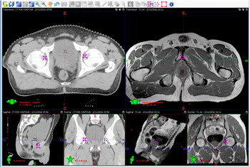
In this study, we have selected three locations for point matching: first is the meeting point of the pubis symphysis, second and third are mid of left and right femur fovea, respectively. These points were chosen as they are rigid landmarks and the prostate is located within these points. Each marker pair is placed separately on MR and CT images in the corresponding location mentioned above using all three planes (refer Figure 1). Once the point selection is done, the matching is performed which results in image fusion with a mean error. The mean error is a geometrical error determined by calculating the error between the distances of point pairs defined in both the image sets. A mean error of less than 2 mm is accepted for image fusion. This registration is termed as REGM.
Point match registration window.

Rigid registration
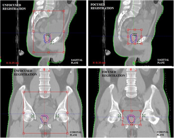
Non-focused registration is a regular registration process where a crude manual matching is done primarily, followed by intensity-based auto-matching, where the region of interest (ROI) is rectilinear to include the entire pelvic area defined by minimum of 2 cm from pubis symphysis inferiorly; L5-S1 junction superiorly; peritoneal wall and pre-sacral area anterioposteriorly and includes the right and left femur laterally. The resultant image registration is termed as REGNF. A gross manual match may be performed to aid the fusion process.
In the focused registration (REGF) process, a crisper ROI was determined to avoid the registration influence of other structures outside the desired area, if any. Figure 2 shows the ROI for focused and unfocused registration.
Selection of ROI for focused and unfocused registrations shown in sagittal and coronal planes.

The crop criteria for REGF are as follows:
-
the inferior and superior border of the femur head is the inferio-superior margin;
-
the anterior and posterior border is adjusted to just remove the rectum and bladder respectively;
-
laterally, the ROI is cropped just inside the femur heads. The focused ROI is to verify whether it could possibly remove the influence of other surrounding structures.
Data analysis
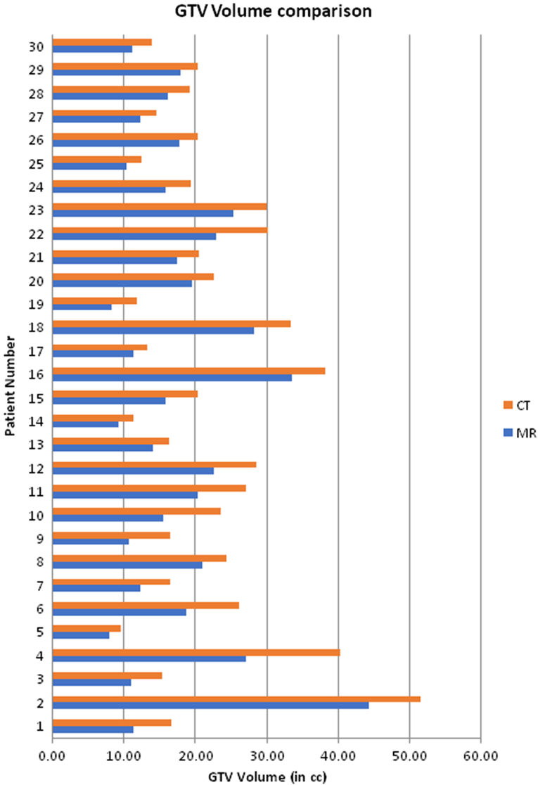
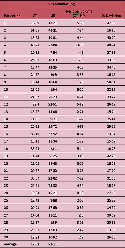
For each patient, GTV is contoured both on CT and MRI individually with a latent period of at least 1 month between both contouring sessions. It is instructed that GTV is clearly the visible tumour and not any microscopic spread. For each CT and MRI and for each patient, the volume of GTV is calculated. On CT and MRI, the location and volume can be identical but the volume can have different shapes. Thus, a residual volume (CT–MRI) is calculated. This residual volume is expressed as a percentage of encompassing volume. Here, the encompassing volume is CT which overestimates all MR-based GTV volumes. Figure 3 shows the chart comparison of GTV volumes based on CT and MRI.
Comparison of CT- and MR-based GTV volumes.

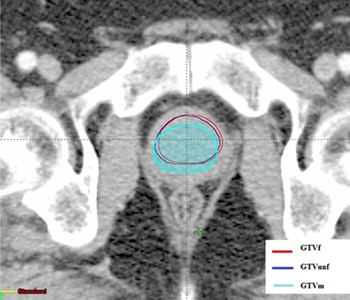
Prostate GTV is contoured in the T2-weighted MR image. After each registration mentioned above, the GTV from MR dataset is copied onto the CT slices in the registration platform itself. GTVM, GTVNF andGTVF are respective GTVs of REGM, REGNF and REGF for each patient considered in this study. Prostate GTV in MRI is done by a single radiation oncologist who has 12 years of experience in PR. Contouring by one radiation oncologist will avoid any observer variation. Since the GTV is contoured based on the MR image, the volume of all GTVs is equal and forms the basis for registration comparison. Contouring each GTV in CT slice based on three different registrations is time-consuming and may result in differential GTV volumes, although contoured by a single radiation oncologist. The MR-based contours can be viewed in Figure 4.
MR-based contours on CT with three different registrations methods.

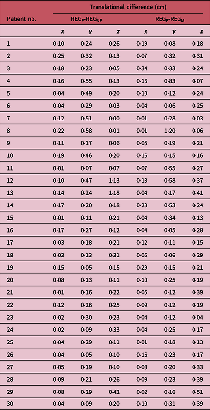
The registration comparison is done by estimating the translation differences of three axes (x, left-right; y, anterior-posterior; z, inferior-superior) by calculating the translation of centre of mass (COM) of each GTV. COM is obtained by aligning three fields (ungrouped) to its corresponding GTV in a dummy plan using external beam planning platform from VMS. During alignment, the field is moved from user origin (CT isocenter) to the new location. Hence, three translation coordinates for each GTV are available. The coordinates of GTVF are taken as baseline value as it avoids the influence of other structures during registration and also it is practised routinely in our department. The difference of each axis (x, y and z) for GTVM and GTVNF is compared against GTVF.
Results
GTV volume
Prostate GTV is contoured in contrast-enhanced CT and on T2-weighted MR images separately. The mean GTV contoured on MR and CT is 17·61 and 22·07 cc, respectively. Volumes of GTV based on MR images are lesser in comparison to CT-based GTV. The deviation of volume reduction ranges from 13·55% to as high as 54·51%. Refer Table 1 for a summary of GTV volumes. A paired student shows that the p-value is less than 0·001.
Summary of GTV volumes

Abbreviations: GTV, gross tumour volume; cc, cubic centimetres; %, percentage.
Registration comparison
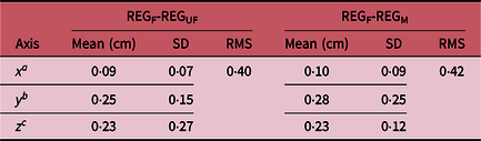
REGF was compared to REGUF and REGM for 30 cases. The registration differences are stated based on the translational differences (Table 2). Mean differences are reported with standard errors and root mean square (RMS) error. They represent the total offset from the COM values and are presented in Table 3. The average difference for REGF-REFNF is ~1 mm (x), 2·5 mm (y) and 2·3 mm (z), and for REGF-REGM, it is 1 mm (x), ~3 mm (y) and 2·3 mm (z). Corresponding RMS error values are 0·40 and 0·42.
Translation differences of center-of-mass values

Abbreviations: REGF, focused registration; REGNF, non-focused registration; REGM, marker-based registration; cm, centimetres.
Statistical summary of registration comparison

a x: left-right.
b y: posterior-anterior.
c z: inferior-superior.
Abbreviations:SD, standard deviation; RMS, root mean square displacement; cm, centimeters.
Discussion
With the advent of image-guided radiotherapy (IGRT), the reduction in toxicity and local control for prostate have been improved. Despite the fact that there is no definite evidence of reduction in side effects, IGRT has become the standard care for PR. Reference Ménard, Paulson and Nyholm4,Reference Moseley, White and Wiltshire10,Reference Murray and Tree11 Cone-beam CT (CBCT) assessment just before the treatment is relatively good in comparison with gold-fiducial-based orthogonal 2D matching. But they are insensitive to the intra-fractional movement of the target and its surrounding structures. Reference Njeh and Parker12 Although treatment of PR with gold fiducial markers or CBCT have their own principal disadvantages, they could be used in combination to overcome the same. Reference Deegan, Owen and Holt13 But the above process cannot account for intra-fractional motions or any deformations of target and adjacent pelvic nodes during the course of the treatment. The impact of IGRT is significant when the target volume is small and hypofractionation is preferred. MRI for radiotherapy planning showed decreased target volume, although very few centres have shown experience of insignificant difference. Reference Henderson, Tree, Harrington and van As14 In the assessment of GTV volumes contoured exclusively on MR and CT (blind to each other), it shows that CT-based GTV overestimates the MR-based GTV and it is statistically significant where the median deviation of volumes is as high as 26·36%. More than half of the patients from the random sample of 30, the deviation percentage is greater than 25 and for two-thirds of the sample, it is more than 20%.
It is well established that MRI reduces the inter-observer variation in target contouring, especially for PR. Reference Deegan, Owen and Holt15,Reference Khalifa, Commandeur and Bachaud16 In this study, we have not attempted any observer study in prostate GTV. Inter-observer study with non-spinal oligo-metastases showed that MR-based contours are significantly small and more accurate in terms of both target and OAR delineation. The ESTRO–ACROP consensus guidelines on CT- and MR-based GTV localisation for the prostate tumour are aimed at improving the consistency and reliability of prostate contours. Reference Salembier, Villeirs and De Bari17 They believe that this is the weakest link in radiation therapy.
Depending on the method of MR–CT image registration, the amount of registration uncertainty varies. Accurate registration of images is challenging as there is a substantial risk of changes in anatomy between two examinations. Here, we have used three different registration techniques, where the focused registration is the most commonly practised and well established. REGF is also reliable as the interference of surrounding structures is avoided. Results show that the registration error is prominent in vertical and longitudinal directions than in the lateral direction. The mean error is approximately 1 mm for both REGF-REGNF and REGF-REGM in the lateral direction. In the vertical direction, it is 0·25 mm for REGF-REGNF and 0·28 mm for REGF-REGM, whereas in the longitudinal direction, it is 0·23 mm for REGF-REGNF and REGF-REGM. The registration based on markers will result in higher dose to the OARs if the target is delineated using MR data and planning is done based on CT data. Hence, it is inferred that an additional PTV margin of ~3 mm and >2 in vertical and longitudinal direction, respectively, is adequately required apart from regular PTV margin if either point-based or unfocused registration is used. As long as both the target and OARs are delineated based on MR data alone, the plan generated on CT data will be consistent.
A point match registration process is based on interactive visual identification of user-defined anatomical landmarks (here is pubis symphysis). Alternatively, this point can be a marker attached to the anatomy (implanted gold markers). In either case, there is a high chance that it may be erroneously displaced from its correct location. This displacement of error is called localisation error and such error may occur in both image spaces used for registration. Reference Prete, Prestidge, Bice, Dubois and Hotchkiss18 This error is not observed directly and is reflected through the registration error. Point-based registration is not used widely and most users are highly reliable on automated rigid registration with a random selection of the region of registration. Thus, we have attempted to find the registration uncertainty which could be error-prone due to the adjacent structures. In the unfocused registration method, the errors are as poor as the point match registration technique in comparison with the REGF. REGNF is poor in terms of both target and OARs since the longitudinal and vertical errors are greater than 2 mm. Altogether, an additional margin of 2–3 mm is mandated for PR apart from the set-up margin irrespective of the registration method used.
Even the REGF does not take into account of the prostate movement. When the target structure is static and the registration uncertainty is unveiled, it is feasible to incorporate the quantified additional margin in the PTV. This is not the scenario for PR and hence MR-only radiotherapy planning will have a big impact on increasing the spatial accuracy of treatment. Reference Murray and Tree11,Reference Arivarasan, Anuradha, Subramanian, Anantharaman and Ramasubramanian19 If accurate and focused registration is not achieved in MR–CT workflow, then MR-only treatment planning will lead to reduction in uncertainty remarkably pertaining to systemic errors. Reference Jonsson, Nyholm and Söderkvist20 Thus, the reduction in overall PTV margin and minimisation of uncertainty during optimisation probability will result in the reduction of exposure of healthy tissue. Reference Sandler, Liu and Dunn21,Reference Pérez-Romasanta, Lozano-Martín and Velasco-Jiménez22 It is ascertained that the prostate visibility at the apex and at the bladder interface is improved with MRI and is difficult to see on CBCT during delivery. Thus, the increased accuracy with MR may allow PTV margin reduction which leads to a decrease in toxicity although the reduction in PTV margin is small (in the order of 2–3 mm).
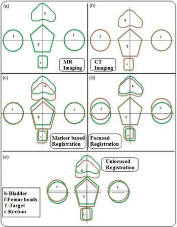
In Figure 5, a and b assume the different position of the target and other structures from MR (green) and CT (brown) procedures, respectively. Figure 5c shows that if the registration is done using the fixed structures (femur heads) meant for routine patient positioning (REGM), then there is a difference in target and critical structures and when planning is done based on CT, there is a high chance that rectum may receive a slightly higher dose. In scenario d, the registration is performed with target volume (REGF), where the structure-based registration is of low quality. Here, the surrounding structures have to be delineated using CT instead of MR for accurate delivery. This is because the surrounding structures are near equidistance from the target volume. Figure 5e illustrates a scenario of unfocused registration which uses both the surrounding structures and the target volume (REGNF) during the registration process. In this scenario, it is not possible to obtain a definite representation of the anatomy of the patient until the patient positioning is done using MR information. Here, the delineation information cannot be consistent taking into account both the imaging modality. Consequently, we need to focus on moving towards the MR-only workflow for PR.
Schematic representation of three different registration scenarios in a MR–CT workflow.

We have limited this study with the ‘eclipse’ software provided with VMS. This study could be extended with automatic registration tools provided with other commercial vendors in radiotherapy planning to come out with a broad data of uncertainty from image registration. We have not considered the geometric distortion associated with the MR imaging in this study. This is because the MR imaging used for registration is of diagnostic quality with moderate 1·5 T and our ROI is centrally located (prostate gland). The geometric distortion for MR imaging is predominant in the periphery of the body and when the field strength is less than 0·8 T and/or if adequate radio frequency coils are not utilised. Reference Walker, Liney, Metcalfe and Holloway23 Nevertheless, characterisation of distortion and monitoring is utmost important to introduce MR-only radiation treatment planning. Reference Adjeiwaah, Bylund, Lundman, Karlsson, Jonsson and Nyholm24,Reference Khoo, Dearnaley, Finnigan, Padhani, Tanner and Leach25
Conclusion
For the treatment of prostate and other diseases, where MR is preferred to CT and if the variations in anatomy are appreciable between two imaging sessions, accurate image registration is challenging and prone to error. From our study, we observe that high-quality registration is not achieved using the bony structures which are routinely used for patient positioning during treatment delivery in a regular MR–CT workflow. Thus, by removing the CT imaging, the additional PTV margin accounted for registration error is eliminated. MR-only workflow will significantly reduce the spatial uncertainty with small target volumes prescribed with high heterogeneous dose distributions.
Acknowledgements
No funding has been raised for this work. Institutional scientific and ethics board has approved this study. Thanks to Dr Subramanian Shanmugam, Chief Medical Physicist, Yashoda Hospitals, Hyderabad, for allowing to carry out this retrospective study. The authors thank Medical Physicist Mr. Anantharaman Ayyalusamy and other supporting staffs for extending their support for submission. The authors would like to thank Dr Shyama Prasanna Satpathy, Consultant Radiation Oncologist for contouring assistance. The authors extend their sincere thanks to Dr Ramasubramanian Velayudham, Professor of Physics, Vellore Institute of Technology, Vellore, for his encouragement and mentoring.
Financial Support
None.
Conflict of Interest
None.