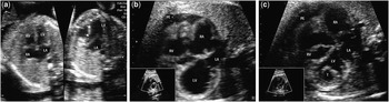
A healthy 35-year-old gravida 2, parity 1, pregnant woman was referred for foetal echocardiography, as she was over 35 years of age, which is a routine practice in Brazil. There was no family history of CHD, maternal metabolic disease, or teratogen exposure. Previous maternal trans-abdominal morphological ultrasound imaging at 22 weeks of gestation had shown a normal foetus with no cardiac abnormality (Fig 1a). Foetal echocardiogram at 28 weeks of gestation showed left ventricular dilatation, severe systolic dysfunction, and a small pericardial effusion (Fig 1b). Follow-up foetal echocardiography at 31 weeks of gestation revealed an apical left ventricular thrombus (Fig 1c). There was no evidence of aortic valve or arch obstruction at any time. There was no apparent intra-ventricular spontaneous contrast before or after the appearance of left ventricular thrombosis. Colour Doppler interrogation of mitral inflow showed abnormal relaxation (E less than A wave), as well as mild mitral and tricuspid regurgitation. Screening for viral pathogens was negative. Subsequently, the foetus did not develop hydrops and was delivered at term. The findings were confirmed by a postnatal echocardiogram, although unfortunately not recorded. Heart failure treatment and anticoagulation therapy were provided, but the baby died on the 3rd day of life due to progressive left ventricular dysfunction. There was no evidence of embolic complications. A postmortem examination was not undertaken. Dilated cardiomyopathy is a rare disease in foetuses and is generally associated with a poor outcome; it may present as an isolated finding or in association with other cardiac or non-cardiac anomalies.Reference Mongiovì, Fesslova, Fazio, Barbaro and Pipitone 1 A normal cardiac assessment in the second trimester does not exclude subsequent development of dilated cardiomyopathy and its consequences, as seen in our case.
( a ) Trans-abdominal morphological ultrasound imaging at 22 weeks of gestation showing a normal foetal heart. Arrows indicate inter-ventricular septum and left ventricular outflow tract. ( b ) Foetal echocardiogram at 28 weeks of gestation demonstrating a dilated LV without thrombus. ( c ) Foetal echocardiogram at 31 weeks of gestation revealing a fixed apical left ventricular thrombus. LA, left atrium; LV, left ventricle; PE, pericardial effusion; RA, right atrium; RV, right ventricle; T, thrombus.

Acknowledgements
None.
Financial Support
This research received no specific grant from any funding agency, commercial, or not-for-profit sectors.
Conflicts of Interest
None.
Ethical Standards
The authors assert that all procedures contributing to this work comply with the ethical standards of the Brazilian guidelines on human experimentation (CONEP) and with the Helsinki Declaration of 1975, as revised in 2008, and has been approved by the ethical committee of our institution.