Dilated cardiomyopathy is a rare but serious disease that can lead to congestive heart failure. The annual incidence of dilated cardiomyopathy in children is ~0.58 cases per 100,000 person-years.Reference Wilkinson, Landy and Colan 1 According to the Pediatric Cardiomyopathy Registry in the United States of America, including more than 1400 children with dilated cardiomyopathy, the one and 5-year rates of transplant-free survival were 69 and 54%, respectively.Reference Wilkinson, Landy and Colan 1 Effective therapeutic strategies for dilated cardiomyopathy in children are limited. The current treatment for children presenting with dilated cardiomyopathy consists mainly of supportive heart failure therapy, and there is no indisputable evidence for therapies targeting the underlying cause.
The causes for dilated cardiomyopathy are wide-ranging. About 65% of dilated cardiomyopathy in children is idiopathic, with an unknown causative agent. The most frequently identified cause for dilated cardiomyopathy is viral myocarditis (16%).Reference Lipshultz, Cochran and Briston 2 Other causes are neuromuscular, genetic, structural, and syndromal, or are related to metabolic disorders.Reference Felker, Thompson and Hare 3 It is important to identify the origin of the disease, as an accurate diagnosis largely determines prognosis and treatment options.
For viral myocarditis, the prognosis is better than for idiopathic dilated cardiomyopathy.Reference Foerster, Canter and Cinar 4 The transplant-free survival for children with viral myocarditis is about 70–75%.Reference Klugman, Berger, Sable, He, Khandelwal and Slonim 5 – Reference Lee, McCrindle and Bohn 8 Data from the Pediatric Cardiomyopathy Registry showed that 53% of patients with myocarditis reached normalisation in echocardiographic measurements of the left ventricle after 3 years, compared with 21% of children with idiopathic dilated cardiomyopathy.Reference Foerster, Canter and Cinar 4
Nevertheless, the diagnosis of viral myocarditis is difficult to establish. It has a variable clinical presentation with poorly defined diagnostic criteria.Reference Kaski and Burch 9 The gold standard for diagnosing viral myocarditis is an endomyocardial biopsy in which inflammation together with viral presence is proven.Reference Martin, Webber and Fricker 10 , Reference Sagar, Liu and Cooper 11 This invasive test is not often performed in children, because of the unfavourable risk–benefit ratio.Reference Pophal, Sigfusson and Booth 12 MRI is becoming an important imaging modality in dilated cardiomyopathy.Reference Friedrich, Sechtem and Schulz-Menger 13 Other diagnostic tools are echocardiography and the detection of viral agents in various extra-cardiac patient materials, but their diagnostic value is limited.Reference Sagar, Liu and Cooper 11 These limitations lead to a group of patients with undefined dilated cardiomyopathy, in which viral myocarditis can neither be proven nor be ruled out.
An effective treatment for new onset dilated cardiomyopathy has not yet been found. The effects of antiviral treatment and immunosuppression have been explored in several studies, none of which could be explicitly associated with a better outcome.Reference Brunetti and DeSantis 14 , Reference Mason, O’Connell and Herskowitz 15 Some positive results have been linked to immunomodulatory therapies, including immunoadsorptionReference Ikeda, Kasai and Izawa 16 and administration of intravenous immunoglobulins. It has been shown that intravenous immunoglobulins have both an antiviral and an anti-inflammatory effect.Reference Maisch, Hufnagel and Kölsch 17 Although the only randomised trial on the use of intravenous immunoglobulins in adults with myocarditis did not show a beneficiary effect,Reference McNamara, Holubkov and Starling 18 a more recent non-randomised prospective trial in children with enteroviral myocarditis and encephalitis has associated intravenous immunoglobulins with greater improvement of left ventricular ejection fraction and with improved survival.Reference Bhatt, Sankar and Kushwaha 19 This finding was supported by various observational studies on intravenous immunoglobulins in children with viral myocarditis and dilated cardiomyopathy.Reference Bhatt, Sankar and Kushwaha 19 – Reference Prasad and Chaudhary 22 More evidence is warranted to definitely establish the effect of intravenous immunoglobulins in children.
In the present study, we aimed to explore the efficacy of intravenous immunoglobulin administration in a population of children with new onset dilated cardiomyopathy, consisting of both myocarditis and idiopathic dilated cardiomyopathy. Second, we provide an overview of the viral tests performed to establish the diagnosis of viral myocarditis in this population.
Material and methods
We performed a retrospective, observational, cohort study. Patients were selected from the Cardiomyopathy Registry Study in the Netherlands, an ongoing multicentre observational registry from seven tertiary paediatric hospitals, consisting of children aged 0–18 years who received the diagnosis of dilated cardiomyopathy after 1 January, 2005. Patients were eligible for the Cardiomyopathy Registry if they presented with at least two of the following characteristics: symptomatic heart failure, impaired systolic function determined by shortening fraction ⩽25% on transthoracic echocardiography, or left ventricular dilatation determined by left ventricle end-diastolic dimension >+2 z-score on transthoracic echocardiography.Reference Den Boer, Meijer and van Iperen 23
From the Cardiomyopathy Registry, we selected children with a discharge diagnosis of viral myocarditis or idiopathic dilated cardiomyopathy who were included from 1 January, 2005, until 31 December, 2012. Patients with dilated cardiomyopathy due to other causes than viral myocarditis were excluded, namely those having gene mutations, familial cardiomyopathy, and structural heart disease, those with metabolic, endocrine, auto-immune, and neuromuscular disorders, as well as those having syndromes associated with dilated cardiomyopathy, drug toxicity, and tachycardiomyopathy.Reference Felker, Thompson and Hare 3 No pre-set diagnostic work-up protocol was used during the study. Information on the aetiology was extracted from the available correspondence from the treating physician. Familial dilated cardiomyopathy is defined as having two or more first- or second-degree relatives with dilated cardiomyopathy.
Patients were categorised into the treatment group if they received intravenous immunoglobulins within 2 weeks after initial presentation. The dosing regimen for intravenous immunoglobulins was consistently maintained at 2 g/kg of body weight.
Baseline characteristics, diagnostic test results, and follow-up data up to the latest record available were collected from medical charts and anonymised.
Transthoracic echocardiographic results were registered at the baseline, at 6 months’ follow-up, and at the end of the study – that is, at the time of recovery or at the latest record available. Shortening fraction was directly copied from the echocardiographic report and left ventricle end-diastolic dimension was transformed into z-scores using body surface area.Reference Sluysmans and Colan 24
We defined two separate primary endpoints. The first primary endpoint was the composite endpoint of heart transplantation or death. The second primary endpoint was the achievement of complete recovery. The latter was defined as the normalisation of echocardiographic measurements – that is, having shortening fraction >25% and left ventricle end-diastolic dimension z-score ⩽2 – and no use of cardiac medication. Secondary endpoints were improvement of systolic function and left ventricle dilatation on echocardiography after 6 months’ follow-up.
Viral tests and diagnosis of viral myocarditis
Viral test results for the detection of cardiotropic viruses were collected. They were performed within 2 weeks of initial presentation with several different patient materials, namely blood, faeces, nasopharyngeal fluid, cerebrospinal fluid, urine, and tissue obtained using endomyocardial biopsy. Positive viral tests were defined as one or more of the following findings: elevated IgM or IgA levels against a specific virus, a positive result from viral polymerase chain reaction, or positive viral culture of any patient material.
We registered the discharge diagnosis for all study participants. Second, we reclassified our population for the diagnosis of viral myocarditis according to the diagnostic criteria presented by Den Boer et al.Reference Den Boer, Meijer and van Iperen 23 In this study, the authors assigned a diagnosis of “definite myocarditis” to those patients with histological or immunohistological evidence of myocarditis on endomyocardial biopsy. “Probable myocarditis” was diagnosed on the basis of detection of enterovirus, adenovirus, parechovirus, or human parainfluenza virus in whole blood or cerebrospinal fluid using cultures or the polymerase chain reaction. A positive polymerase chain reaction from blood samples for parvovirus B19, human herpesvirus 6, cytomegalovirus, or Epstein–Barr virus had to be accompanied by serological proof of recent infection – that is, a positive result for IgM or IgA – in order to be classified as “probable myocarditis”. “Possible myocarditis” was assigned to those patients with virus detected in the faeces or nasopharyngeal fluid, or through seroconversion for enterovirus, adenovirus, parechovirus, or human parainfluenza virus. Myocarditis was “unlikely” in case of negative test results, or when parvovirus B19, human herpesvirus 6, cytomegalovirus, or Epstein–Barr virus was detected in faeces, nasopharyngeal fluid, or blood without serological evidence of a recent infection. According to this stratification we divided our patients into two groups, namely “unlikely or possible myocarditis” and “probable or definite myocarditis”.
Statistical methods
Continuous data are expressed as mean with standard deviation (±) or as median with interquartile range. Categorical variables are expressed as numbers and percentages. The percentage of baseline characteristics was calculated by dividing the number of cases with the characteristic by the total number of cases in which information on the characteristic was known; cases with missing values were not taken into account. Differences between treatment groups were tested using Pearson’s χ 2 test for nominal data, or using the Mann–Whitney U test for continuous data. For the primary endpoints, survival curves and cumulative incidences with 95% confidence intervals were calculated using Kaplan–Meier analysis with a log-rank test for comparison. A Cox regression analysis was performed for the primary endpoints with correction for possible confounders. Because of the limited number of events, we selected three factors for Cox regression analysis, based on the most important differences between treatment groups at the baseline. Alterations in shortening fraction and left ventricle end-diastolic dimension z-score at 6 months’ follow-up were compared between treatment groups using a Student t-test for independent samples. Statistical significance was defined as two-sided p<0.05.
Statistical analyses were performed using IBM SPSS statistics, version 22 (IBM Corp., Armonk, New York, United States of America). Graphs were compiled in Prism 6 (GraphPad, La Jolla, California, United States of America).
Results
Patient characteristics
A total of 152 children with new onset dilated cardiomyopathy were included in the Cardiomyopathy Registry Study up to 31 December, 2012. Of these, 18 patients (12%) had familial or genetic dilated cardiomyopathy, 10 patients (7%) had cardiac arrhythmias associated with cardiomyopathy, in nine patients (6%) the cardiomyopathy was caused by anthracycline treatment, and seven patients (5%) had a metabolic disease. Further, three children (2%) had a true non-compaction cardiomyopathy, two children (1%) were diagnosed with Alström syndrome, one with Carvajal syndrome, and one with systemic vasculitis. In six patients (4%) the correspondence was unclear about the aetiology of the dilated cardiomyopathy, but pointed towards either a familial, metabolic, or arrhythmic disease. These 58 patients (38%) were excluded from further analysis.
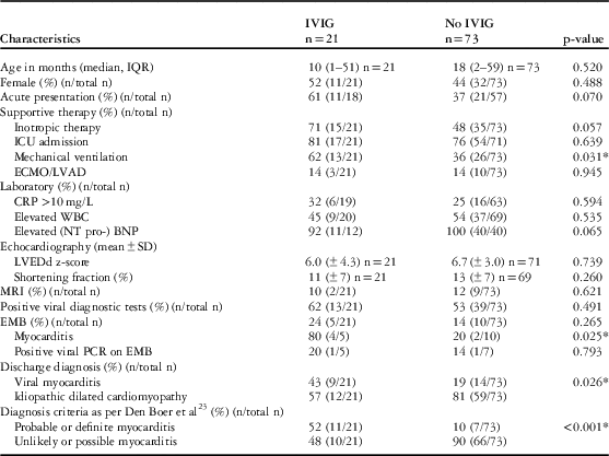
Consequently, a total of 94 children with new onset dilated cardiomyopathy, consisting of viral myocarditis and idiopathic dilated cardiomyopathy, were included in the study, and were followed during a median period of 33 months. Of those, 21 children (22%) received intravenous immunoglobulins. Baseline characteristics are summarised in Table 1. The median age was 15 months (interquartile range 2–58) and gender was evenly distributed. Almost half of the patients (43%) had an acute onset of symptoms (<1 week). Most patients were admitted to the intensive care unit (77%) and needed inotropic therapy (53%). At baseline, 62% of children in the treatment group needed mechanical ventilator support, compared with 36% (p=0.031) in the non-treatment group. In those patients who underwent testing for brain natriuretic peptide or N-terminal-pro-brain natriuretic peptide, the level was elevated above the normal upper limit. On echocardiography, the left ventricle was overall severely dilated with a mean left ventricle end-diastolic dimension z-score of 5.8±3.3 and had a diminished systolic function with a mean shortening fraction of 13±7%. All patients underwent viral diagnostic tests and 55% had at least one positive test result. Some children tested positive for various viruses. The few endomyocardial biopsies (15/94; 16%) revealed six cases of histopathological myocarditis (6/15; 40%), of which one presented a positive result from the polymerase chain reaction for parvovirus B19. Another positive polymerase chain reaction result for parvovirus B19 on endomyocardial biopsy was not accompanied by histopathological proof of myocarditis.
Baseline characteristics.

BNP=brain natriuretic peptide; CRP=C-reactive protein; ECMO=extra corporal membrane oxygenation; EMB=endomyocardial biopsy; IQR=interquartile range; IVIG=intravenous immunoglobulins; LVAD=left ventricular assist device; LVEDd=left ventricle end-diastolic dimension; MRI=magnetic resonance imaging; PCR=polymerase chain reaction; WBC=white blood cell count
Values are presented as % (n/total n), mean±SD or median (interquartile range). The percentage is calculated by dividing the number of cases with the characteristic by the total number of cases in which information on the characteristic was known. The latter is indicated as “n=…” or “total n”
* p-value has reached significance (<0.05)
When applying the criteria of Den Boer et alReference Den Boer, Meijer and van Iperen 23 a total of 18 patients (19%) had “probable or definite viral myocarditis” and 76 patients (81%) had “unlikely or possible myocarditis”. This led to the reclassification of four out of 71 patients (6%) discharged with the diagnosis of idiopathic dilated cardiomyopathy and of nine out of 23 patients (39%) discharged with the diagnosis of viral myocarditis.
Significantly more patients treated with intravenous immunoglobulins were diagnosed with viral myocarditis at discharge (43%) as opposed to those in the non-treatment group (19%). This difference was consistent after reclassification based on the criteria of Den Boer et alReference Den Boer, Meijer and van Iperen 23 : 52 versus 10% for “probable or definite myocarditis”; p<0.001.
Viral diagnostic tests
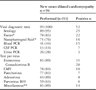
The types of patient material collected and the selection of viruses investigated in these patient materials varied considerably between hospitals, and were dependent on the timing during the 7-year course of this retrospective overview (Table 2). Overall, serology tests were performed in almost all cases (95%), followed by tests of faeces (83%) and nasopharyngeal fluid (76%) using polymerase chain reaction or culture. Cerebrospinal fluid was only evaluated in cases of suspected encephalitis (14%). Almost all patients were tested for the presence of enterovirus (n=83; 89%). This was also the most frequently detected virus, and was found in 31 children. Out of those, 20 cases were further identified to be Coxsackie B virus. Other frequently detected viruses were cytomegalovirus, adenovirus, parechovirus, and parvovirus B19, which were detected in 10, 8, 7, and 6 children, respectively. The viruses were encountered in various patient materials, but most frequently using serology for IgM or IgA or using faeces polymerase chain reaction or culture.
Viral diagnostic tests.

CMV=cytomegalovirus; CSF=cerebrospinal fluid; EMB=endomyocardial biopsy; PCR=polymerase chain reaction
Positive test results are shown as absolute numbers, and not in percentages, because the tests were not performed in all children, which could have led to missed cases. Some patients tested positive on multiple patient materials and for multiple viruses
* Faeces and nasopharyngeal fluid were tested using PCR or viral culture
** Miscellaneous consists of human herpesvirus 6, herpes simplex virus 1-2, influenza virus, and parainfluenza virus
Transplant-free survival and complete recovery
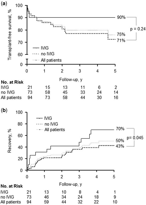
In our population of 94 children with new onset dilated cardiomyopathy, the overall transplant-free survival within 5 years was 75% (95% CI: 63–87; Fig 1a) and the recovery rate within 5 years was 50% (95% CI: 36–64; Fig 1b).
( a ) Transplant-free survival in patients with or without intravenous immunoglobulin treatment. A Kaplan–Meier curve showing the association between intravenous immunoglobulins and long-term transplant-free survival. ( b ) Recovery in patients with or without intravenous immunoglobulin treatment. A Kaplan–Meier curve showing the association between intravenous immunoglobulins and complete recovery. IVIG=intravenous immunoglobulins

In children treated with intravenous immunoglobulins, the transplant-free survival of 90% (95% CI: 86–100) within 5 years was comparable to 71% (95% CI: 58–86) in the non-treated patients (log rank=0.236); however, significantly more children in the treatment group recovered completely within 5 years (70%; 95% CI: 46–94, versus 43%; 95% CI: 33–57; log rank=0.045).
The endpoints were assessed using a Cox regression analysis, including the potential predictors for intravenous immunoglobulin treatment, “probable or definite myocarditis”, and mechanical ventilator support (Supplementary Material Table 1). There was no significant effect of intravenous immunoglobulins on mortality or heart transplantation (hazard ratio: 0.53; 95% CI: 0.11–2.60; p=0.432). The hazard ratio for complete recovery was 2.13 (95% CI: 0.98–4.61) for the treatment group, but did not reach significance, with p=0.056. The diagnosis of “probable or definite myocarditis” did not have significant influence on either mortality or heart transplantation (hazard ratio: 0.41; 95% CI: 0.08–2.07; p=0.278) or on recovery (hazard ratio: 1.39; 95% CI: 0.53–3.61; p=0.501). Mechanical ventilator support was associated with a higher rate of mortality or heart transplantation (hazard ratio: 2.77; 95% CI: 1.11–6.92; p=0.029).
We evaluated the influence of the diagnosis of viral myocarditis on survival and recovery within a sub-group of those treated with intravenous immunoglobulins (n=21). Transplant-free survival and complete recovery were compared between the patients diagnosed with “probable or definite myocarditis” (n=11; 52%) and those with “unlikely or possible myocarditis” (n=10; 48%).
Of the six patients with histopathologically proven myocarditis, “definite myocarditis”, four were treated with intravenous and two weren’t. A single patient died in each treatment group within the first month after diagnosis. Of the four survivors, the echocardiographic parameters ameliorated during the follow-up period, with a median shift in left ventricle end-diastolic dimension z-score of −1.3 (interquartile range: −4.6 to 1.13) and a median increase of 18% (interquartile range: 3–18) in the shortening fraction. Finally, three patients recovered completely.
Echocardiography after 6 months’ follow-up
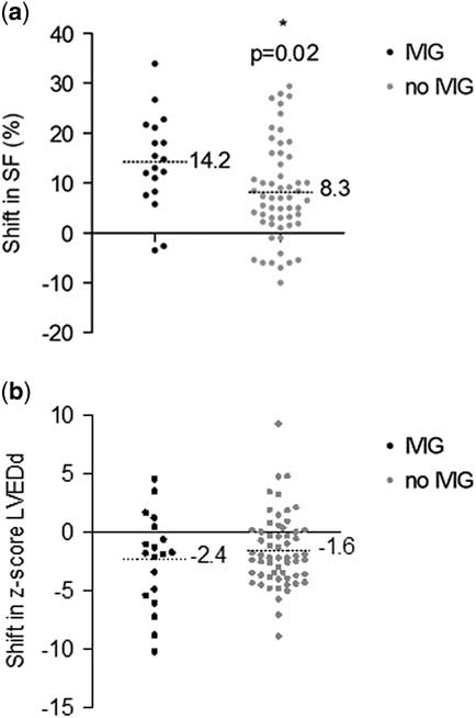
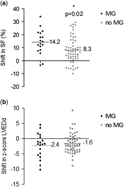
At baseline, the left ventricle was dilated in the whole population with a mean z-score of left ventricle end-diastolic dimension of 5.8±3.3, and the systolic function was impaired with a mean shortening fraction of 13±7%. After 6 months, the mean shortening fraction improved to 26±10% – an increase of 14±10% – in the group treated with intravenous immunoglobulins, and to 22±8% – an increase of 8±10% – in the non-treatment group (p=0.023) (Fig 2a; Supplementary Material Table 1). The decrease in mean left ventricle end-diastolic dimension z-score was comparable for both treatment groups (p=0.369), with a z-score of 3.3±2.8 – a decrease of 2.4±4.0 – in the treatment group and a z-score of 4.0±3.0 – a decrease of 1.6±3.1 – in the non-treatment group after 6 months (Fig 2b; Supplementary Material Table 1).
( a ) Shift in shortening fraction at 6 months’ follow-up. A scatter plot showing the shift in shortening fraction from baseline to 6 months’ follow-up. The shortening fraction improved by 14±10% in the treatment group, and by 8±10% in the non-treatment group. ( b ) Shift in z-score of left ventricle end-diastolic dimension at 6 months’ follow-up. A scatter plot showing the shift in z-score of the left ventricular end-diastolic dimension from baseline to 6 months’ follow-up. The z-score decreased by 2.4±4.0 points in the treatment group and by 1.6±3.1 in the non-treatment group. The difference was not significant. IVIG=intravenous immunoglobulins; LVEDd=left ventricle end-diastolic dimension.
Discussion
In this study we present a large cohort of children with the rare condition of new onset dilated cardiomyopathy. In this population, administration of intravenous immunoglobulins did not affect transplant-free survival. It was, however, associated with a higher recovery rate and improved shortening fraction as a surrogate marker of the systolic left ventricle function, compared with children not treated with intravenous immunoglobulins. The clinical presentation of the patients was in general severe, with frequent admissions to the intensive care unit, but had an overall favourable outcome with a transplant-free survival of 75% in 5 years. Viral tests were performed in all children, but the diagnosis of viral myocarditis remained difficult to establish. These findings contribute to existing evidence on the efficacy of intravenous immunoglobulins in children with new onset dilated cardiomyopathy.
Bhatt et al carried out the only prospective controlled trial on this subject in children.Reference Bhatt, Sankar and Kushwaha 19 They demonstrated reduced mortality with a relative risk of 0.17 and improved left ventricle ejection fraction following intravenous immunoglobulin administration, compared with standard treatment alone in 83 children with acute encephalopathy syndrome and myocarditis of probable viral origin. The interpretation of the effect of intravenous immunoglobulins in myocarditis, however, is distorted by the comorbidity of encephalitis in these children, with secondary causes such as intracranial pressure and systemic failure resulting in some of the deaths. Further, there are a small number of observational cohort studies comparing intravenous immunoglobulins with supportive treatment alone. Haque et alReference Haque, Bhatti and Siddiqui 20 were unable to detect beneficial effects of intravenous immunoglobulins on left ventricular ejection fraction, but did show a higher survival in 12 children treated with intravenous immunoglobulins (survival 92%) compared with 13 children without this treatment (survival 54%) with acute myocarditis. In an older study of 47 children with acute dilated cardiomyopathy, carried out by Drucker et alReference Drucker, Colan and Lewis 21 , administration of intravenous immunoglobulins was associated with significantly greater normalisation of left ventricular function 1 year after initial presentation, and there was a trend towards a beneficial effect on the survival rate at 1 year. Similarly, in adults, a few uncontrolled studies on intravenous immunoglobulins in idiopathic or inflammatory dilated cardiomyopathy showed improved left ventricular ejection fraction and/or NYHA classificationReference Dennert, Velthuis and Schalla 25 – Reference Goland, Czer and Siegel 28 , but these trials are of limited value because of the lack of a control group. The only randomised placebo-controlled trial was conducted in 62 adult patients with acute dilated cardiomyopathy (<6 months), demonstrating neither a difference in the survival rate nor an improvement in left ventricular ejection fraction between the treatment arms.Reference McNamara, Holubkov and Starling 18 This contradiction of the effect of intravenous immunoglobulins on adults with dilated cardiomyopathy in comparison with the children might be explained by a presumed higher efficacy of intravenous immunoglobulins in children, in whom the acquired immune system is still less developed than in adults.
The results of our study are in line with the abovementioned paediatric trials, especially regarding the recovery of systolic left ventricular function. We observed a significantly greater improvement in shortening fraction in those children who were treated with intravenous immunoglobulins, compared with that in untreated patients. Shortening fraction is an alternative to left ventricular ejection fraction for measurement of the left ventricular systolic function, and is easily obtained using M-mode echocardiography.Reference Chen 29 Both left ventricular ejection fraction and shortening fraction are subject to limitations introduced by the experience of the echocardiographist and the assumption of an elliptical left ventricle.Reference Slama and Maizel 30 More reliable measurements of left ventricular function are strain and strain-rate imaging, which quantify the rate of segmental myocardial deformation. These measurements are especially appropriate for patients with regional cardiac disease, such as those with ischaemic heart disease and hypertrophic cardiomyopathy.Reference Dragulescu and Mertens 31 Shortening fraction, however, is a good estimate of the systolic function in symmetric cardiac disease, which is generally the case in dilated cardiomyopathy.
When evaluating the viral diagnostic tests that were performed in this population of children who presented with dilated cardiomyopathy during this 7-year study period in the Netherlands, we conclude that there is little consistency. Although viral tests were performed in all children, the type of patient material and the type of viruses evaluated differed significantly, depending on the treating hospital, the physician, and the timing of presentation during the course of the study.
According to a consensus document from the European Cardiology Society in 2013, only polymerase chain reaction tests for blood samples are of potential diagnostic value, besides the gold standard of viral detection and histopathological evidence of myocarditis from endomyocardial biopsies.Reference Caforio, Pankuweit and Arbustini 32 A more extended diagnostic proposal was recently published, advocating performing polymerase chain reaction using blood for the detection of cardiotropic viruses in all children presenting with new onset dilated cardiomyopathy, accompanied by serology for parvovirus B19, human herpesvirus 6, cytomegalovirus, and Epstein–Barr virus.Reference Den Boer, Meijer and van Iperen 23 If the polymerase chain reaction result from blood is negative, a polymerase chain reaction can be performed using other patient materials, such as faeces and nasopharyngeal fluid, but this would not lead to a high probability of the diagnosis of viral myocarditis. Endomyocardial biopsy and cerebrospinal fluid evaluation should be performed only when clinically relevant, because of the high risks from these invasive tests.
An important limitation of our study is the lack of a definite diagnosis for dilated cardiomyopathy at presentation. The distinction between viral myocarditis and idiopathic dilated cardiomyopathy is difficult to make, because of the lack of well-defined diagnostic criteria. We have tried to pursue a more consistent diagnosis in the whole population, by reclassifying the patients according to the recently proposed diagnostic criteria for viral myocarditis.Reference Den Boer, Meijer and van Iperen 23
A definite diagnosis is of importance, as the prognosis differs for various causes of dilated cardiomyopathy. The outcome is more favourable for viral myocarditis compared with idiopathic dilated cardiomyopathy.Reference Lipshultz, Cochran and Briston 2 In addition, intravenous immunoglobulins are supposed to be especially effective in the event of proven viral myocarditisReference Maisch, Hufnagel and Kölsch 17 , although we were unable to demonstrate this with a sub-group analysis. To correct for the effect of the different diagnosis on outcome, we performed a Cox regression analysis, showing a hazard ratio of 2.1 for recovery with intravenous immunoglobulin treatment, but this value did not reach significance; therefore, we should interpret the positive association between intravenous immunoglobulin treatment and recovery, demonstrated using the Kaplan–Meyer analysis, with caution.
The retrospective nature of the study has yielded only limited evidence. The absence of consistency in diagnosis, evaluation, or in treatment provided at the time of presentation makes the interpretation of the results quite difficult. These are limitations that most studies on intravenous immunoglobulins in dilated cardiomyopathy have encountered because of the observational design; however, we were able to study a large cohort of children with dilated cardiomyopathy using data from seven tertiary paediatric hospitals in the Netherlands. To our knowledge, this is the largest cohort of children with the rare disease of new onset dilated cardiomyopathy in which the efficacy of intravenous immunoglobulins has been studied. Our findings support the concept that children with new onset dilated cardiomyopathy might benefit from intravenous immunoglobulin administration. Although the transplant-free survival was comparable for both treatment groups, we did demonstrate an association between intravenous immunoglobulin treatment and a greater improvement in systolic left ventricular function as well as a higher recovery rate. Hopefully, in the future, the confirmation of our findings will be sought through a prospective controlled trial, considering the different causes of dilated cardiomyopathy.
Acknowledgments
The authors would like to thank the collaborators of the Cardiomyopathy Registry Study for sharing their data on children with dilated cardiomyopathy in the Netherlands.
Financial Support
This research received no specific grant from any funding agency, commercial, or not-for-profit sectors.
Conflicts of Interest
None.
Supplementary Material
To view supplementary material for this article, please visit https://doi.org/10.1017/S1047951117001561