




Carney complex is a rare multiple neoplasia and lentiginosis syndrome hallmarked by multiple endocrine and non-endocrine tumours, abnormal skin pigmentation, myxomas (heart, breast, bone, skin, and mucosae), melanotic schwannoma, and a predisposition to various cancers, especially in the thyroid, ovaries, pancreas, and liver. Reference Zaidi, Hutchins and Rajpurkar1,Reference Pitsava, Zhu, Sundaram, Mills and Stratakis2 Cardiac myxomas occur most commonly in one of the atria (75% in left atrium, 19% in the right atrium) and rarely in the right or left ventricle. Reference Burke and Virmani3 With right ventricular myxomas, patients are monitored for inflow and/or outflow obstruction and potential thromboembolic events (pulmonary arterial or systemic embolic events, if there is a concomitant atrial septal defect or patent foramen ovale). Reference Ma, Wang, He, Zhang, Zhou and Ying4 While embolism occurs in 30 to 40 percent of patients with myxomas, evidence of clinically significant embolic events from right-sided tumours is uncommon. Reference Reynen5
Case
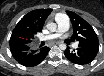
A 14-year-old female presented to the hospital after two days of shortness of breath and pleuritic chest pain. Her oxygen saturation was normal, but she was tachycardic for her age. An elevated D-dimer of 8,000 ug/L prompted chest CT, which showed large bilateral pulmonary emboli (Image 1). Echocardiogram demonstrated a large mass (3×3×7 cm) in the right ventricle extending through right ventricular outflow tract, right ventricular dilation, and normal right ventricle function (Image/Video 2). Additional laboratory results showed a normal white blood cell count, haemoglobin, and platelet count. Inflammatory markers were elevated with a C-Reactive protein of 10 mg/dL and fibrinogen of 810 mg/dL. Systemic heparin was started, and she was transferred to our paediatric cardiac ICU.
CT scan at the level of the right ventricular outflow tract showing a pulmonary embolus of the distal right pulmonary artery (red arrow).

Echocardiogram 2-dimensional image/clip in a parasternal long axis view tilted towards the tricuspid valve and right ventricle (RV) inflow. There is a large mobile right ventricular pedunculated mass measuring 3 × 3 × 7 cm from the RV free wall originating beneath the tricuspid valve annulus and extending to the outflow tract and abutting a portion of the pulmonary valve.

On physical examination, she had multiple abnormal cutaneous and mucosal lentigines. Further review of past medical history reveled that she had a pituitary adenoma resected 3 years prior. She was conceived by donor egg thus maternal family history was unknown and paternal family history was unrevealing.
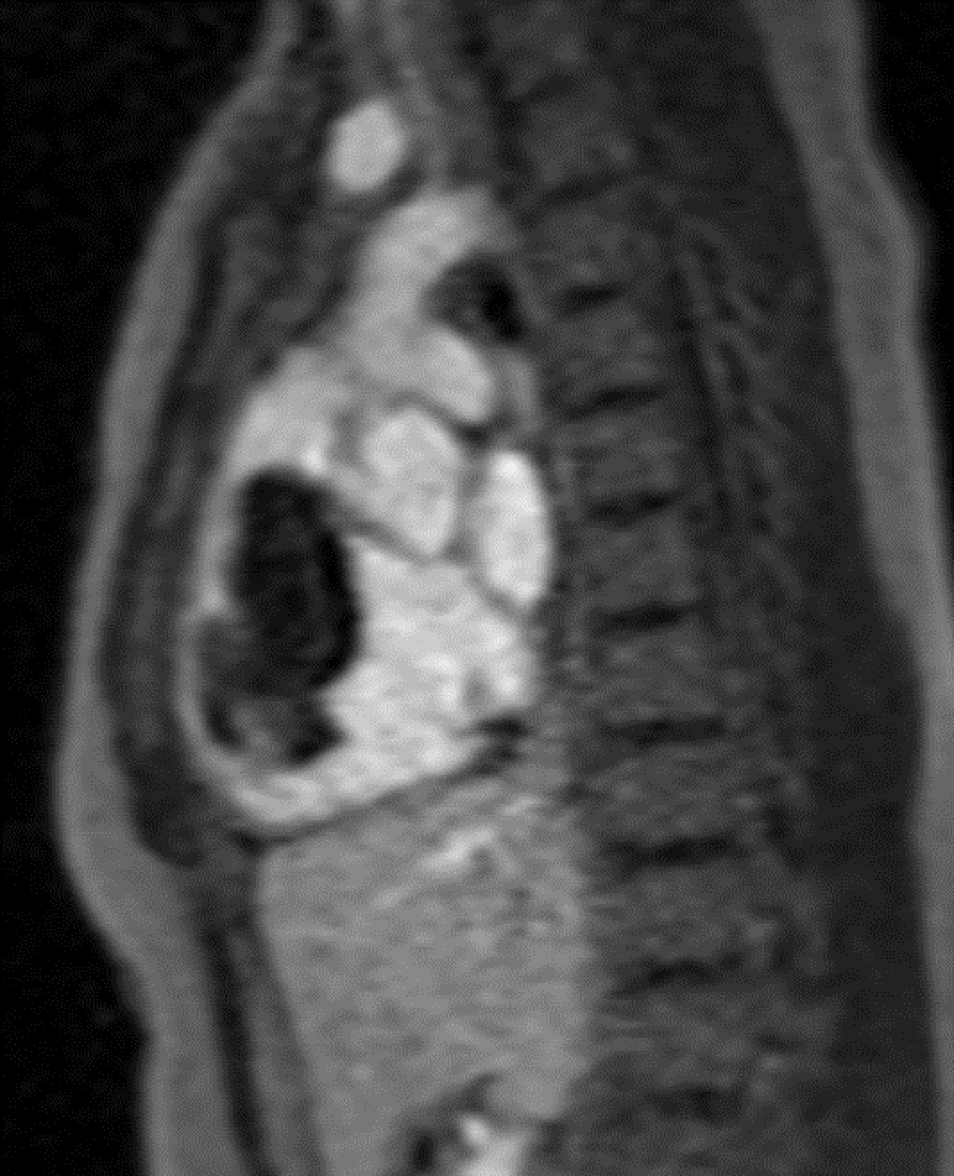
Cardiac MRI was obtained the day after presentation to further characterise the type and location of the mass and showed a large, mobile right ventricle mass with attachment to the mid-cavity wall/tricuspid support apparatus and a mass in the right pulmonary artery bifurcation with extension to the left pulmonary artery (Image 3). Imaging characteristics on MRI, including +SSFP, +T1, +T2, – fat saturation, - first-pass perfusion, – late gadolinium, were consistent with a myxoma. She was taken to the operating room the following day for right ventricular myxoma resection and bilateral pulmonary embolectomy (Image 4). Pathology was able to confirm the mass as a cardiac myxoma and thromboembolized cardiac myxoma into the pulmonary arteries. She did well postoperatively and was discharged home on hospital day 4 with the new diagnosis of Carney complex (multiple endocrine neoplasia, lentiginosis syndrome, cardiac myxomas, and endocrine/non-endocrine tumours). Genetic testing was sent for PRKAR1A gene sequencing and deletion/duplication studies that returned for an intronic variant of unknown significance (c.502 + 3A > C) consistent with a pathogenic variant. She continues to do well and is followed closely by paediatric endocrinology, genetics, and cardiology.
MRI image in right ventricle (RV) inflow/outflow view showing a mobile, heterogeneous cardiac mass connecting to a stalk arising from mid body of RV cavity measuring 6.2 × 2.9 × 3.2 cm.

6.2 × 4.8 × 2.4 cm dark-brown, haemorrhagic, myxoid soft tissue mass with resection margin removed during right ventricular myxoma resection and bilateral pulmonary embolectomy. Sectioning demonstrates a markedly myxoid cut surface with 90% haemorrhage.

Discussion
Primary cardiac tumours are rare and range from 0.017% to 0.19% based on autopsy studies with a combined frequency of 0.02%. Reference Reynen6 Cardiac myxomas have an incidence of 0.5–1 per million per year. Unlike cardiac tumours with high rates of recurrence, cardiac myxomas may appear in younger children. Reference Sarjeant, Butany and Cusimano7 In patients with Carney complex, cardiac myxomas occur at a much higher incidence than in the general population and can be found in up to 20–40% of cases. Reference Espiard and Bertherat8 In a recent cohort of patients with Carney complex and cardiac myxoma, there was a recurrence rate of 44%, which is much higher than prior estimates of 10 to 22%. Reference Burke and Virmani3 Thus, children with Carney complex and history of excised cardiac myxoma should be followed with biannual echocardiograms for 20 years, then by yearly echocardiograms through adulthood. Reference Pitsava, Zhu, Sundaram, Mills and Stratakis2 Most cases of Carney complex are caused by a pathogenic mutation in the PPKAR1A gene on chromosome 17q24 as was demonstrated in our patient. Reference Sarjeant, Butany and Cusimano7
Conclusion
Paediatric pulmonary embolism due to cardiac myxoma is rare with most cardiac myxomas occurring in the atria. To our knowledge, this is the first paediatric case of Carney syndrome presenting with bilateral pulmonary embolism. A high index of suspicion, careful examination, and early diagnostic imaging resulted in a clear diagnosis and an excellent outcome.
Supplementary material
The supplementary material for this article can be found at https://doi.org/10.1017/S1047951124000684.
Financial support
This research received no specific grant from any funding agency, commercial, or not-for-profit sectors.
Competing interests
None.



 Open access

