Introduction
Adenoidal tissue is part of Waldeyer's lymphoid ring and is located in the uppermost part of the nasopharynx, next to the Eustachian tube orifice and the choana.Reference Hellings, Jorissen and Ceuppens1, Reference Gates, Muntz and Gaylis2 It has 12–15 shallow crypts and is covered with columnar ciliated respiratory epithelium with numerous goblet cells. The lymphoid tissue consists of lymph follicles with germinal centres containing B-cells and interfollicular areas containing mainly T-cells.Reference Bykova and Satdykova3
Adenoid hypertrophy or chronic adenoiditis is usually considered a surgical problem, and adenoidectomy is considered once there is persistent nasal obstruction.Reference Koca, Erdem and Bayindir4 Associated cor pulmonale, pulmonary hypertension and alveolar hypoventilation make the decision for surgery straightforward.Reference Glade, Pearson, Zalzal and Choi5, Reference Brown, Manning and Ridenour6 However, treatment with topical nasal steroids has been proposed for mild or moderate enlargement of the adenoids.Reference Chan, Au, Lam, Lee, Wing and Li7 Steroids have anti-inflammatory and lymphopenic effects that may shrink the adenoidal tissue and improve the nasal airway.Reference Butler and van Der Voort8–Reference Sobhy11
Mometasone furoate nasal spray has been used in children aged more than two years to control the symptoms of allergic rhinitis, but is also used as a medical treatment for adenoid hypertrophy.Reference Chohan, Lal, Chohan, Chakravarti and Gomber12, Reference Penagos, Compalati, Tarantini, Baena-Cagnani, Passalacqua and Canonica13 No study to date has examined the histological effects induced by topical steroids on adenoidal tissue. This study aimed to investigate histopathological changes in adenoidal lymphoid tissue after four weeks’ use of topical mometasone furoate aqueous nasal spray (Nasonex®).
Materials and methods
This prospective study was performed in the Otolaryngology Department, Suez Canal University Hospital (Ismailia, Egypt), from June 2016 to March 2018. The study protocol was approved by the local ethics committee and written informed consent was obtained from all patients’ relatives.
Twenty-six children, aged six to nine years, diagnosed with adenoid hypertrophy grade 3 (more than 75 per cent choanal obstruction) and scheduled for adenoidectomy after one month, were included in the study. Children with craniofacial abnormalities, such as cleft palate and choanal atresia, and those with otitis media with effusion, were excluded from the study.
All patients were subjected to complete head and neck evaluation. Nasal endoscopy, either flexible or rigid, was conducted, and children who had 75 per cent or more of the choanal space occupied by the adenoidal tissue (adenoid hypertrophy grade 3) were included in the study.Reference Kurien, Lepcha, Mathew, Ali and Jeyaseelan14
The 26 patients were randomly divided into 2 equal sized groups: a mometasone furoate group and a control group. Randomisation was conducted through a computer-generated table of random numbers; if the first digit of the random number was 0–4, the patient was assigned to the mometasone group, and if the first digit was 5–9, assignment was to the control group.
The mometasone group (n = 13) received mometasone furoate (Nasonex; 100 mcg/day) in the form of nasal spray (50 mcg/puff), two puffs per nostril once daily for four weeks. The minimum age of use for Nasonex is two years; the children in the study were six to nine years old. We used the dose recommended for treating seasonal allergic and perennial allergic rhinitis nasal symptoms: one spray (50 mcg of mometasone furoate in each spray) in each nostril, once daily for one month.
The control group (n = 13) received nasal normal saline (0.9 per cent) once daily, also for four weeks. The first dose was administered in the clinic to show the caregiver how to use it.
After four weeks, all patients underwent adenoidectomy, and the resected adenoidal tissue was processed for light microscopy. Tissue specimens were fixed in 10 per cent neutral buffered formalin, dehydrated in a graded alcohol series, cleared in xylene and embedded in paraffin wax. Paraffin sections of 5 µm thickness were then stained with haematoxylin and eosin, and examined under the light microscope. Ten sections were examined for each patient, under high power field magnification (×40). The pathologist examined for infiltration with inflammatory cells, lymphoid follicles architecture and congested blood vessels in each section. The two groups’ findings were compared. All slides were examined by the same pathologist.
Results
Twenty-six children (10 males and 16 females), with a mean age of 7.1 years, were included in the study, all of whom were scheduled to undergo adenoidectomy after 1 month. Endoscopic examination of the nasopharynx revealed grade 3 adenoid enlargement (more than 75 per cent choanal obstruction) in all patients.
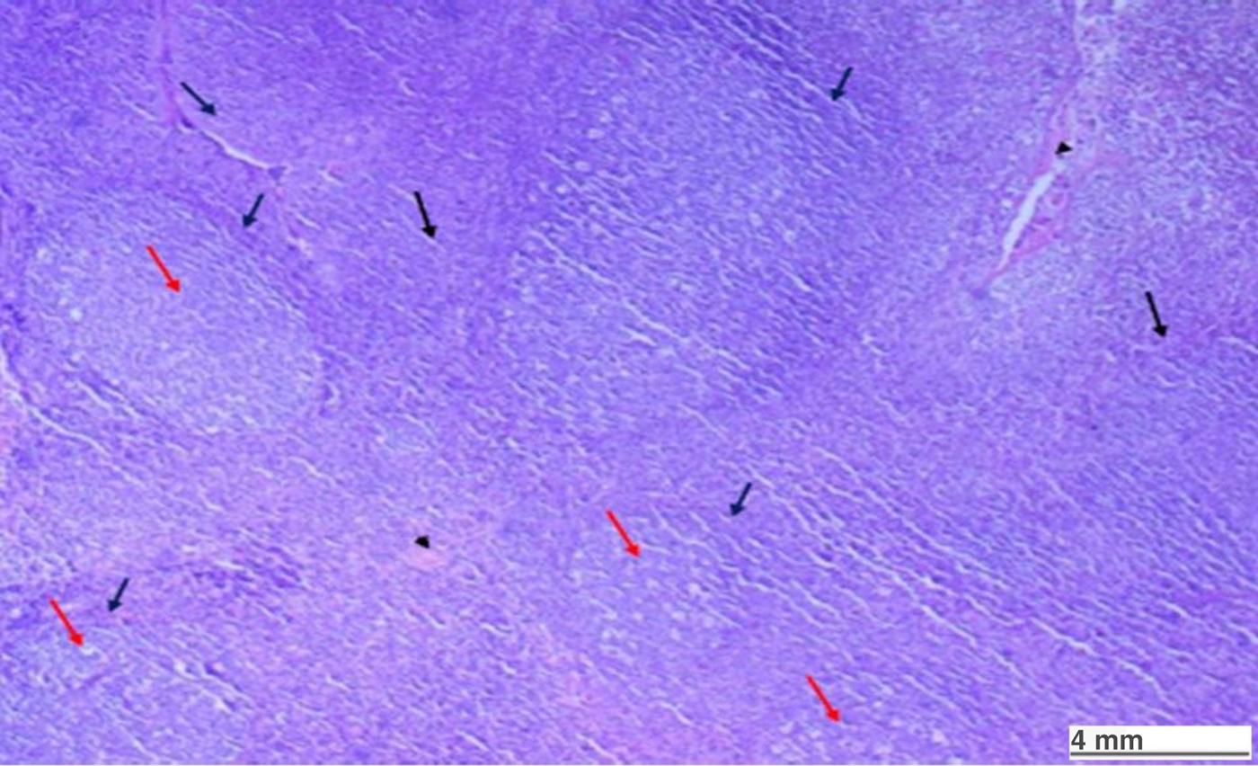
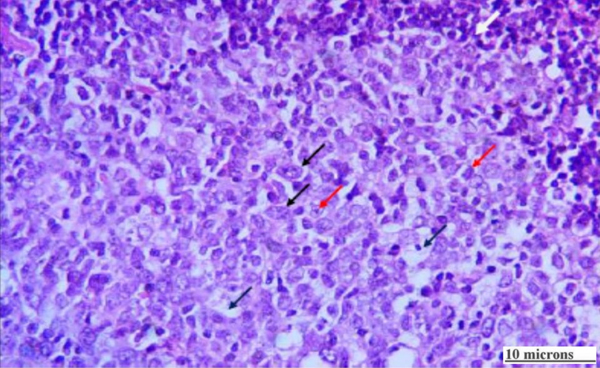
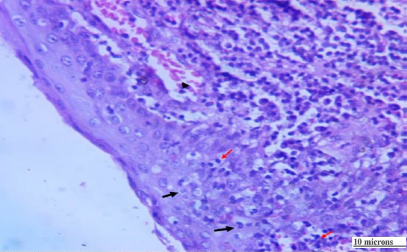
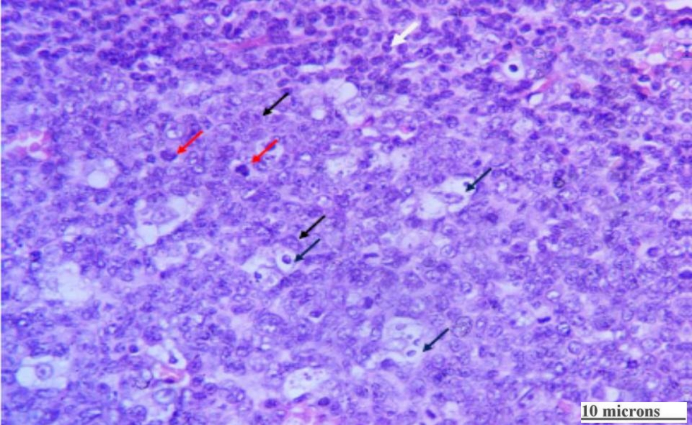
The adenoidal tissue from the control group showed proliferating, variable sized and variable shaped, lymphoid follicles, with marked spongiosis. Few of the follicles were primary; the majority were secondary follicles. Germinal centres were composed of small cleaved centrocytes, large non-cleaved centroblasts, a few tingible body macrophages and many mitotic figures. Interfollicular areas showed proliferated lymphocytes and scattered congested blood vessels. Epithelial and subepithelial tissue infiltration with chronic inflammatory cells was clearly seen in this group (Figures 1–3).
Control group showed marked spongiosis and infiltration with chronic inflammatory cells, with well-defined uniform germinal centres (red arrows). These had sharply defined margins and were surrounded by a mantle zone layer (blue arrows). Interfollicular areas showed uniform lymphocytes (black arrows), with small congested capillaries (arrow heads). (H&E; ×4)

Control group showed germinal centres surrounded by a well-defined mantle zone (white arrow), composed of small cleaved centrocytes (red arrows), large non-cleaved centroblasts (black arrows) and tingible body macrophages (blue arrows). (H&E; ×40)

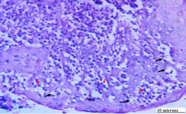
Control group showed marked spongiosis (black arrows) and infiltration with chronic inflammatory cells (red arrows). A congested blood vessel is seen (arrow head). (H&E; ×40)

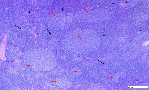
Adenoidal tissue from the mometasone group showed more uniform lymphoid follicles, with less spongiosis and less activity in the germinal centres (more centrocytes than centroblasts, fewer mitotic figures and more tingible body macrophages). Interfollicular zones had fewer congested blood vessels. Epithelial and subepithelial tissue had less infiltration with chronic inflammatory cells (Figures 4–6).
Study group showed less spongiosis and less infiltration with chronic inflammatory cells, with well-defined uniform germinal centres (red arrows). These had sharply defined margins and were surrounded by a mantle zone layer (blue arrows). Interfollicular areas showed uniform lymphocytes (black arrows) with small congested capillaries (arrow heads). (H&E; ×4)

Study group showed germinal centres (white arrow), fewer centrocytes (red arrows), fewer centroblasts (black arrows) and tingible body macrophages (blue arrows). (H&E; ×40)

Study group showed spongiosis (black arrows), with less infiltration with chronic inflammatory cells (red arrows). (H&E; ×40)

No significant side effects of the mometasone furoate (Nasonex) were reported by the children or their parents. A few children, however, experienced a drug taste in the throat.
Discussion
Histopathological evaluation of the adenoids after four weeks of topical mometasone furoate (Nasonex) administration demonstrated less reactive lymphoid follicles, less spongiosis and fewer congested blood vessels compared with the control group. This supports the efficacy of topical nasal steroids in reducing immunological and inflammatory reactions in the adenoidal tissue and the potential for reducing its size.
The adenoids, which have complex microanatomy, are essential for the uptake and presentation of airborne antigens, and for the formation of local immunity. Lymphocytes isolated from the adenoidal germinal centres exhibit ultrastructural features of centroblasts and centrocytes, and they differentiate into immunoglobulin-secreting cells. Those isolated from follicular mantles are small mature B-lymphocytes and have a condensed chromatin pattern. The greater the antigenic stimulation, the more activity in the germinal centres and the larger the adenoidal tissue. Cytokines from dendritic cells maintain this follicular arrangement in separate domains (i.e. mantle zone and germinal centre).Reference Van Kempen, Rijkers and Van Cauwenberge15
The proliferative process within the germinal centres goes hand in hand with apoptotic mechanisms, as is evident from the number of tingible body macrophages. Tingible bodies are the nuclear chromatin of apoptotic cells that can be made clearer with immunostaining.Reference Tabe, Kawabata, Koba and Homma16
As a child grows, apoptotic mechanisms become more dominant and the adenoids regress in size. This effect could also be induced with corticosteroids, either systemic or topical. Different topical steroids have been used in children with nasal obstruction due to large adenoids. Criscuoli et al. were able to detect improvement in patients’ symptoms and significant decreases in adenoid size after two weeks of topical beclomethasone nasal spray administration.Reference Criscuoli, D'Amora, Ripa, Cinquegrana, Mansi and Impagliazzo17 Zhang et al. also demonstrated that topical corticosteroids significantly improved children's nasal symptoms, and reduced severely hypertrophic adenoids to a moderate size.Reference Zhang, Mendoza-Sassi, Cesar and Chadha18
Chadha et al. reviewed 7 studies of topical steroid use in a total of 493 patients with adenoidal hypertrophy. The authors found improvements in nasal symptom scores and a reduction in adenoid size in six of these studies. The treatment seemed to be safe and well-tolerated.Reference Chadha, Zhang, Mendoza-Sassi and Cesar19
Of the several commercially available nasal steroid sprays, we selected mometasone furoate (Nasonex) because it has no adverse effects on growth or the hypothalamic pituitary adrenal axis, and has very low bioavailability compared to other steroids.Reference Chohan, Lal, Chohan, Chakravarti and Gomber12
Similar to our study, Berlucchi et al. evaluated the use of mometasone furoate aqueous nasal spray in children with adenoidal hypertrophy over a short period, and found it effective in reducing adenoid size and symptom score.Reference Berlucchi, Valetti, Parrinello and Nicolai20 Rezende et al. used mometasone with saline nasal irrigation, and found that nasal saline douches could improve the nasal symptoms, but the addition of mometasone to the regimen was needed in order to reduce adenoidal tissue size.Reference Rezende, Silveira, Barbosa, Menezes, Ferriani and Rezende21
Several mechanisms, such as a direct effect on lymphoid follicles and an anti-inflammatory effect, may explain how steroids decrease adenoid volume and improve nasal obstructive symptoms.
Our study is the first to report light microscopic changes in adenoidal tissue induced by mometasone furoate (Nasonex) nasal spray administration. However, immunohistochemistry was not conducted. This would have made it possible to better identify cellular and architectural changes in the adenoidal tissue.
We decided to use fibre-optic nasal endoscopy (FNE) to assess adenoid size clinically and to grade the choanal obstruction. Our focus, however, was on the light microscopic changes in adenoidal tissue after one month of topical mometasone furoate (Nasonex) use.
Our study aimed to report the clinical effects of mometasone furoate on patients’ symptoms and to determine the effects of Nasonex nasal spray on adenoidal tissue. The children included in the study had large adenoids (grade 3 adenoid hypertrophy). One month of treatment with the minimum dose of Nasonex nasal spray is not sufficient to obviate the need for surgery in the presence of large adenoids. A further study covering a longer treatment period is needed to determine whether surgery can be avoided in children with a lesser degree of adenoid enlargement.
• Topical nasal steroids improved adenoidal hypertrophy symptoms
• In this study, 13 children received mometasone spray and 13 received normal saline spray for 4 weeks
• Adenoidal tissue from the mometasone group had fewer reactive germinal centres, less subepithelial spongiosis and fewer congested blood vessels than the control group
• Intranasal mometasone furoate nasal spray reduced adenoidal tissue reactive cellular changes and its vascularity
Conclusion
Intranasal mometasone furoate aqueous nasal spray (Nasonex) used for one month reduced adenoidal tissue reactive cellular changes and its vascularity. This is, however, a pilot study; a further study with a longer treatment period is needed to assess the effect of mometasone furoate on adenoidal size.
Competing interests
None declared