Introduction
Cochlear implantation is a widely used treatment for children with sensorineural hearing loss (SNHL). Although it is a safe surgical procedure, facial nerve injury and consequent facial nerve paralysis can occur. Surgeons should be aware that an aberrant facial nerve coincident with inner-ear anomalies can contribute to facial nerve injury by complicating access to the round window and impeding electrode implantation.Reference Raine, Hussain, Khan and Setia 1 , Reference Sennaroglu 2 Although pre-operative identification of a facial nerve anomaly decreases the possibility of intra-operative nerve injury, there are insufficient reports on this topic. Pre-operative facial nerve imaging can increase the surgeon's confidence to plan cochlear implantation and perform the procedure.Reference Song, Park, Jang, Lee, Oh and Chang 3
This study aimed to evaluate the shape and incidence of facial nerve anomalies, determine their concurrence with inner-ear anomalies in paediatric cochlear implant candidates, and suggest a convenient imaging method for their prediction.
Materials and methods
A total of 48 congenital SNHL patients who were admitted to Uludağ University Faculty of Medicine between 2012 and 2014 were retrospectively enrolled into the study. These patients were evaluated by temporal high-resolution computed tomography (CT) and magnetic resonance imaging (MRI). High-resolution CT was performed using the bone algorithm and a 0.6-mm contiguous section thickness, and multiplanar images were generated (Siemens Somatom Definition; Siemens, Forchheim, Germany). Magnetic resonance imaging was performed with a 3.0 T system (Philips Achieva, Philips Medical Systems, Best, Netherlands). Axial and coronal T1- and T2-weighted images and axial balanced fast field echo images were obtained; no contrast medium was used. Inner-ear anomalies were identified using both temporal high-resolution CT and MRI. Facial nerve anomalies coincident with inner-ear anomalies were investigated with temporal high-resolution CT. Inner-ear anomalies were classified according to Sennaroglu as total labyrinthine aplasia, common cavity anomalies, cochlear aplasia, cochlear hypoplasia, or incomplete partition types 1 and 2 anomalies.Reference Sennaroglu and Saatci 4 The relationship between the inner-ear and facial nerve anomalies was determined, and the facial canal width was calculated.
Results
In all, 11 of the 48 patients had inner-ear anomalies (23 per cent): 4 children had bilateral wide vestibular aqueducts; 3 had bilateral incomplete partition type 1 anomalies; 1 had bilateral incomplete partition type 2 anomalies; 1 had complete labyrinthine aplasia on the right side and an incomplete partition type 1 anomaly on the left side; 1 had cochlear aplasia on the right side and an incomplete partition type 1 anomaly on the left side with bilateral semicircular canal malformations; and 1 had vestibular aplasia on the right side and a common cavity anomaly on the left side with bilateral semicircular aplasia. No external ear or middle-ear anomalies were found. These 11 children were aged between 10 months and 12 years (mean 5.3 years).
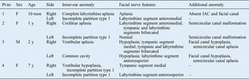
Four of the 11 children with inner-ear anomalies (8 per cent of the total) had a facial nerve anomaly coincident with the inner-ear anomalies. In these four children, the inner-ear and accompanying facial nerve anomalies were evaluated as separate sides (i.e. as eight ears). Facial nerve abnormalities were present in seven of the eight ears (Table I). The four children with facial nerve anomalies were aged between 10 months and 7 years (mean 2.7 years). No facial nerve anomalies were found in children with normal inner-ear structures.
Patient characteristics

Pt no = patient number; F = female; mon = months; IAC = internal auditory canal; y = year(s); M = male
In a child with a bilateral incomplete partition type 1 anomaly, the vestibule on the right side was hypoplastic (Table I, patient 4). The tympanic segment of the facial nerve was medially replaced secondary to this condition, while the labyrinthine segment had an anterosuperior extension on the left side.
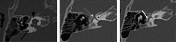
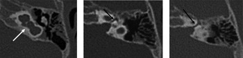
In a child with vestibular aplasia on the right side and a common cavity anomaly on the left side, the bilateral osseous facial canals were narrower than normal and both facial nerves were hypoplastic (<0.7 mm; Table I, patient 3). On the right side, the first genu of the facial nerve was absent and labyrinthine and tympanic segments were bifurcated; on the left side (with vestibular aplasia), the tympanic segment of the nerve was medially displaced (Figure 1). The labyrinthine segment of the facial nerve had an anteromedial extension on the left side with a common cavity anomaly (Figure 2).
Temporal high-resolution computed tomography images of patient 3, showing right side vestibular aplasia (thick black arrow) with a normal cochlea (black arrowhead). The osseous facial canal is narrower than normal and the facial nerve is hypoplastic. The first genu of the facial nerve is absent (thin white arrow) and the labyrinthine and tympanic segments are bifurcated (thin black arrow). The tympanic segment of the facial nerve is medially displaced at the vestibular aplasia side (thick white arrow).

Temporal high-resolution computed tomography images of patient 3, showing a common cavity anomaly in the left ear (white arrow). The labyrinthine segment of the facial nerve is anteromedially displaced (black arrows).

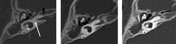
In a child with cochlear aplasia on the right side, the internal auditory canal was narrow and the facial nerve was anteromedially displaced (Table I, patient 2). The first genu of the facial nerve was absent and the labyrinthine and tympanic segments were bifurcated (Figure 3). No facial nerve anomaly accompanied the left-sided incomplete partition type 1 anomaly in this child.
Temporal high-resolution computed tomography images of patient 2, showing right side cochlear aplasia (thick white arrow) with normal vestibule (thin white arrow). The internal auditory canal is narrow (black arrowhead) and the labyrinthine segment of the facial nerve is anteromedially displaced (thin black arrows). The first genu of the facial nerve is absent and the labyrinthine and tympanic segments are bifurcated (thick black arrow).

In a child with complete labyrinthine aplasia on the right side and an incomplete partition type 1 anomaly on the left side, the right internal auditory canal, facial canal and facial nerve were absent (Table I, patient 1). The facial nerve was anteromedially located on the left side.
Discussion
Although some researchers are convinced that various cochlear malformations cause facial nerve anomalies, the relationship between the type of inner-ear malformation and type of facial nerve abnormality is not well understood, which makes it difficult to estimate the risk from the facial nerve anomaly pre-operatively. The frequency of facial nerve anomalies in normal ears is reported to be 0.3 per cent.Reference Sennaroglu and Saatci 4 Facial nerve anomalies coincident with vestibulocochlear anomalies was reported to be 16 per cent by Papsin, 16 per cent by Hoffman et al. and 0.7 per cent by Song et al.Reference Song, Park, Jang, Lee, Oh and Chang 3 , Reference Papsin 5 , Reference Hoffman, Downey, Waltzman and Cohen 6 The rate was 8 per cent in the present study. These differences might result from the different patient numbers in each study. No facial nerve anomalies were found in normal ears in the present study nor in the reviewed studies.
Very few reports on aberrant facial nerve extensions in cochlear implant candidates were found. Facial nerve anomalies increase the facial nerve injury risk, especially when using the transmastoid facial recess approach (a standard technique). The labyrinthine segment of the facial nerve was shown to be displaced anteriorly by Raine et al., anteromedially by Hoffman et al. and anteroinferiorly by Huang et al.Reference Raine, Hussain, Khan and Setia 1 , Reference Hoffman, Downey, Waltzman and Cohen 6 , Reference Huang, Lin and Wu 7 Such facial nerve extension anomalies prevent visualisation of the round window and the facial recess. Sennaroglu, alone and with Saatci, showed that anteromedial displacement of the labyrinthine segment of the facial nerve enclosed the oval and round windows of patients with inner-ear anomalies.Reference Sennaroglu 2 , Reference Sennaroglu and Saatci 4 They also reported that cochlear aplasia itself might cause anterior dislocation of the labyrinthine segment. In their patient series, Song et al. reported anteromedial displacement of the labyrinthine segment to be the most common aberrant extension and bifurcation of the labyrinthine and tympanic segments to be the second most common.Reference Song, Park, Jang, Lee, Oh and Chang 3 Raine et al. described four patients with anteriorly located labyrinthine segments and one with a bifurcated labyrinthine segment.Reference Raine, Hussain, Khan and Setia 1 In the present study, anteromedial displacement of the labyrinthine segment was observed in three children and bifurcation of the labyrinthine and tympanic segments was observed on two of the eight sides (i.e. ears) of the four children with facial nerve anomalies. These findings are compatible with the published data. In one child, the labyrinthine segment extended anterosuperiorly. Cochlear anomalies were present in all children with anteromedially and anterosuperiorly extended labyrinthine segments. The genu of the facial nerve canal was absent in two children with bifurcated labyrinthine and tympanic segments: one child had cochlear aplasia and the other had vestibular aplasia.
Cochlear anomaly increases the risk of anteromedial displacement of the facial nerve. Published reports have linked anteromedial displacement of the labyrinthine segment of the facial nerve to cochlear malformation (especially common cavity and incomplete partition type 1 anomalies, and cochlear aplasia and hypoplasia). However, Romo and Curtin reported that abnormal facial nerve extension does not coincide with an incomplete partition type 2 anomaly, explaining that the latter may occur during normal cochlear development.Reference Romo and Curtin 8
The relationship between inner-ear and facial nerve anomalies is thought to have an embryological aetiology.Reference Prashant, Sugoto and Douglas 9 , Reference Fishman 10 While the facial nerve begins to develop in the third week of gestation from the facioacoustic primordium, the facial canal develops from the primordial capsule and Reichert's cartilage from the second branchial arch. Facial nerve development ends in the fifth week. The facial canal is a deep sulcus in the canalicular portion of the primordial otic capsule at 10 weeks, and at that developmental stage, the otic capsule is a cartilaginous structure. The cartilaginous portion of the labyrinthine and tympanic segments of the facial canal develops from Reichert's cartilage. The osseous facial canal is mainly closed by bone; this process continues until late fetal development. The tympanic and mastoid segments of the facial nerve derive from the second branchial arch. Abnormal extension of these segments might be associated with abnormal development of the first and second branchial arch derivatives, such as the bony wall of the facial nerve canal, the stapes, the styloid process and/or the external auditory canal. Reichert's cartilage is the cartilage analogue of the second branchial arch, and bony structures derived from this probably affect the nerve course through the temporal bone. Durcan et al. were the first to report anterior displacement of the tympanic and mastoid segments of the facial nerve.Reference Durcan, Shea and Sleecchx 11 Al-Mazrou et al. and others reported an association between conductive congenital hearing loss and an anomalous facial nerveReference Al-Mazrou, Alorainy, Al-Dousary and Richardson 12 ; Romo and Curtin made a similar suggestion about the labyrinthine segment of the facial nerve.Reference Romo and Curtin 8 Based on the embryonic development of the facial nerve and cochlea, the latter authors postulated that cochlear malformation could cause anteromedial displacement of the labyrinthine segment of the facial nerve canal. The facioacoustic primordium is formed by the VIIth and VIIIth cranial nerves and is located near to the otocyst, from which the cochlear duct develops. The geniculate ganglion is located between the semicircular canal and the cochlea proper, above the basilar turn of the cochlea. Romo and Curtin reported that anomalous facial nerve canal segments were likely to develop alongside inner-ear anomalies because a delay in rostral growth of the cochlea may cause the facial nerve to escape anteriorly and be displaced anteromedially.Reference Romo and Curtin 8 These authors hypothesised that a normal cochlea may behave similarly to Reichert's cartilage derivatives, which prevent migration of the facial nerve and tympanic facial nerve canal anteriorly. Certain inner-ear malformations are the most important factors influencing facial canal location and may therefore cause aberrant localisation of the facial nerve canal, making its course more complicated.
Vestibular anomalies were classified as a distinct group by Sennaroglu, but they are a very rare subtype of inner-ear anomalies.Reference Sennaroglu 2 Association of a facial nerve anomaly with vestibular anomalies has not been previously reported. However, medial displacement of the tympanic segment of the facial nerve coincided with vestibular aplasia and hypoplasia in two patients in the present series. Thus, medial displacement of the facial nerve and facial canal might be a secondary effect of abnormal bone development caused by hypoplastic or aplastic vestibular structures. If correct, this is supportive evidence that inner-ear malformations and malpositions may influence the development of facial nerve anomalies, which are usually associated with the malposition of vestibular structures and the cochlea.
In the present series, both facial canals were thinner than normal in one child and the internal auditory canal and facial canal were absent in another child with complete labyrinthine aplasia. Complete labyrinthine aplasia requires careful management. In the absence of the cochleovestibular nerve, brain stem implantation is the only possible option for auditory rehabilitation. However, surgery may be indicated for some children with infectious complications such as chronic otitis media or cholesteatoma; knowledge of the facial nerve course is therefore essential. The facial nerve and facial canal or any segment of these structures may be either normal or absent in complete labyrinthine aplasia patients. If middle-ear structures are normal, then the facial nerve follows a nearly normal course, with separate tympanic and mastoid portions; however, if an anomaly is present, the facial canal is also anomalous.Reference Zarandy, Kouhi, Kashany, Rabiei, Hajimohamadi and Rabbani-Anari 13 In addition, the labyrinthine segment is not affected. One child in the present series with complete labyrinthine aplasia had no osseous facial canal.
Middle-ear and ossicular anomalies were reported to accompany inner-ear anomalies in patients with facial nerve anomalies in a previous series.Reference Song, Park, Jang, Lee, Oh and Chang 3 , Reference Heman-Ackah, Ronald, Haynes and Waltzman 14 , Reference Ahn and Lee 15 Most of these patients had craniofacial syndromes such as coloboma, heart defect, atresia choanae, retarded growth and development, genital abnormality and ear abnormality (‘CHARGE’), DiGeorge or Treacher Collins syndrome. In the present series, a patient with complete labyrinthine aplasia had Klinefelter syndrome. This patient did not have a craniofacial syndrome or middle-ear and ossicular anomalies.
Anomalies in the facial nerve course increase the facial nerve injury risk in cochlear implant surgery, especially when using the standard transmastoid facial recess approach.Reference Mylanus, Rotteveel and Leeuw 16 – Reference Chadha, James, Gordon, Blaser and Papsin 20 Various methods are reported to prevent facial nerve injury during cochlear implantation in patients with inner-ear anomalies. For cochleostomy, Sennaroglu and Saatci used a combined anteroposterior approach through the ear in which the facial nerve crossed over the oval and round windows.Reference Sennaroglu and Saatci 4 Song et al. reported that the facial nerve may be mobilised and rerouted, and that transcanal cochleostomy may be required.Reference Song, Park, Jang, Lee, Oh and Chang 3 Papsin stated that it is necessary to dissect the promontory anteriorly to enter the cochlea.Reference Papsin 5 He added that the facial nerve may be split and a second anteriorly displaced nerve may be found in addition to the one in the normal position (posterior to the facial recess). McElveen et al. described a transmastoid labyrinthotomy approach to common cavity malformations to minimise the injury risk for an aberrant facial nerve.Reference McElveen, Carrasco, Miyamato and Linthicum 21
Limitations of the present study were that the patient series was small and aberrant extension of the facial nerve was not confirmed by surgery. Despite the small series, sufficient patients were included to allow the detection of inner-ear abnormalities. However, multicentre studies are required to increase the cohort size.
In the present series, anteromedial displacement of the labyrinthine segment was the most common aberrant facial nerve extension, and bifurcation of the labyrinthine and tympanic segments was the second most common and may coincide with cochlear anomalies. The facial nerve and facial canal might also be displaced medially secondary to abnormal bone development caused by vestibular structure anomalies. This finding supports the theory that inner-ear malformations and malpositions (usually of vestibular structures and the cochlea) can influence the development of facial nerve anomalies.
-
• Cochlear implantation is widely used to treat children with sensorineural hearing loss
-
• Although considered a safe procedure, facial nerve injury and consequent facial nerve paralysis is possible
-
• An aberrant facial nerve often coincides with inner-ear anomalies
-
• Patients with inner-ear anomalies should undergo pre-operative radiological evaluation of the facial nerve course and shape
In the present study, patients with no inner-ear abnormalities (as shown by MRI) did not have facial nerve abnormalities (as shown by temporal high-resolution CT). It is therefore recommended that children with SNHL should undergo MRI before cochlear implantation to evaluate inner-ear and vestibulocochlear nerve structure. If MRI shows inner-ear anomalies, then temporal high-resolution CT can be used to trace the facial nerve course to reveal a possible relationship between facial canal bone and inner-ear structures. However, CT uses radiation and children are highly sensitive to radiation (more so than adults). Therefore, temporal high-resolution CT evaluation of the facial nerve should be avoided in this patient group if no inner-ear anomalies are detected before cochlear implantation.
Conclusion
The possibility of an aberrant facial nerve course should be considered for every patient with inner-ear anomalies, and the facial nerve course and shape should be carefully evaluated pre-operatively with radiological imaging. An awareness of this anomaly will reduce the facial nerve injury risk during surgery by helping to determine the best surgical technique, thus avoiding major complications. Surgeons should be aware that the facial nerve damage risk is higher in patients with inner-ear malformations. Children with SNHL should be investigated by MRI before cochlear implantation to detect inner-ear abnormalities. If these are present, temporal high-resolution CT should then be used to trace the facial nerve course and determine the relationship between facial canal bone structures.