Iron is an essential element for cell growth and division but is also a strong pro-oxidant, so cells and whole organisms have developed sophisticated mechanisms for keeping iron levels within the physiologically optimum range. Either too little or too much iron can have significant clinical consequences. The main cause of low iron levels in the body is inadequate dietary intake, and iron deficiency is one of the world's most significant nutritional deficiencies (Ref. Reference Zimmermann and Hurrell1). In contrast, body iron overload usually results from mutations in genes encoding proteins that regulate body iron intake. Although less widespread than iron deficiency, iron-loading syndromes represent an important class of human diseases, particularly in populations where iron nutrition is adequate. One of these disorders, HFE-related hereditary haemochromatosis (HFE refers to the affected protein), is the most common disease of humans that shows autosomal recessive inheritance (Ref. Reference O'Neil and Powell2). There have been important advances in understanding these diseases in recent years, and their analysis has taught us much about the mechanisms of body iron homeostasis.
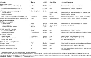
If the total body iron load increases, the extra iron must come from external sources. There are basically two such sources: the diet and blood transfusions. Iron-loading disorders characterised by defects in proteins directly involved in body iron acquisition are known as primary iron-loading diseases. Under these conditions, intestinal iron absorption is dysregulated such that the body continues to accrete iron in the presence of adequate or even enhanced iron stores (Ref. Reference Lee and Beutler3). Secondary iron loading usually results from blood transfusions. Patients who receive transfusions to treat haemoglobinopathies, bone marrow failure or haemolytic syndromes accumulate iron in their tissues after the iron derived from the transfused red cells is released in macrophages following erythrophagocytosis (Ref. Reference Gattermann4). Haematological conditions where erythropoiesis is markedly ineffective might also be associated with increased intestinal iron absorption, and this compounds their iron loading (Ref. Reference Rechavi and Rivella5). Thus the distinction between primary and secondary iron loading is often blurred. A summary of the various forms of iron loading is provided in Table 1.
Classification of some iron-loading disorders

aPredicted levels based on current knowledge of hepcidin regulation.
Abbreviations: ALAS2, delta aminolevulinate synthase 2; CP, ceruloplasmin; FANC, Fanconi anaemia complementation; FPN, ferroportin; FXN, farataxin; HAMP, hepcidin; HBB, haemoglobin beta; HJV, haemojuvelin; OMIM, Online Mendelian Inheritance in Man (ID); PKLR, pyruvate kinase, liver and red blood cell; SLC40A1, solute carrier family 40 (iron-regulated transporter), member 1; TF, transferrin; TFR2, transferrin receptor 2.
Normal iron metabolism
The adult human male contains approximately 4–5 g of iron (Ref. Reference Forth and Rummel6). Most of this iron is used for metabolic functions (in haemoglobin, myoglobin and a myriad of enzymes that require iron as a cofactor), but approximately 1 g is storage iron that is in excess of immediate requirements and can be accessed in times of need. In women the total body iron content is somewhat lower and iron stores average around 300 mg (Ref. Reference Brittenham, Brock, Halliday and Pippard7). In subjects with iron overload, body iron stores can expand enormously and can reach 20–40 g or even higher (Ref. Reference Powell, Jazwinska, Halliday, Brock, Halliday and Pippard8).
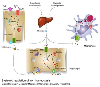
The body iron content is determined through the regulation of iron transport across enterocytes of the proximal small intestine (Fig. 1). Most dietary iron is in the oxidised or ferric (Fe3+) form and must be reduced to ferrous iron (Fe2+) before it can be utilised by the enterocytes. A candidate ferric reductase is the brush-border enzyme Dcytb (CYBRD1) (Ref. Reference McKie9), but other reductases undoubtedly exist. The reduced iron is transported across the apical membrane of the enterocyte by divalent-metal-ion transporter 1 [DMT1 (SLC11A2)] (Refs Reference Fleming10, Reference Gunshin11). Iron within the enterocyte that is not immediately required by the body is stored within ferritin and is ultimately lost when the enterocyte is sloughed into the lumen at the end of its life. Iron that is needed is transported across the enterocyte basolateral membrane and into the circulation through the iron exporter ferroportin 1 [FPN1 (SLC40A1)] (Refs Reference Abboud and Haile12, Reference Donovan13, Reference McKie14). For this process to occur efficiently, FPN1 must act in conjunction with the iron oxidase hephaestin (HEPH) (Ref. Reference Vulpe15). Physiological (Refs Reference Conrad and Crosby16, Reference Conrad, Weintraub and Crosby17) and molecular (Refs Reference Frazer18, Reference Chen19) studies have shown that the basolateral transport of iron is rate limiting for iron absorption.
Systemic regulation of iron homeostasis. Iron is reduced at the brush-border membrane of duodenal enterocytes (Dcytb is a candidate ferric reductase) and transported across the membrane by DMT1. Excess iron is stored in ferritin and the remainder is transported across the basolateral membrane and into the circulation by the iron exporter FPN1. Excess iron can also suppress the ARNT–HIF-2α complex, which normally induces DMT1 and Dcytb expression. Iron that enters the bloodstream is bound to circulating plasma TF. TF delivers iron to various tissues, including hepatocytes where it is either stored in ferritin or utilised for cellular metabolism. Iron that is not bound to TF (NTBI) can also enter hepatocytes and contributes to the labile iron pool. Excess iron is stored in ferritin and its degradation product haemosiderin. When required, iron can be removed from ferritin and exported from hepatocytes. Senescent red blood cells are engulfed by reticuloendothelial macrophages. Hb is broken down, and iron is either released into the circulation or stored in ferritin. The liver-derived peptide hepcidin blocks cellular iron release by binding to FPN and facilitating its removal from the membrane, thereby regulating iron entry into the plasma. Iron stores and inflammation positively regulate hepcidin levels, whereas hypoxia and increased erythropoiesis downregulate it. Abbreviations: ARNT, aryl hydrocarbon receptor nuclear translocator; DMT1, divalent-metal-ion transporter 1; FPN1, ferroportin; Hb, haemoglobin; HIF-2α, hypoxia-inducible factor 2α; NTBI, non-transferrin-bound iron; TF, transferrin; TFR1, transferrin receptor 1.

Iron that enters the bloodstream is bound to circulating plasma transferrin (TF) and is delivered throughout the body to sites where it can be utilised or stored (Ref. Reference Huebers and Finch20) (Fig. 1). Transferrin binds to transferrin receptor 1 [TFR1 (TFRC)] on the plasma membrane of most body cells and the iron is delivered internally by receptor-mediated endocytosis (Ref. Reference Huebers and Finch20). TFR1 has a particularly high affinity for diferric (or holo) transferrin, but it can also bind monoferric transferrin with lower affinity. Cells that have very high iron requirements express particularly high levels of TFR1. Such cells include immature erythroid cells (which must synthesise large amounts of haemoglobin) and rapidly dividing cells, such as the crypt enterocytes of the intestine. The recycling of haemoglobin-derived iron from senescent red cells is carried out by macrophages (Ref. Reference Anderson21). This is quantitatively the most important iron-trafficking pathway within the body and accounts for approximately 80% of body iron movement (Ref. Reference Brittenham, Brock, Halliday and Pippard7). After iron is released from haem by haem oxygenase, the iron is exported from the macrophages by FPN1 (Ref. Reference Donovan22) (Fig. 1).
Both cellular and body iron trafficking are tightly regulated to ensure an adequate supply of iron during times of increased demand, and to prevent the accumulation of unnecessary and potentially toxic iron. Body iron homeostasis is regulated by a small peptide known as hepcidin (HAMP) (Ref. Reference Nemeth and Ganz23). Hepcidin is synthesised predominantly in the liver and has a crucial role in communicating the body's iron requirements to the intestine (and other cells) (Fig. 1). Its main function is to control the amount of iron released into the circulation and it does this by binding to the iron exporter FPN1 and facilitating its internalisation and degradation (Ref. Reference Nemeth24). When body iron requirements are high (e.g. under iron-deficient conditions or when erythropoiesis is elevated), hepcidin levels are reduced and enterocytes, macrophages and other cells are free to donate iron to the plasma. In contrast, when iron demands are reduced, high levels of hepcidin mean a reduction in iron transport to the plasma. Thus there is a close inverse relationship between hepcidin levels and iron absorption. An understanding of the biology of hepcidin is critical for understanding the pathogenesis of iron-loading disorders, and is considered in detail below.
Primary iron-loading disorders
Primary iron loading in most cases results from increased absorption of dietary iron by the small intestine, although in some cases there might also be a redistribution of iron between tissues. These diseases are collectively referred to as haemochromatosis.
HFE-related (type 1) haemochromatosis
The most common type of primary iron overload is HFE-related haemochromatosis (Ref. Reference O'Neil and Powell2). This autosomal recessive disorder is usually caused by a homozygous mutation within the HFE gene on chromosome 6, leading to the C282Y substitution in the membrane protein HFE, although several other mutations have also been described (Ref. Reference Lee and Beutler3). HFE-related haemochromatosis is essentially restricted to populations of northern European origin, in which the prevalence of homozygosity for the main mutation that predisposes to iron loading can be 1 in 200 or even higher (Ref. Reference O'Neil and Powell2). However, although the great majority of such individuals accumulate excess iron in their body, only a minority progress to significant disease. A recent population study demonstrated that approximately 30% of males but only 1% of females develop clinical disease (Ref. Reference Allen25). The reasons for this are not entirely resolved, but a range of environmental and genetic factors are likely to be involved. In the case of women, menstrual blood loss and childbirth are significant contributors to the loss of body iron and probably the main reason why the disease is far less common in women. However, studies in Hfe-knockout mice demonstrate that multiple pregnancies do not reduce iron stores (Ref. Reference Neves26). Recently, genome-wide association studies have shown that polymorphisms in the transferrin gene in combination with C282Y mutation in the HFE gene can explain 40% of the genetic variation in serum transferrin levels (Ref. Reference Benyamin27). Polymorphisms in other genes that contribute to genetic variation of the iron status have been reported extensively; however, they are beyond the scope of this review.
Like other primary iron-loading disorders, HFE-related haemochromatosis is characterised by reduced hepcidin levels (Fig. 2), increased intestinal iron absorption and the subsequent deposition of iron in a range of tissues, including the liver, heart and pancreas (Ref. Reference O'Neil and Powell2). Iron acts as a strong pro-oxidant and is able to catalyse reactions leading to the production of reactive oxygen species (Ref. Reference Galaris and Pantopoulos28). These in turn are able to damage a wide range of biological macromolecules, and this underlies the tissue damage that is associated with body iron loading (Ref. Reference Ramm and Ruddell29). Many of the clinical manifestations of the disease reflect hepatic iron accumulation. Iron initially deposits in the hepatocytes surrounding the portal tracts, but in advanced cases iron deposition can be panlobular. Resident macrophages of the liver (Kupffer cells) are characteristically low in iron in haemochromatosis patients but might contain iron in advanced cases.
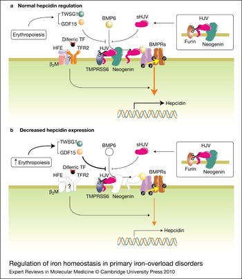
Regulation of iron homeostasis in primary iron-overload disorders. (a) Normal hepcidin regulation. The presence of HFE, TFR2, HJV and neogenin on the hepatocyte surface is required for normal hepcidin expression and regulation. Neogenin interacts with and stabilises HJV, preventing its cleavage by TMPRSS6 and furin. TMPRSS6 cleaves HJV and thus reduces hepcidin expression in hepatocytes. TWSG1, GDF15 and soluble HJV inhibit binding of BMP6 to BMPRs and regulate hepcidin negatively. The nature of the interaction between HFE and TFR2 remains unclear. (b) Decreased hepcidin expression. Mutations in, or the absence of, HFE, TFR2, HJV, neogenin or BMP6 result in a decrease in hepcidin expression, increased intestinal iron absorption and body iron overload.

TFR2-related (type 3) haemochromatosis
This is a rare form of primary iron loading associated with mutations in the transferrin receptor 2 (TFR2) gene (Ref. Reference Camaschella30). TFR2 is a homologue of TFR1, but it has a much more restricted tissue distribution with predominant expression in the parenchymal cells of the liver (hepatocytes) where it is expressed at levels several times higher than TFR1 (Ref. Reference Chloupkova, Zhang and Enns31). Like TFR1, TFR2 can bind diferric transferrin and facilitate its internalisation (Ref. Reference Kawabata32). However, the primary role of TFR2 does not appear to be iron acquisition because the livers of patients with TFR2 mutations (or Tfr2-knockout mice) readily accumulate iron (Refs Reference Camaschella30, Reference Fleming33). Rather, TFR2 has an important role in regulating the expression of the iron-regulatory peptide hepcidin (Refs Reference Kawabata34, Reference Nemeth35) (Fig. 2). This is considered in more detail below. Patients with TFR2 mutations show a histological pattern of iron deposition similar to that seen in HFE-related disease, and the consequences of iron loading are essentially the same (Ref. Reference Pietrangelo36). Examination of the small number of clinical cases suggests that the rate of iron accumulation in type 3 haemochromatosis may be somewhat greater than HFE-related iron loading, and hence the disease is of slightly greater severity (Ref. Reference Pietrangelo36). This has been borne out in studies using knockout mice where animals lacking TFR2 accumulate greater amounts of iron than those lacking HFE (Ref. Reference Wallace37).
Juvenile (type 2) haemochromatosis
Severe, early-onset iron loading is known as juvenile haemochromatosis (Ref. Reference De Gobbi38). Symptoms in affected patients are usually seen in the second decade of life, and by the third decade organ damage may occur. Cardiac dysfunction is far more prevalent in juvenile haemochromatosis than in patients with mutations in HFE or TFR2 (Ref. Reference De Gobbi38). Overall, however, the pattern of iron deposition is similar to that seen in HFE-related haemochromatosis and is consistent with greatly increased dietary iron absorption. Almost all cases of juvenile haemochromatosis can be attributed to mutations in one of two genes, those encoding haemojuvelin [HJV (HFE2)] (type 2a haemochromatosis) (Ref. Reference Papanikolaou39) or hepcidin (type 2b haemochromatosis) (Ref. Reference Roetto40). Mutations in HJV are more common, but both conditions are considered rare. Like subjects with mutations in HFE or TFR2, hepcidin levels are reduced in juvenile haemochromatosis (Fig. 2), but in this condition the levels are negligible or undetectable (Refs Reference Papanikolaou39, Reference Roetto40). This explains the uninhibited iron absorption under these conditions and the rapid iron deposition in the tissues.
Ferroportin disease (type 4 haemochromatosis)
Mutations in the gene encoding FPN1 are also able to lead to body iron loading (Ref. Reference De Domenico41). This form of iron overload, which is inherited in an autosomal dominant manner, is often known as ferroportin disease and shows a relatively complex pathogenesis. In some cases, plasma iron levels are increased and iron accumulates predominantly in hepatic parenchymal cells, consistent with increased intestinal iron absorption; however, in most cases reticuloendothelial iron deposition is more prominent and plasma iron levels are low (Ref. Reference De Domenico41). The difference in the iron-loading mechanisms between these two variants of ferroportin disease depends on the nature of the underlying mutation. Most mutations lead to an FPN1 protein that is defective in iron transport. In these cases the total body iron load is not increased as intestinal iron transport is reduced. In contrast, iron recycling through macrophages is also defective, and this means that the normal internal recycling of iron is impeded. Consequently, iron accumulates in reticuloendothelial macrophages and plasma iron levels are low. However, some mutations do not impair the iron-transport capacity of FPN1 but affect its ability to bind hepcidin. Hepcidin ‘resistance’ mutations mean that the body is unable to limit its iron absorption effectively and an iron-loading phenotype results, which is similar to that of patients with HFE, TFR2, HJV or hepcidin mutations. Many mutations in FPN1 have been described, and ferroportin disease is the most frequently reported form of non-HFE primary iron overload (Ref. Reference Lee and Beutler3). Unlike HFE-related haemochromatosis, ferroportin disease does not show a strong founder effect and cases have been identified worldwide.
Secondary iron-loading disorders
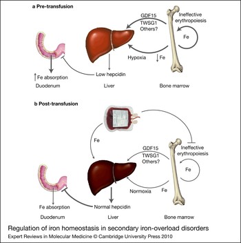
The term secondary iron loading can be applied to a range of iron-overload disorders where the pathway regulating intestinal iron absorption is intact (Ref. Reference Gattermann4). The excess iron in these conditions comes from two main sources: either from increased intestinal iron absorption as a consequence of some underlying pathology or from repeated blood transfusions (transfusional siderosis). Often both routes are involved. The best-characterised disorders of this class are certain diseases of the red blood cell (Ref. Reference Gattermann4). These include those associated with ineffective erythropoiesis (e.g. β-thalassaemia, sideroblastic anaemia), chronic haemolytic anaemias (e.g. pyruvate kinase deficiency, sickle cell anaemia) or hypoplastic anaemias (e.g. chronic renal failure, aplastic anaemias). Repeated blood transfusions are usually the main source of the iron in these conditions, but in those associated with ineffective erythropoiesis, iron absorption is often markedly elevated (Ref. Reference Rechavi and Rivella5) (Fig. 3). Indeed, in some disorders, such as β-thalassaemia, heavy iron loading can occur even in the absence of transfusion.
Regulation of iron homeostasis in secondary iron-overload disorders. (a) Pre-transfusion. Ineffective erythropoiesis in various red blood cell disorders leads to an increase in iron recycling in the bone marrow, an increase in the expression of the erythropoiesis-related factors TWSG1 and GDF15, and a decrease in serum iron and oxygenation. These factors combine to reduce hepcidin expression in the liver and increase iron absorption in the duodenum. (b) Post-transfusion. Transfusion of blood leads to suppression of ineffective erythropoiesis, lower levels of TWSG1 and GDF15, and relative normoxia and serum iron levels. It also contributes to higher iron stores in the liver. These factors combine to increase hepcidin levels and restore iron absorption to normal levels. Abbreviations: GDF15, growth differentiation factor 15; TWSG1, twisted gastrulation 1.

In patients with various iron-loading anaemias, iron enters the reticuloendothelial system as defective erythrocytes are cleared from the marrow or from the circulation. The same fate befalls transfused red cells as they age and are processed by tissue macrophages. In the early stages of the disease, the iron released from these erythrocytes is recycled back to the bloodstream and is reutilised as part of the enhanced erythropoiesis associated with these conditions. However, because humans do not possess a mechanism for the active secretion of iron (Ref. Reference McCance and Widdowson42), the extra iron derived from transfusions accumulates. Initially, iron accumulation is observed in macrophages, but over time deposition in parenchymal cells develops as well. In secondary iron-loading disorders, cardiac toxicity tends to be more prominent compared with the predominantly hepatic manifestations associated with primary iron loading (Ref. Reference Gattermann4). Nevertheless, hepatic and endocrine dysfunction can also occur in transfusional siderosis. Patients with secondary iron loading cannot be treated with phlebotomy therapy, so the excess iron is removed by chelation therapy (Ref. Reference Gattermann4). For many years the most commonly used chelator was desferrioxamine, which required prolonged subcutaneous infusion. However, in recent years the orally active and highly effective chelator deferasirox has come into widespread use (Refs Reference Yang, Keam and Keating43, Reference Vichinsky44). An intriguing recent finding is that administration of transferrin (either apo or holo protein) was able to ameliorate many of the pathological features of β-thalassaemia in a murine model (Ref. Reference Li45). How transferrin exerts these effects is unclear, but alterations in marrow iron supply and effects on hepcidin synthesis could both be involved.
In addition to the disorders described in this and the previous section, there are several miscellaneous forms of iron overload of varying aetiologies, but most of these are uncommon to rare. These include medicinal iron overload (a classical, but rare, form of secondary iron loading) (Ref. Reference Wheby46), African iron overload (which appears to have both genetic and environmental components) (Ref. Reference Gordeuk47), rare diseases of plasma protein synthesis, such as congenital atransferrinaemia (Ref. Reference Hamill, Woods and Cook48) and aceruloplasminaemia (Ref. Reference Morita49), and neonatal haemochromatosis (Ref. Reference Whitington50). In each of these situations severe iron loading can develop. Less substantial iron loading is associated with chronic liver disease (e.g. alcoholic liver disease, fatty liver disease, dysmetabolic iron-loading syndrome) (Ref. Reference Deugnier and Turlin51) and porphyria cutanea tarda (Ref. Reference Sarkany52). A full discussion of each of these disorders is beyond the scope of this review, but disturbances in hepcidin regulation have an important role in a number of them.
Mechanisms of body iron loading: central role of hepcidin
A common feature of primary iron-loading disorders is increased intestinal iron absorption. Under these conditions, the normal mechanism that limits the absorption of excess iron when the body is iron replete is dysfunctional and absorption continues in the face of adequate or even elevated iron stores (Ref. Reference Hutchinson53). In subjects with inherited erythroid disorders characterised by iron loading, iron absorption is also increased (Ref. Reference Rechavi and Rivella5). In these cases, however, the high absorption is a normal physiological response to the accelerated erythropoiesis associated with these conditions. Despite the importance of this process, few studies have examined iron absorption per se in primary haemochromatosis and essentially all the work that has been done in humans has been carried out in HFE-related haemochromatosis. An important study by McLaren et al. showed that for a given level of body iron, the amount of iron absorbed by the small intestine is several times higher in patients with HFE-related haemochromatosis than in normal individuals (Ref. Reference McLaren54). Interestingly, the normal reciprocal relationship between body iron stores and iron absorption (i.e. higher iron stores means lower iron absorption) was still observed in such patients, and the slope of the correlation line was very similar to that of controls. However, the entire curve was shifted to the right, such that a given amount of iron repressed iron absorption less in subjects with HFE-related haemochromatosis than in normal controls. This shows that such patients are able to respond to increases in body iron load, but that the sensitivity of the intestinal response to increased body iron is reduced. Although it has not been examined directly, a similar response might be expected in patients with TFR2 mutations. However, in patients with much more severe iron loading, such as subjects with HJV or HAMP mutations (juvenile haemochromatosis), it is unlikely that significant regulation of iron absorption remains (Ref. Reference Cazzola55).
As described above, the primary systemic regulator of intestinal iron absorption is the liver-derived peptide hepcidin (Ref. Reference Nemeth and Ganz23). This is best demonstrated in primary iron-loading disorders. Human subjects with HAMP mutations have very low or no circulating hepcidin (Ref. Reference Roetto40), and hence iron absorption continues unimpeded. In a normal individual, hepcidin levels would increase as body iron accumulates, and this would feed back to the gut to limit further iron absorption. It comes as no surprise that other forms of primary iron loading are also characterised by disruption of the hepcidin–FPN axis (Fig. 2). Patients with mutations in HFE, TFR2 and HJV also have low or negligible hepcidin expression (Refs Reference Kawabata34, Reference Papanikolaou39, Reference Bridle56), implicating these molecules in the regulation of hepcidin and explaining why they cannot limit their iron intake. Further evidence has recently come from studies in mice: targeted ablation of HFE in liver recapitulated the haemochromatosis phenotype, whereas ablation of HFE in intestine did not (Refs Reference Spasic57, Reference Vujic Spasic58). This, in conjunction with the lower hepcidin levels seen in Hfe-knockout mice, clearly demonstrates that HFE has a role in the regulation of hepcidin expression in the liver rather than directly regulating iron absorption in the intestine. In those subjects with ferroportin disease resembling classical primary iron overload, the mutations in FPN interfere with hepcidin binding to the exporter and effectively render hepcidin inactive (Ref. Reference Lee and Beutler3).
BMP–SMAD signalling and hepcidin regulation
The low hepcidin levels in patients with HFE, TFR2 and HJV mutations show that these proteins are upstream regulators of hepcidin. The dissection of how HJV alters hepcidin expression has been the most revealing to date. HJV is a glycosylphosphatidylinositol (GPI)-linked membrane-associated protein that acts as a co-receptor for the bone morphogenetic protein (BMP) signalling pathway (Fig. 4), and when signalling through this pathway is increased, hepcidin expression also increases (Ref. Reference Babitt59). The BMPs are members of the transforming growth factor β (TGF-β) superfamily of proteins, and they bind to a complex of type I and type II BMP receptors on the plasma membrane (Ref. Reference Chen, Zhao and Mundy60). The type II receptors then phosphorylate the type I receptors, and this activated complex in turn phosphorylates the transcriptional regulators SMAD1, -5 and -8 (small mothers against decapentaplegic homologues) in the cytoplasm. HJV binds to the BMPs and enhances their effectiveness in activating the SMAD signalling pathway (Ref. Reference Babitt59). In hepatic cells, BMPs 2, 4 and 6 appear to be the endogenous ligands for HJV (Ref. Reference Xia61). Interestingly, the absence of HJV leads to a reduction in basal hepcidin expression rather than a change in the responsiveness of HAMP to BMPs (Ref. Reference Truksa62). Activated SMAD1/5/8 binds to SMAD4, and this complex translocates to the nucleus where it stimulates the expression of a range of target genes, including HAMP (Refs Reference Babitt59, Reference Chen, Zhao and Mundy60) (Fig. 4). The finding that mice lacking SMAD4 have negligible hepcidin expression and develop body iron loading (Ref. Reference Wang63) provides very strong evidence that the BMP–SMAD signalling system has a central role in hepcidin regulation. Two other SMADS, SMAD6 and SMAD7, are able to antagonise SMAD1/5/8 binding to SMAD4, and recent data have shown that SMAD7 inhibits HAMP gene expression (Ref. Reference Mleczko-Sanecka64). Thus the SMAD–BMP pathway has emerged as a central player in the regulation of hepcidin.
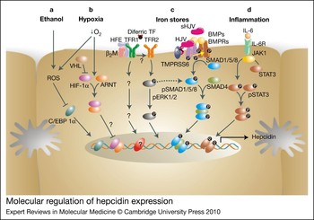
Molecular regulation of hepcidin expression. In hepatocytes, hepcidin can be regulated by ethanol, hypoxia, iron stores and inflammation. (a, b) C/EBPα binds to the promoter and stimulates hepcidin expression. Ethanol and hypoxia induce ROS, downregulate C/EBPα binding and thus lead to suppression of hepcidin. (b) Under hypoxic conditions, HIF-1α is stabilised, forms a heterodimer with ARNT, translocates to the nucleus and downregulates hepcidin expression. VHL destabilises HIF-1α by marking it for ubiquitination and degradation. (c) Diferric transferrin stimulates hepcidin expression, probably through its interactions with TFR2 and the HFE–TFR1 complex on the cell surface. Signalling through the ERK pathway appears to be involved. BMPs, notably BMP6, bind to BMPRs and the co-receptor HJV to induce signalling through the SMAD pathway to modulate hepcidin expression. TMPRSS6 downregulates hepcidin expression by cleaving HJV. (d) Inflammatory cytokines, such as IL-6, bind to their receptors (IL-6R) and stimulate STAT3 to induce hepcidin expression. Abbreviations: ARNT, aryl hydrocarbon receptor nuclear translocator; BMPRs, BMP receptors; BMPs, bone morphogenetic proteins; C/EBPα, CCAAT/enhancer-binding protein alpha; pERK1/2, phosphorylated extracellular-signal-regulated kinase 1/2; HIF-1α, hypoxia-inducible factor 1α; HJV, haemojuvelin; IL-6, interleukin-6; JAK1, Janus kinase 1; β2M, β2-microglobulin; ROS, reactive oxygen species; SMAD, small mothers against decapentaplegic homologue; STAT, signal transducer and activator of transcription; TF, transferrin; TFR, transferrin receptor; TMPRSS6, transmembrane protease, serine 6; VHL, von Hippel–Lindau.

Signalling through the BMP–SMAD pathway can be modulated through a range of upstream events, and thus the regulation of hepcidin is potentially quite complex. BMPs 2, 4, 5, 6, 7 and 9 have all been shown to be able to stimulate HAMP expression (Refs Reference Babitt59, Reference Wang63, Reference Babitt65), but recent studies have suggested that BMP6 is physiologically the most important (Figs 2 and 4). In the absence of BMP6, mice develop systemic iron loading and hepcidin is barely detectable (Refs Reference Andriopoulos66, Reference Meynard67), suggesting that the other BMPs cannot compensate for the loss of BMP6. In addition, the administration of exogenous BMP6 increases hepcidin expression (Ref. Reference Andriopoulos66). BMP6 itself is regulated by iron (Ref. Reference Kautz68), and this provides at least one mechanism by which body iron stores may be communicated to the hepcidin-regulatory machinery. When the body iron load is high, BMP6 levels increase, leading to enhanced BMP–SMAD signalling and increased hepcidin expression. Intriguingly, BMP6 does not appear to be regulated by HFE, TFR2 or HJV because knockout mouse studies show that Bmp6 expression correlates with iron stores in these animals as normal (Refs Reference Lee69, Reference Zhang70, Reference Du71). This implies that HFE and TFR2 regulate hepcidin expression in a BMP6-independent manner. Because HJV acts as a BMP co-receptor, it would not be expected to affect Bmp6 expression, but instead potentiates the signal produced when BMP6 binds to its receptor (Ref. Reference Du71).
Of additional interest is the potential role of BMP9 in the regulation of hepcidin. Tissue culture studies have shown it to be the most potent stimulator of hepcidin expression of all the BMPs tested and it is almost exclusively expressed in the nonparenchymal cells of the liver (Refs Reference Kautz68, Reference Lin, Goldberg and Ganz72). However, it does not appear to bind HJV and is not regulated by iron stores (Ref. Reference Andriopoulos66), implying that BMP9 induces hepcidin expression by a mechanism different from that of BMP6. Whether BMP9 has a role in regulating hepcidin expression in vivo remains to be determined. It is likely that other iron-related signals also influence hepcidin expression, and these are considered in more detail below.
It is clear that the GPI-linked, membrane-associated form of HJV acts as a BMP co-receptor to stimulate hepcidin expression. However, a soluble form of HJV (sHJV) has been identified in the circulation and it has been shown to antagonise signalling through the BMP–SMAD pathway, presumably by competitively inhibiting the binding of membrane-bound HJV to BMPs (Ref. Reference Lin, Goldberg and Ganz72) (Fig. 2). The source of sHJV is unclear. Although HJV is expressed at reasonable levels in the hepatocyte, the site of expression of most components of the hepcidin-regulatory pathway, it is much more strongly expressed in skeletal and cardiac muscle (Ref. Reference Papanikolaou39). Muscle has a particularly high iron requirement for the production of myoglobin; hence it is possible that muscle-derived sHJV reflects the iron requirements of this tissue and communicates it to the liver. However, this remains speculation at present. The release of sHJV requires the protease furin (Ref. Reference Silvestri, Pagani and Camaschella73) (Fig. 2). Furin levels can be increased by iron depletion and hypoxia and this can explain why levels of sHJV increase under iron-deficient conditions (Ref. Reference Silvestri, Pagani and Camaschella73), a situation conducive to reduced HAMP expression. It has been reported that the release of sHJV also requires the membrane protein neogenin (Ref. Reference Zhang74), but more recent studies have suggested that neogenin stabilises GPI-linked HJV and inhibits its secretion (Ref. Reference Lee69) (Fig. 2). This is consistent with the observation that neogenin is required for HJV-mediated BMP signalling (Ref. Reference Zhang70) and that neogenin-knockout mice have low hepcidin levels and develop iron overload (Ref. Reference Lee69).
The most recent member of the hepcidin-regulatory network to be identified is a membrane-bound serine protease known as matriptase-2 (TMPRSS6). The link between this enzyme and iron homeostasis was first made through two mouse models of anaemia with impaired matriptase-2 function (Refs Reference Du71, Reference Folgueras75), and has been consolidated with the finding that many cases of refractory iron deficiency in humans can be ascribed to mutations in the TMPRSS6 gene (Refs Reference Finberg76, Reference Guillem77). Significantly, several recent genome-wide association studies in humans seeking genetic determinants of iron status have all identified matriptase-2 as a strong candidate (Refs Reference Chambers78, Reference Ganesh79, Reference Tanaka80). The physiological substrate of matriptase-2 appears to be membrane-associated HJV (Ref. Reference Silvestri81) (Fig. 2). When HJV is cleaved, it can no longer act as a BMP co-receptor and hepcidin levels are reduced (Ref. Reference Finberg82). This implies that the normal function of this protease is to repress hepcidin production through the BMP–SMAD pathway. Overall, these data suggest that matriptase-2 has a crucial role in maintaining basal hepcidin expression.
Hepcidin regulation by erythropoiesis and inflammation
Strongly enhanced erythropoiesis is a major stimulus for iron absorption and an important suppressor of hepcidin expression (Ref. Reference Frazer83) (Fig. 3). This is particularly relevant to understanding the iron loading associated with a range of haematological disturbances where erythropoiesis is ineffective and the erythropoietic drive is very strong. There are several potential mechanisms for the suppression of HAMP by erythropoiesis. First, increased erythropoiesis creates a high demand for iron and reduced systemic iron levels could feed back to alter HAMP transcription (Ref. Reference Nemeth and Ganz23). The iron-related regulation of hepcidin could occur through the modulation of BMP6 or FURIN expression, or through HFE and TFR2 as discussed in the following section. Second, a reduction in the level of circulating haemoglobin can lead to systemic hypoxia, and this could also reduce hepcidin expression (Ref. Reference Peyssonnaux84). The HAMP gene contains two hypoxia-response elements, and it has been demonstrated that HIF-1α (HIF1A) signalling is able to reduce HAMP transcription (Ref. Reference Peyssonnaux84) (Fig. 4). In addition to iron- and oxygen-related signals, there is evidence that the erythroid-specific factors growth differentiation factor 15 (GDF15) and twisted gastrulation (TWSG1) are also involved in hepcidin regulation. Their expression is increased when erythropoiesis is stimulated, and each has been shown to act on hepatic cells to reduce hepcidin expression (Refs Reference Tanno85, Reference Tanno86) (Fig. 2). However, one study failed to find a correlation between GDF15 and hepcidin during the recovery of erythropoiesis following bone marrow transplantation (Ref. Reference Kanda87). The expression of GDF15 and TWSG1 is particularly high in β-thalassaemia where hepcidin levels are low and iron absorption is strongly upregulated (Refs Reference Tanno85, Reference Tanno86). Precisely how these proteins exert their effects has yet to be determined, but it is likely that they interfere with BMP signalling (Refs Reference Chang88, Reference Ross89).
Inflammation is a strong inducer of hepcidin (Ref. Reference Nicolas90), and the hypoferraemia that accompanies inflammation can be explained by increased plasma hepcidin levels. The anaemia associated with a range of chronic inflammatory conditions, known as the anaemia of chronic disease (ACD), can be explained by increased hepcidin levels, and therefore antagonism of the hepcidin–ferroportin axis (Refs Reference Nemeth and Ganz23, Reference Agarwal and Prchal91) represents an important therapeutic avenue for these conditions. Inflammation stimulates the synthesis of a range of pro-inflammatory cytokines such as interleukin-6 (IL-6), and it is these that enhance hepcidin expression by signalling through the JAK–STAT pathway (Ref. Reference Wrighting and Andrews92) (Fig. 4). There appears to be crosstalk between this pathway and the BMP–SMAD signalling pathway as a functional SMAD-binding site in the HAMP promoter is necessary for the IL-6 stimulation of hepcidin expression (Ref. Reference Wang63).
Role of HFE and TFR2 in sensing body iron levels
How the iron-regulatory system, and specifically hepcidin, responds to changes in body iron status remains unclear. As noted above, BMP6 and furin are regulated by body iron levels and provide some potential mechanisms for relaying body iron requirements to this regulatory system. However, it is widely believed that HFE and TFR2 are also involved in the iron-sensing process. How do they do this and what do they sense? There are several lines of evidence to suggest that circulating diferric transferrin is an important signal in regulating hepcidin expression (Refs Reference Frazer83, Reference Wilkins93) (Fig. 4). Under normal physiological conditions, almost all the iron in the blood is bound to transferrin (Ref. Reference Huebers and Finch20). Transferrin contains two iron-binding sites and these may be unoccupied (apotransferrin), partly occupied (monoferric transferrin) or fully occupied (diferric transferrin). Diferric transferrin is the preferred substrate for TFR1, and thus is the major form in which iron is delivered to cells (Ref. Reference Huebers94). Diferric transferrin levels increase with iron loading and decrease with iron deficiency, and a positive correlation between the concentration of diferric transferrin and hepcidin expression has been demonstrated (Ref. Reference Frazer83). Mice that lack transferrin expression show reduced hepcidin and high iron absorption even in the presence of a high hepatic iron load (Refs Reference Buys95, Reference Weinstein96). This is consistent with a regulatory role for diferric transferrin, but the interpretation is complicated in this case because of the strongly enhanced erythropoiesis and hypoxia in these mice, which could independently regulate hepcidin.
A further link between diferric transferrin and hepcidin regulation comes from the analysis of TFR2 and HFE. TFR2 binds diferric transferrin, and this binding can stabilise the receptor (Ref. Reference Johnson and Enns97); hence it has the potential to act as a sensor of body iron levels. In cell culture studies it has been shown that the TFR2-mediated induction of HAMP requires diferric transferrin (Refs Reference Gao98, Reference Ramey, Deschemin and Vaulont99). HFE itself does not bind diferric transferrin, but is able to bind to TFR1, with the binding site overlapping with one of the diferric-transferrin-binding sites on TFR1 (Ref. Reference West100). Thus, the HFE–TFR1 complex could also act in an iron-sensing role, with diferric transferrin displacing TFR1-bound HFE and leaving it free to initiate a signal to increase hepcidin synthesis. Consistent with such a mechanism is the demonstration that mice engineered to prevent the HFE–TFR1 association showed enhanced hepcidin expression (Ref. Reference Schmidt101). Despite considerable work in this area, much of it using proteins overexpressed in cell lines, we still do not have a clear picture of this iron-sensing mechanism, nor indeed whether diferric transferrin provides the key regulatory signal. For example, some studies have suggested that diferric transferrin will not stimulate HAMP expression in hepatocytes isolated from Hfe- or Tfr2-knockout mice (Ref. Reference Gao98), whereas other studies have suggested that HFE is dispensible (Ref. Reference Ramey, Deschemin and Vaulont99).
Irrespective of the mechanisms by which iron status is assessed by hepatocytes, how do HFE and TFR2 direct changes in HAMP expression? The short answer is that we do not know. It has been proposed that HFE, TFR2, HJV and the BMP–receptor complex combine to form a ‘super’ complex that regulates hepcidin expression through the SMAD pathway (Ref. Reference Andrews102). However, the natural history of primary iron-loading disorders seems to argue against a mechanism where HFE and TFR2 are part of the same pathway (at least the same proximal pathway), and suggests that they perform independent, yet equivalent, roles. Mutations in the genes encoding HFE and TFR2 lead to adult-onset forms of iron overload – much milder disease than that due to mutations in HJV or HAMP (Ref. Reference Lee and Beutler3). In addition, when HFE is mutated (and possibly TFR2), iron absorption can still be regulated, albeit with a reduced sensitivity compared with unaffected individuals (Ref. Reference McLaren54). Furthermore, recent studies have shown that mice lacking both HFE and TFR2 have much lower hepcidin levels and develop more iron loading than animals lacking either gene alone (Ref. Reference Wallace37). These observations suggest that HFE and TFR2 do not act as part of the same pathway, but act independently to modulate hepcidin levels.
It is presumed that HFE and TFR2 activate signal transduction pathways to alter HAMP expression, and TFR2 accumulates in lipid rafts as many signalling proteins do (Ref. Reference Calzolari103). Data from several sources now indicate that HFE and TFR2 can signal through the mitogen-activated protein kinase (MAPK) pathway to increase the phosphorylation of ERK1/2 [extracellular-signal-regulated kinase 1/2 (MAPK1/MAPK3)] (Fig. 4). Levels of phosphoERK are reduced in Hfe- and Tfr2-knockout mice (Ref. Reference Wallace37), and in primary mouse hepatocytes (Ref. Reference Ramey, Deschemin and Vaulont99) or K562 (erythroleukaemia) cells (Ref. Reference Calzolari103) diferric transferrin can increase ERK1/2 phosphorylation. Furthermore, inhibition of ERK can block HAMP stimulation by diferric transferrin (Ref. Reference Ramey, Deschemin and Vaulont99). There are some data that suggest HFE and TFR2 feed into the BMP–SMAD pathway, and reduced phosphorylation of SMAD1/5/8 has been reported in Hfe- and Tfr2-knockout mice (Ref. Reference Wallace37). Mice lacking either HJV or SMAD4 do not show an increase in hepcidin after iron administration (Refs Reference Wang63, Reference Niederkofler, Salie and Arber104), indicating that an intact BMP–SMAD pathway is essential for hepcidin to respond to iron.
Other regulators of hepcidin expression
In addition to the predominantly iron-related regulatory mechanisms, hepcidin expression can also be modified by seemingly unrelated stimuli. The role of hepcidin in iron homeostasis was originally discovered using a mouse model that had upstream stimulatory factor 2 (USF2), a transcription factor involved in carbohydrate metabolism, knocked out. These animals were found to express very low levels of hepcidin and, consequently, develop severe iron loading (Ref. Reference Nicolas105). It was subsequently shown that USF1 and USF2 form heterodimers with MYC/MAX and bind to E-boxes within the promoter region of hepcidin, regulating expression in both a cis and trans manner (Ref. Reference Bayele, McArdle and Srai106). This provides tantalising evidence of a link between iron metabolism and the metabolism of carbohydrate and fat, where USF1 and USF2 are known to play an important role.
Alcohol consumption has been reported to increase body iron stores and can exacerbate the iron loading and morbidity associated with haemochromatosis. Ethanol has been shown to suppress hepcidin expression both in vivo and in vitro (Refs Reference Bridle107, Reference Harrison-Findik108, Reference Flanagan, Peng and Beutler109). It appears that ethanol-induced hypoxia leads to the formation of reactive oxygen species that downregulate hepcidin expression by reducing the DNA-binding activity of CCAAT/enhancer-binding protein alpha (C/EBPα) to the hepcidin promoter. This effect of ethanol on hepcidin expression can be prevented by antioxidants (Ref. Reference Harrison-Findik108) (Fig. 4).
Finally, polymorphic variations in the hepcidin promoter have been reported to regulate hepcidin expression and might have a role in determining the level of iron loading that occurs in primary haemochromatosis (Refs Reference Bayele and Srai110, Reference Andreani111).
Integrating the regulatory network
How these multiple signals integrate to regulate hepcidin expression in health and disease is incompletely understood, although an intact BMP–SMAD pathway seems essential for normal HAMP regulation, irrespective of the stimulus. Some of the components are likely to be involved in maintaining basal HAMP expression, whereas others may be involved in the adaptation to changes in body iron demand. Various arms of the system may respond differently to acute and chronic stimuli. For example, it has been demonstrated that in HFE-related haemochromatosis the acute response to iron administration is missing (Ref. Reference Piperno112) but subjects still respond to chronic iron loading (Ref. Reference van Dijk113). The cellular site of this iron regulatory network also requires further analysis. Hepatic parenchymal cells express all the key components of the system and are probably the main regulatory site, but the finding that important players such as BMP6 and HJV are primarily expressed in other cell types (hepatic stellate cells and myocytes, respectively) (Refs Reference Papanikolaou39, Reference Knittel114) suggests a complex cellular interplay. There is also a growing body of evidence that hepcidin can be synthesised locally in a range of tissues (Ref. Reference Theurl115), with macrophages being a potential source of the peptide in these situations. Finally, it should be pointed out that many of the studies to delineate pathways of hepcidin regulation have been carried out in cell culture models, often using exogenous protein overexpression. Hence these findings need to be validated in vivo.
Clinical implications and applications
Advances in our molecular understanding of the pathogenesis of iron-loading disorders have brought the promise of novel therapeutic approaches for these diseases. The recognition that hepcidin has a central role in regulating body iron intake has meant that most interest has centred on modulating hepcidin levels. However, as yet, none of these approaches has reached the clinic.
Modulating hepcidin levels
Because iron-loading diseases are characterised by reduced hepcidin expression, the administration of hepcidin or its analogues holds great promise in limiting iron loading. When mice overexpressing hepcidin are crossed with those deficient in HFE, iron loading in the Hfe-knockouts is prevented (Ref. Reference Nicolas116), confirming in principle the utility of raising hepcidin levels. Several studies have also shown that administering hepcidin to mice will reduce iron levels (Refs Reference Laftah117, Reference Chung118), and more recently it has been shown that short forms of hepcidin based on the N-terminal portion of the peptide can have similar effects (Ref. Reference Nemeth119). These small-sized or ‘minihepcidins’ have potential as oral therapeutic agents. Hepcidin administration is equally applicable to both primary and secondary iron-loading disorders because hepcidin levels are reduced in each case. Increasing hepcidin levels will reduce or prevent the accumulation of additional iron, but will not necessarily lead to a reduction in existing body iron overload. Hence, it is likely that such therapies will be applied in conjunction with iron chelation to remove existing excess iron and prevent further iron accumulation. To remove accumulated iron, the newer-generation oral iron chelators such as deferasirox are likely to be the agents of choice (Ref. Reference Paradkar120).
It is also possible to increase hepcidin expression by enhancing signalling through the BMP–SMAD pathway. BMP6 is a clear candidate here and injection of Bmp6 has been shown to stimulate hepcidin expression in mice (Ref. Reference Andriopoulos66). Along similar lines, antagonists of matriptase-2 could be used to enhance BMP signalling. In either case, however, the possibility of off-target effects will need to be thoroughly investigated. Gene therapy approaches for specific disorders might also be valuable in the future. For example, the virus-mediated delivery of Hfe and Tfr2 transgenes into hepatocytes has been recently shown to partially correct the defects in Hfe- and Tfr2-knockout mice, respectively (Ref. Reference Gao121). In these mice, serum hepcidin levels increased and transferrin saturation and hepatic iron levels decreased. A similar approach might also be suitable to correct HJV or hepcidin insufficiency. Although these possibilities are worth considering, the most practical solution to ameliorating iron loading in the foreseeable future is to directly administer hepcidin or hepcidin agonists.
Although increasing the hepcidin levels is appropriate for ameliorating iron overload, in other situations it may be desirable to reduce hepcidin levels or to interfere with its action. The anaemia of inflammation or chronic disease is associated with increased hepcidin and can ultimately lead to iron-deficient erythropoiesis, so reducing hepcidin to enhance iron supply could be potentially beneficial. In a murine model of the anaemia of inflammation (Brucella abortus infection), a neutralising monoclonal antibody against hepcidin was shown to reduce the anaemia when used in combination with erythropoietin treatment (Ref. Reference Sasu122). Serum iron levels were returned to near normal. Such approaches could also be valuable in treating patients with chronic kidney disease and those with refractory iron deficiency anaemia (which is frequently caused by matriptase-2 mutations). If the anaemia is secondary to an infection, increasing the serum iron levels by blocking hepcidin needs to be employed cautiously because iron is an important virulence factor for a range of pathogens.
Modulating iron transport
Therapeutics targeting the hepcidin–FPN axis are potentially valuable for limiting body iron intake by reducing the basolateral transport step of iron absorption. It may also be possible to block absorption using oral agents that interfere with iron transport across the brush-border membrane by DMT1, but this has not yet been achieved. Interestingly, however, L-type calcium-channel-blocking drugs such as nifedipine have been reported to modulate the activity of DMT1 systemically. They mobilise liver iron in primary and secondary iron-loading disorders and enhance the urinary excretion of iron (Ref. Reference Ludwiczek123). Other L-type calcium-channel blockers such as amlodipine and verapamil have also been shown to reduce iron-overload cardiomyopathy (Refs Reference Oudit124, Reference Sugishita125) and improve heart function and patient survival.
Hepcidin measurement as a diagnostic tool
Because hepcidin is a central controller of body iron trafficking, its measurement (in plasma or urine) has great potential as a clinical indicator of body iron status and it is almost certain that hepcidin evaluation will be part of a standard haematological/iron workup in the future. Unfortunately, the hepcidin peptide is a difficult antigen to work with and few robust antibodies to the peptide have been generated. Consequently, at the time of writing there is no reliable commercially available immunoassay for the peptide, although several groups have published hepcidin immunoassays as research tools (Refs Reference Ganz126, Reference Grebenchtchikov127, Reference Busbridge128). Despite this limitation, there has been considerable success in developing mass spectrometry (MS)-based methods for measuring hepcidin in biological fluids (Refs Reference Ward129, Reference Kemna130, Reference Murphy131, Reference Li132, Reference Kroot133); several laboratories around the world offer the service of MS-based hepcidin measurement and a growing number of studies using these measurements are appearing (Refs Reference Iqbal134, Reference Peters135, Reference van Deuren, Kroot and Swinkels136). A commercially available assay for the hepcidin precursor prohepcidin is available, but it has been shown to have limited utility (Ref. Reference Young137). Prohepcidin levels do not correspond well with iron absorption, and there is a strong inverse correlation with the levels of the mature peptide.
Research in progress and outstanding research questions
Defining the mechanisms of body iron homeostasis and their relationship to disorders of iron metabolism, particularly iron-loading disorders, is an extremely active field at present and new discoveries are regularly being published. Despite these recent advances, much more remains to be learned in this important field. Perhaps the most significant unresolved question in our understanding of the biology of hepcidin is how HFE and TFR2 modulate expression of the protein. Do they act independently or together? Do they feed into the BMP–SMAD signalling pathway? Do they respond to the same systemic signals? Some of the other significant issues requiring attention are summarised in Box 1. Studies over the past decade have clearly shown the potential of hepcidin-related diagnosis and therapies for iron-overload disorders. Translating this into clinical practice will be the focus of the next decade, and this in turn will be guided by elucidating the unresolved research questions listed above.
Box 1. Outstanding research questions
• How does the hepatocyte sense body iron requirements? Diferric transferrin certainly makes sense as a candidate, but what are the roles of BMP6 and the iron-dependent regulation of furin?
• The concentration of diferric transferrin in the plasma is sufficiently high to seemingly saturate TFR1 and TFR2. Under these circumstances, how can it act to signal tissue iron requirements?
• How does iron regulate BMP6 expression? Does it respond to intracellular iron or extracellular cues?
• What role do cell types other than hepatocytes play in hepcidin regulation? For example, BMP6 appears to be most strongly expressed in hepatic stellate cells.
• How are all the signals that modulate hepcidin expression modulated at the whole-body level? Much of our detailed knowledge at present is derived from cell culture studies, so the in vivo relevance of these findings needs to be tested.
• What is the role of HJV in cardiac and skeletal muscle?
• What are the identities of the genes that modify disease penetrance during iron overload, and particularly in HFE-related haemochromatosis?
• How aggressively should patients with iron overload be treated by phlebotomy? The benefits of removing excess iron are clear, but too much iron depletion could reduce hepcidin and stimulate absorption.
• What is the role of BMP9 in inducing hepcidin? Does it respond to any systemic stimuli that regulate iron homeostasis?
Abbreviations: BMP, bone morphogenic protein; HJV, haemojuvelin; TF, transferrin; TFR, transferrin receptor.
Acknowledgements and funding
G.J.A. is supported by a Senior Research Fellowship from the National Health and Medical Research Council of Australia. We thank the reviewers for their comments and suggestions.