Resources searched: We searched AMED, BNI, EMBASE, HMIC, PsycINFO, MEDLINE from PubMed, CINAHL using the phrase ‘sporadic cjd’.ti,ab, resulting in 723 articles. This search was combined with phrase ‘old’.ti,ab, resulting in 74 articles. The relevant review articles and the reference list of these articles were reviewed.
Introduction
Creutzfeldt-Jakob disease (CJD), one of the most common human prion diseases, can only be diagnosed with certainty after post-mortem examination has confirmed the characteristic spongy appearance of brain tissue, hence the label of spongiform encephalopathy. It is a rare form of adult dementia and is invariably fatal. CJD was first described in the 1920s and there are now four recognized types of the disease: sporadic (85%), genetic (15%), iatrogenic (1%) and variant.Reference Johnson1 Variant CJD tends to affect young people, whereas sCJD, the most common form, usually affects the middle-aged and elderly. It is a rare disorder, arising from a one-in-a-million spontaneous conversion of normal cellular protein into an abnormal three-dimensional configuration. This abnormal molecule, a ‘prion protein’ (PrP), arises either from mutation of the PrP gene (PRNP) or from unidentified environmental prion exposure. This is thought to instigate the changes in cells, leading to aggregates of deposits within the central nervous system (CNS) and resulting in irreversible neuronal degeneration.
Epidemiology
Sporadic CJD is a naturally occurring condition and can be found all over the world. It has no identifiable geographical or ethnic tendency. Each year within the UK, sCJD affects about one in every million people and is responsible for 60–70 deaths. Although sCJD has been known to occur as early as adolescence, it is most common after 45 years of age and the mean age of onset is 60–65 years.Reference Brown, Gibbs, Rodgers-Johnson, Asher, Sulima and Bacote2–Reference Will and Matthews3 Whilst there is a recent case report in a 98-year-old patient,Reference Buganza, Ferrari, Cecchini, Orrico, Monaco and Zanusso4 the age-specific incidence of 400 cases from the UK National Surveillance Unit (studied between 1980 and 1993) fell from 2.28 cases/million/year between 65 and 69 years, to 0.69 cases/million/year between 80 and 84 years, suggesting that there is no evidence of increasing incidence in old age.Reference de Silva, Findlay, Awad, Harries-Jones, Knight and Will5 Men and women are equally affected.Reference Kretzschmar6, Reference Ravilochan and Tyler7
Pathophysiology and pathology
Prion proteins are encoded by a single-copy gene known as PRNP that is present on the short arm of chromosome 20. Except for the codon 129 polymorphism of the PRNP gene, there are no well-established risk factors for sCJD. The function of the normal cellular form of the PRNP gene (PrPc) is unknown, but a high concentration of PrPc is found in neurons. A modified form, prion protein scrapie (PrPsc), aggregates to produce rod-like particles (prion rods). PrPsc is insoluble and relatively protease resistant and accumulates in tissues forming amyloid structures.Reference Knight and Will8 These particles are identified on immunostaining of brain tissue of animals and humans with prion diseases. The mechanism by which the PrPc is modified to the pathogenic form of PrPsc, the method by which PrPsc replicates, and the way PrPsc accumulates in brain leading to neuronal degeneration, is not known.
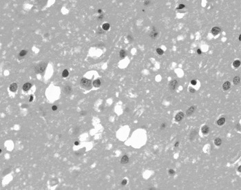
The pathological findings in CJD are limited to the brain, especially the basal ganglia, cerebellum and thalamus. The brainstem and spinal cord are usually spared. There is neuronal loss and vacuolization within the cell bodies and dendrites that gives a spongiform appearance to the cortex.Reference Johnson1 There is hypertrophy and proliferation of giant cells and absence of inflammation and white matter involvement (Figure 1).
High power section of cerebral cortex in sCJD, showing spongiform change (vacuolation) in grey matter and reduction in number of neurons.

The mode of infection for sCJD remains unknown. Exposure to people with the illness, including even intimate exposure, does not appear to increase the risk of contracting the illness. Some case-control studies have suggested a dietary cause, including eating brain, but these may be influenced by recall bias.Reference Bobowick, Brody, Matthews, Roos and Gajdusek9, Reference Davanipour, Alter, Sobel, Asher and Gajdusek10 Furthermore, sCJD has been found in lifelong vegetarians, casting doubt on the role of diet as a mode of infection.Reference Matthews and Will11 Earlier data suggested that the CJD agent may be acquired by inoculation through injury or during surgery and perhaps on certain absorbable sutures of animal origin.Reference Davanipour, Alter, Sobel, Asher and Gajdusek12 Some believe that sCJD may follow exogenous infection, but not many indications of the potential route of entry have been found.Reference Johnson1 However, sCJD can be transmitted among humans by medical procedures.Reference Johnson1
Subtypes of sCJD
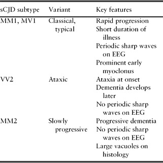
There is marked genetic susceptibility in sCJD, most cases occurring in homozygotes at codon 129 of PRNP, where methionine or valine may be coded. Based on the genotype at codon 129 on both PRNP alleles and the size of the protease-resistant PrPSc fragments, six subtypes have been described, each with distinct clinical and neuropathological features. The most common sCJD subtype (sCJD MM1) and the sCJD MV1 subtype are associated with the classic CJD clinical phenotype.Reference Castellani, Colucci, Xie, Zou, Li and Parchi13 The various molecular subtypes with their respective clinical phenotypes are shown in Table 1.
Subgroups of sCJD

Clinical features
Sporadic CJD typically presents with rapidly progressive dementia (developing over weeks) and myoclonus, with associated cerebellar, pyramidal and extrapyramidal features. A meta-analysis of studies published between 1996 and 2006 identified age of onset as a predictor of presenting symptoms.Reference Appleby, Appleby and Rabins14 Young age (<50 years) predicted symptoms of affective illness, sleep disturbance, poor concentration, cerebellar/gait disturbance, visual/oculomotor disturbance, sensory disorder, vertigo/dizziness, and headache. However, there was no molecular classification, so it is possible that some reported sCJD cases were actually genetic CJD cases, falsely lowering the age of onset in the sCJD group. Furthermore, different molecular subtypes of sCJD may present differently and have varying age of onset and survival times,Reference Hill, Joiner, Wadsworth, Sidle, Bell and Budka15, Reference Appleby, Appleby, Crain, Onyike, Wallin and Rabins16 but the CJD cases included in the meta-analysis did not have genotypes examined.
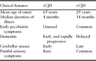
All studies suggest considerable clinical heterogeneity at all ages.Reference Parchi, Giese, Capellari, Brown, Schulz-Schaeffer and Windl17 Thus initial symptoms may include systemic disturbances such as fatigue, decreased appetite, slowness in thinking, difficulty concentrating, mood changes, emotional lability, hallucinations and sleep disturbances. In some cases the onset of illness is so abrupt as to suggest a diagnosis of stroke. Clinical features correlate with the involvement of specific regions of the brain (occipital, thalamic and cerebellar type). Visual disturbances are noted in 40–60% of cases. Less common are sensory, auditory, olfactory or gustatory symptoms of central origin. Progressive insomnia along with psychomotor agitation and visual hallucination may be seen in addition to autonomic disturbances. Epileptiform fits occur in 10–20% of cases. The most common neurological sign is myoclonus, occurring in more than 90% of cases and provoked by startle. With disease progression, an extrapyramidal syndrome often develops and pyramidal signs occur in half of cases. Given the recent interest in vCJD, it is useful to compare the clinical features of the sporadic and variant forms (Table 2).
Clinical features of sCJD and vCJD

The median duration of illness in sCJD is four months. Progression of disease results in death within one year in 90% of cases. A few patients have lived for five years, but there are no reports of any recoveries from the disease.Reference Ravilochan and Tyler7, Reference Prusiner18–Reference Brown, Cathala, Castaigne and Gajdusek20
Diagnostic approach
The diagnosis of sCJD is primarily made on clinical grounds, supported by the results of investigations. The first step is recognizing the possibility of the diagnosis. Barriers to this are the heterogeneity of presentations, the non-specific symptoms and the rarity of the disease. Sporadic CJD should always be considered in the older patient presenting with a short history of progressive dementia, where more common conditions have been excluded. Some authors suggest that the apparent lack of increasing incidence of sCJD in patients aged over 70 years may partially be due to under-diagnosis in older patients with multiple co-morbidities.Reference Cohn, Crimmins, Heard and Rose21
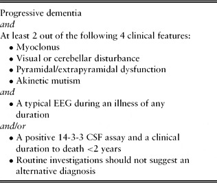
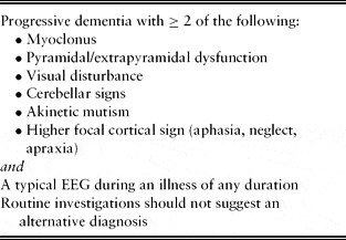
In a suspected case, the diagnostic approach is aimed at ruling out differential diagnoses. This involves detailed review of the case history and then careful selection of investigations, which should be interpreted in the light of the clinical presentation. Diagnostic criteria for ‘possible’ and ‘probable’ CJD have been proposed by the World Health Organisation (WHO) and are shown in Table 3. The WHO criteria are widely accepted, but have significant limitations. They require either a typical electroencephalograph (EEG) during an illness or a positive 14-3-3 CSF assay and a clinical duration to death <2 years. The EEG is not a very sensitive test for CJD, particularly in the early stages.Reference Collins, Sanchez-Juan, Masters, Klug, van Duijn and Poleggi22 Akinetic mutism occurs very late in the disease. Also, WHO criteria combine cerebellar and visual symptoms, yet there is no evidence that they co-occur. To overcome these diagnostic limitations, University of California San Francisco (UCSF) criteria have been developed.Reference Geschwind, Josephs, Parisi and Keegan23 They separate out visual and cerebellar symptoms, add focal cortical symptoms (e.g. aphasia, neglect, apraxia, acalculia), and replace the CSF 14-3-3 test with a brain magnetic resonance imaging (MRI) scan (Table 4).Reference Shiga, Miyazawa, Sato, Fukushima, Shibuya and Sato24–Reference Zerr, Kallenberg, Summers, Romero, Taratuto and Heinemann26 A definitive diagnosis of sCJD can only be made based on histopathological findings.
WHO diagnostic criteria for probable sCJD

UCSF diagnostic criteria for probable sCJDReference Geschwind, Josephs, Parisi and Keegan23

Differential diagnosis
Differential diagnosis is dependent on the presenting symptoms. Early, non-specific symptoms can lead to speculation about a wide variety of disorders that could be psychiatric, infective, inflammatory or neoplastic. The onset of cognitive decline may raise the suspicion of commoner causes of dementia, such as Alzheimer's disease, Lewy-body dementia, fronto-temporal dementia and vascular dementias. Careful review of history, particularly the rapid decline and the presence of atypical features, can help refute these possibilities. If focal neurological signs are present, structural brain lesions might be considered.
As the disease evolves and initial investigations prove negative, the possibility of CJD may become more apparent. Familial, iatrogenic and even sCJD then need to be considered. Review of family history and past exposures, and ultimately genetic analysis, may be helpful.
Investigations
Investigations remain of greatest use in excluding differential diagnoses. In sCJD, all routine blood tests, electrocardiographs and chest X-rays are normal, as are virology, immunology and auto-antibody screening. Computerized tomography (CT) of the head is unremarkable in the majority of cases, but after six months or more from symptom onset it may show subtle cerebral or cerebellar atrophy.Reference Galves and Carter27 CT is always useful to exclude structural brain disease and obvious cerebral atrophy associated with other forms of dementia in early disease. Characteristic EEG appearances and cerebrospinal fluid (CSF) findings in sCJD are recognized in the WHO diagnostic criteria for ‘probable sCJD’. MRI changes in sCJD are recognizedReference Shiga, Miyazawa, Sato, Fukushima, Shibuya and Sato24–Reference Zerr, Kallenberg, Summers, Romero, Taratuto and Heinemann26 and are included in UCSF, but not WHO diagnostic criteria.
EEG
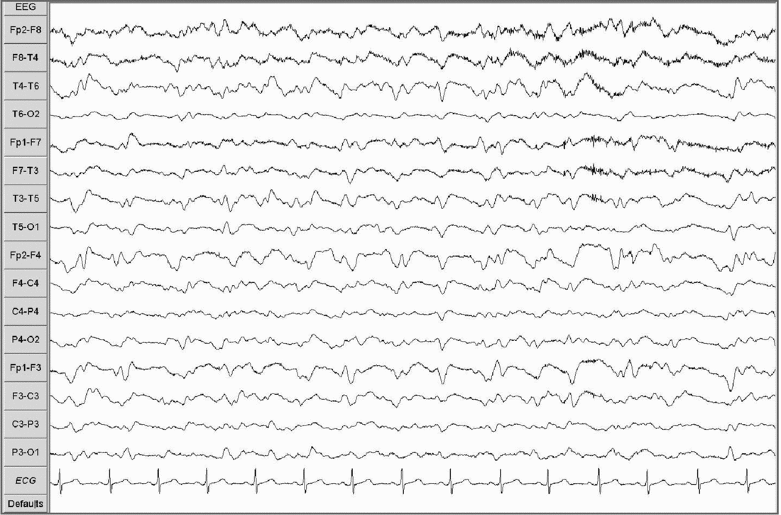
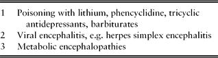
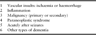
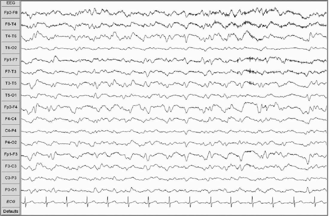
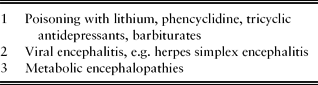
The typical EEG pattern is of generalized bi-, tri- or polyphasic periodic sharp wave complexes (PSWC), with duration of 200–600 ms, occurring at a frequency of 1–2 per second (Figure 2). This finding has a reported sensitivity of 67% and specificity of 86% for sCJD.Reference Steinhoff, Racker, Herrendorf, Poser, Grosche and Zerr28 In cases where no PSWCs are found, there may be non-specific degradation of normal complexes. Other conditions where PSWCs may be found are listed in Table 5.
EEG showing periodic sharp wave complexes 1–2 per second (settings: sensitivity 100 μV/cm, time constant 0.30 s, filter 70 Hz).
Differential diagnosis of PSWCs on EEG
CSF
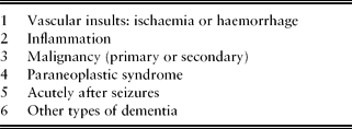
CSF microscopy, biochemistry, cytology and virology are usually normal, although pleocytosis has been noted in a minority of cases. There is no growth on cultures. Routine CSF examination is primarily undertaken to exclude intracranial infection or inflammatory diseases. CSF protein analysis can be performed at specialist laboratories such as the National CJD Surveillance Unit in Edinburgh. The most useful marker is the 14-3-3 protein, a normal neuronal protein that is released acutely from damaged neurons. It is therefore non-specific to sCJD. It contributes to the diagnosis only where the clinical picture is compatible and other conditions in which raised 14-3-3 can occur can be excluded (Table 6). In the correct clinical context it has a reported sensitivity and specificity of around 95%. Additionally, sensitivity varies in subtypes of sCJD, with higher sensitivity in patients with classical molecular features of sCJD than non-classical CJD subtypes.Reference Castellani, Colucci, Xie, Zou, Li and Parchi13 Other protein markers that are potentially useful for diagnosis are C100B, NSE, S-100 and tau protein. None of these has such a high sensitivity and specificity as the detection of 14-3-3 protein, so they are of little diagnostic value at the present time.
Differential diagnosis of 14-3-3 protein in CSF
MRI
The commonest MRI feature is hyperintensity in the caudate head and putamen. Basal ganglia hyperintensity has been reported as 67% sensitive and 93% specific for sCJD, though this is likely to be observer dependent.Reference Schroter, Zerr, Henkel, Tschampa, Finkenstaedt and Poser29 MRI changes may occur before EEG changes.Reference Onofrj, Fulgente, Gambi and Macchi30, Reference Tartaro, Fulgente, Delli Pizzi, Bonomo, Bocola and Onofrj31 Diffusion-weighted MRI can detect characteristic lesions in the majority of patients with CJD, irrespective of the presence of PSWCs.Reference Shiga, Miyazawa, Sato, Fukushima, Shibuya and Sato24, Reference Young, Geschwind, Fischbein, Martindale, Henry and Liu25 Other reported changes include apparent accentuation of hyperintensity of the anterior half of the putamen relative to the posterior half, and an increase in the size and number of Virchow-Robin spaces.Reference Collie, Sellar, Zeidler, Colchester, Knight and Will32
Histology
Tissue for histological examination can be obtained either on brain biopsy or at post-mortem examination. Typical findings are spongiform changes, astrocyte gliosis and neuronal loss. The identification of the PrPsc in brain tissue by immunocytochemisty clinches the diagnosis.
Management
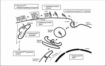
There is no proven treatment of sCJD,Reference Korth and Peters33 but recent major advances in potential pharmacological therapies, some of which are undergoing clinical evaluation, seem promising. In order to understand the mode of action of some of these therapies, one needs to understand the ‘prion life cycle’ (Figure 3).
Schematic drawing of the life of the prion protein (PrP) inside the cell, its conversion to infectious prions, and cell biology-based possibilities of pharmacological intervention. Reproduced with kind permission from Korth et al. Arch Neurol 2006; 63: 497–501 (copyright © 2006, American Medical Association. All rights reserved).

Normal PrP derives from a gene that is expressed by most cells of the body, with particularly high levels in the CNS and lymphatic organs. After protein synthesis, PrP is post-translationally processed before it is secreted to the plasma membrane where it sorts into cholesterol-rich detergent-resistant membrane (CR-DRM) domains. It is this anchored PrP that can adopt a pathological and infectious conformation by a mutation in its amino acid sequence. It is thought that a spontaneous switch into the ‘wrong’ conformation occurs in sCJD and that simultaneously cellular or subcellular clearance mechanisms that would normally degrade these mis-folded proteins are impaired.Reference Korth and Peters33
Once a critical amount of abnormal PrP is present it induces normal PrP molecules to adopt an abnormal conformation.Reference Pruisner34 Signals involving yet unidentified pathways are transduced, resulting in neuronal dysfunction and cell death. This process is thought by some, however, not to be irreversible and that targeted pharmacological anti-prion therapies may be of benefit.Reference Mallucci, Dickinson, Linehan, Klohn, Brandner and Collinge35
Some examples of these potential therapies are:
1. Protection and shielding of normal PrP from conversion to the abnormal form by antibodies. This is a powerful strategy in cell and animal models. The problem arises in producing antibodies that can cross the blood–brain barrier.Reference White, Enever, Tayebi, Mushens, Linehan and Brandner36
2. Intactness of the CD-DRM domains is mandatory for PrP conversion. Its destabilization is thought to prevent conversion and abnormal PrP replication. However, interference with CD-DRMs that execute essential cellular functions may cause adverse effects.Reference Korth and Peters33 This is thought to be the anti-prion effect of quinacrine (a previously used anti-malarial drug). There are currently several ongoing trials investigating its use as a potential treatment.Reference Korth and Peters33
3. Inactivating or shielding the abnormally produced PrP such that it is rendered conversion-incompetent seems to be the most obvious strategy. Many agents, or polyanions (e.g. congo red, dextran, heparin sulphate, pentosan polysulfate) have anti-prion activity in cell and animal models. The presumed mechanism of action is thought to be due to direct action on the abnormally produced PrP.Reference Korth and Peters33 Because pentosan polysulfate is used in the treatment of interstitial cystitis and other urological disorders, this could be a likely agent.Reference Stewart, Rydzewska, Keogh and Knight37 However, problems still arise from these agents’ lack of blood–brain barrier permeability.
4. The neurotoxic effects of the abnormal PrP ultimately lead to the clinical symptoms of CJD. This is thought to be the mechanism by which flupirtine (a N-methyl-D-aspartate antagonist) functions. In a recent double-blind placebo-controlled trial, CJD patients treated with this agent showed some cognitive improvements, but no prolongation of survival times.Reference Otto, Cepek, Ratzka, Doehlinger, Boekhoff and Wiltfang38
It seems that, in the quest for potential future pharmacological treatments, the two aspects that will become paramount are developing strategies to counteract abnormal PrP-induced neurotoxicity and developing diagnostic tests for early or asymptomatic CJD.Reference Korth and Peters33
Future developments
Most of the advances in the field of CJD have occurred in diagnostic and therapeutic areas, as described above. The accumulation of abnormal prion protein (PrPsc) in the brain is a pathological event unique to prion diseases.Reference Pruisner34 Hence PrPsc remains the main target for in vivo tests for CJD. The presence of PrPsc in the urine of patients with familial and sCJD has been reported.Reference Shaked, Shaked, Kariv-Inbal, Halimi, Avraham and Gabizon39 However, subsequently other studies failed to confirm PrPsc in urine samples of sCJD.Reference Head, Kouverianou, Taylor, Green and Knight40 Ritchie et al. have demonstrated the use of a modified technique using the paraffin-embedded tissue blot (PET blot) in diagnosis of sCJD.Reference Ritchie, Head and Ironside41 Zanusso et al. isolated PrPsc from the neuroepithelium of olfactory mucosa in nine cases of sCJD, raising hopes for the use of nasal biopsy as a diagnostic method.Reference Zanusso, Ferrari, Cardone, Zampieri, Gelati and Fiorini42, Reference Tyler43
During the past 10 years, significant progress has been made in improving the sensitivity and specificity of tests using brain tissues to identify CJD-infected individuals, but most of the tests are related to identification of vCJD. The search for a blood test is still in its infancy and requires further research.Reference Cervenakova and Brown44
Conflicts of interest
The authors report no conflicts of interest.