Introduction
The infection of a host with the nematodes of the genus Trichinella follows either predation of an infected animal or scavenging of an infected carcass (Campbell, Reference Campbell and Campbell1983). In Papua New Guinea (PNG), Trichinella papuae circulates among wild and sometimes domestic pigs and farmed saltwater crocodiles (Crocodilus porosus) (Pozio et al., Reference Pozio, Owen, La Rosa, Sacchi, Rossi and Corona1999, Reference Pozio, Owen, Marucci and La Rosa2005; Owen et al., Reference Owen, Sims, Wigglesworth and Puana2000). At present, this infection is known to occur only in two PNG regions, Kikori, Gulf Province, where wild pigs and farmed saltwater crocodiles were infected (Pozio et al., Reference Pozio, Owen, Marucci and La Rosa2005), and Morehead District, Western Province, where this parasite was documented in pigs (both domestic and wild) by digestion of muscle tissues (Owen et al., Reference Owen, Sims, Wigglesworth and Puana2000) and in humans by serology (Owen et al., Reference Owen, Gomez Morales, Pezzotti and Pozio2005). The only predators of pigs are humans and possibly crocodiles. This means that a pig-to-pig cycle occurs in these localities through scavenging, as PNG has no large native terrestrial carnivores.
Trichinella papuae is a non-encapsulated species, lacking the thick-walled collagen capsule that characterizes many other species of the genus Trichinella (Pozio & Murrell, Reference Pozio and Murrell2006). The presence of a capsule gives a degree of protection and extends the survival time of larvae of encapsulated species in host carcasses (Campbell, Reference Campbell and Campbell1983; Stewart, Reference Stewart and Campbell1983; Von Köller et al., Reference Von Köller, Kapel, Enemark and Hindsbo2001). The larvae of non-encapsulated species, such as T. papuae, are more vulnerable to adverse conditions found in a decaying carcass (Stewart et al., Reference Stewart, Kennedy and Larsen1990; Von Köller et al., Reference Von Köller, Kapel, Enemark and Hindsbo2001).
Environmental temperature and humidity play an important role in the transmission of Trichinella sp., because they influence the survival of infective larvae in host carcasses (Pozio & Murrell, Reference Pozio and Murrell2006). It was of interest, therefore, to determine how long T. papuae larvae remain infective in a pig carcass under the influence of the environmental conditions occurring in PNG.
Materials and methods
As it was not possible to undertake the investigation in one of the two endemic regions, it was carried out under simulated natural conditions at the National Veterinary Laboratory, Port Moresby. The laboratory and Bula Plain lie on approximately the same latitude (9°S), 650 km apart. Both localities are a few metres above sea-level, and have a similar dry savannah environment with distinct wet and dry seasons.
A weaned piglet (weight 10 kg), purchased from a commercial piggery, was infected with 1500 larvae of T. papuae (isolate code ISS 1491) collected from a laboratory rat, previously infected with larvae obtained from a naturally infected wild pig from Bula Plain, Western Province. Twenty-one weeks later, coinciding with the dry season (October), the pig (weighing 136 kg) was killed and eviscerated. Owing to the size of the pig carcass and the need to protect it from scavengers, the body was cut into three sections. Two strong, fine-mesh wire baskets were used to enclose, separately, the fore and mid sections; the hind section was destroyed. The baskets were placed on a platform 2 m above the ground in an open, fenced area.
A continuous recording thermometer probe was placed inside one carcass section, while thermometers placed under shade nearby recorded maximum and minimum environmental temperatures. The relative humidity (RH) was obtained from a meteorological station located 3 km from the study site.
On the day of slaughtering, 10.0 g samples of each of eight porcine tissues (tongue, masseter, diaphragm, intercostal, back, neck, upper fore and hind limb muscles) were digested, following the standard procedure (Gamble et al., Reference Gamble, Bessonov, Cuperlovic, Gajadhar, van Knapen, Noeckler, Schenone and Zhu2000), and the recovered larvae were counted.
On days 4, 7, 9, 11 and 14 after slaughtering, from 18.5 to 30.0 g muscle samples, comprising of two or three different pig tissues, were minced, digested, and the larvae, placed in phosphate-buffered solution, were given by a gavage needle to laboratory mice. The degeneration of the carcass tissues meant that on day 14 post slaughtering (p.s.), the only available samples were from shoulder and back muscles. After day 14 p.s., all muscle tissues had disintegrated and liquefied and sampling was not possible.
The mice that received larvae from digested pig tissues collected at different days p.s. were killed from 72 to 76 days post infection. Each mouse carcass was skinned, eviscerated and digested. The total number of larvae collected from each mouse, as well as the reproductive capacity index (RCI, number of larvae collected/number of larvae given) was determined.
Results
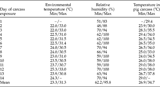
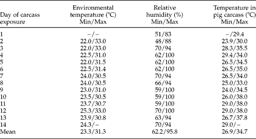
The temperature probe in the carcass recorded a mean maximum temperature of 34.7°C (range 29.4–38.0°C) and a mean minimum temperature of 26.9°C (range 23.9–29.4°C) (table 1). Although RH reached 100% on several days at the meteorological station, no rain fell at the trial site.
Environmental temperature and relative humidity in the locality of the experiment and temperature in the pig carcass.
Of the ten pig tissues digested on the day of slaughter, the diaphragm had the highest number of larvae, followed in descending order by neck, tongue, masseter, intercostal, upper hind leg, back and upper fore leg muscles.
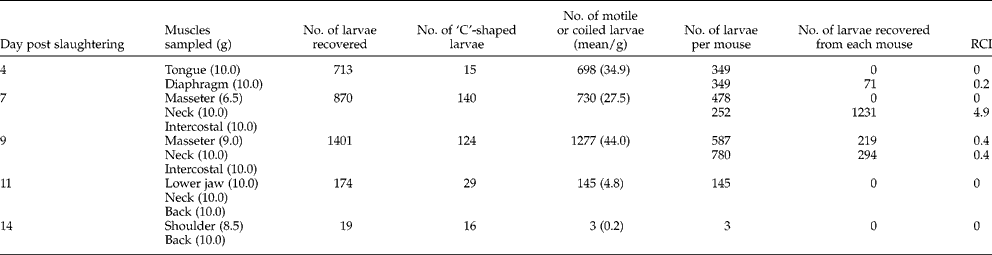
Larval counts carried out on pig tissues on days 4, 7, 9, 11 and 14 p.s., as well as data on the number of larvae given to mice and the number of larvae recovered from the mice, are shown in table 2. It was assumed that tightly coiled larvae and those showing movement were viable, while those in a ‘C’ shape were dead.
Trichinella papuae larval counts on tissues of a pig carcass exposed to natural conditions in Papua New Guinea and the infectivity of larvae derived from the pig tissues in mice (RCI, reproductive capacity index).
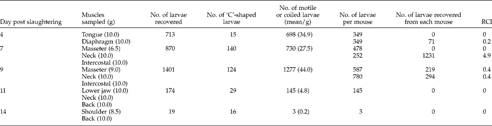
Although T. papuae larvae collected from decaying porcine muscles were infective to laboratory mice up to 9 days p.s., the RCI of larvae in laboratory mice was low (range 0–4.9) and without any relationship with the time elapsed in the decaying tissues (table 2). There was no difference observed between the RCI of larvae recovered from the pig carcass and larvae collected at each passage in infected mice. No larva collected from decaying porcine muscles at 11 and 14 days p.s. was infective to laboratory mice.
Discussion
An important adaptation of Trichinella sp., which facilitates its transmission, is the physiological mechanism utilized by muscle larvae to promote its survival in decaying carcasses. The longer the larvae remain viable, the higher the probability of being ingested by a scavenging host. In spite of the larva-induced angiogenic process that develops around the nurse cell after larval penetration of the muscle cell, larval metabolism is basically anaerobic (Despommier, Reference Despommier1990), which favours its survival in decaying tissues – longer for the encapsulated than for the non-encapsulated species (Pozio & Murrell, Reference Pozio and Murrell2006). The persistence of larvae in putrefying flesh is also determined by the environment: high humidity and low temperatures favour survival. This condition has been proposed as the environment of the ‘free-living’ stage, resembling the egg stage of most other nematode species (Madsen, Reference Madsen and Kim1974).
The results show that T. papuae larvae survived and remained infective to mice at least up to 9 days in a pig carcass maintained at an average temperature of 30.0°C with a peak of 35.5°C and at an average relative humidity of 79.0% with peaks of 100% for several days.
The low RCI of T. papuae in laboratory mice (0–4.9%) is not surprising, because Swiss mice do not appear to be a suitable host for T. papuae. In fact, a RCI of only 5.3 was reached at the ninth passage in Swiss mice for the isolate code ISS572 (Pozio et al., Reference Pozio, Owen, La Rosa, Sacchi, Rossi and Corona1999). The use of Swiss mice to evaluate the infectivity of larvae, without their immunodepression with a drug to facilitate the development of the parasite, could have been a bias preventing the development of larvae collected from porcine muscles at the eleventh and fourteenth days p.s.
Data presented by Kapel et al. (Reference Kapel, Webster and Gamble2005) show that for both pigs/wild boars and horses, the tongue base had more Trichinella pseudospiralis larvae than the tongue tip. The same was found in the pig infected with T. papuae. Also, different muscles of the neck of the pig had markedly different levels of infection, which explains the greater number of larvae recovered from pig tissues sampled on day 9 than on day 7 p.s. (table 2).
Stewart et al. (Reference Stewart, Kennedy and Larsen1990), studying the non-encapsulated T. pseudospiralis, reported larvae obtained up to 15 days p.s. surviving in mouse carcasses kept at 24°C but they were not infective after day 10 p.s. Webster et al. (Reference Webster, Malakauskas and Kapel2002) found T. papuae larvae to be infective to mice after 9 days' exposure at 20°C in fox tissues, but none was detected after 2 weeks. The current work shows that T. papuae can survive and remain infective in a pig carcass for a similar number of days (9 p.s.) and at higher temperatures.
In PNG, wild pigs are hunted by villagers with bow and arrow, and with shotguns when ammunition is available. Unwanted parts of a carcass may be left behind and, inevitably, a wounded animal occasionally escapes to die of its wounds undiscovered. These, and pigs that die of natural causes, will be scavenged by pigs and, over a period of at least 9 days, could be a source of infection for many. It is possible that if an infected animal died in the shade of the forest, where the temperature is lower, larvae would remain infective in the carcass for more days, as Webster et al. (Reference Webster, Malakauskas and Kapel2002) found that larvae of T. papuae in fox muscle kept at a temperature of 5°C under laboratory conditions survived for up to 40 days p.s.
Several species of goannas or monitor lizards (Varanus spp.) occur in southern PNG and, being scavengers, are potential reservoir hosts. Pozio et al. (Reference Pozio, Marucci, Cassuli, Sacchi, Mukaratirwa, Foggin and La Rosa2004) have demonstrated experimentally that the African monitor (V. exanthematicus) can be infected with T. papuae; also, the Nile monitor (V. niloticus) in Zimbabwe is naturally infected with T. zimbabwensis, a species closely related to T. papuae (Pozio et al., Reference Pozio, Foggin, Gelanew, Marucci, Hailu, Rossi and Gomez Morales2007). So far, however, no goanna has been found to be infected in PNG.
Acknowledgements
We are most grateful to Columba Awui for technical assistance and to Edoardo Pozio for constructive comments on the manuscript. The work was supported, through the National Agriculture Quarantine and Inspection Authority (NAQIA), by the Australian Centre for International Agricultural Research (ACIAR) project ASI/2001/554 covering zoonotic diseases in the Pacific Region.