Pulmonary veno-occlusive disease in newborns presents with two major distinct pathologies: the most common of these congenital defects is total anomalous pulmonary venous connection and the second is atresia of the common pulmonary vein. In 1962, Lucas et alReference Lucas, Woolfrey and Anderson 1 first recognised and defined common pulmonary vein atresia as absence of a connection of the pulmonary venous confluence to either the heart or any major systemic veins. This distinguishes it from total anomalous pulmonary venous connection. These patients typically have a presentation similar to those with obstructed total anomalous pulmonary venous connection; however, the presence of spontaneous pneumothorax and/or pulmonary lymphangiectasia should increase suspicion for common pulmonary vein atresia.Reference Anderson, Hlavacek and Shirali 2 This review of common pulmonary vein atresia aims to shed light on the clinical presentation and management options for this rare entity. We present three recent cases from our institution.
Case 1
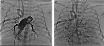
A full-term newborn female was noted to be cyanotic and tachypnoeic with mild retractions an hour after birth. Her oxygen saturation ranged between 30 and 40% on room air. The initial chest X-ray showed uniformly hazy lung fields and a poorly visualised cardiac silhouette. She developed rapidly progressive respiratory distress and acidosis, despite complete support. A transthoracic echocardiogram suggested obstructed total anomalous pulmonary venous connection, but without demonstrable pulmonary venous confluence. She was placed on venous-arterial extracorporeal membrane oxygenation. Cardiac catheterisation confirmed a diminutive pulmonary venous confluence with no connection to the heart or any major systemic vein. The individual pulmonary veins were also noted to be hypoplastic and draining into the splanchnic system (Fig 1). In the absence of a significant common pulmonary venous confluence, surgical repair was not considered feasible. A decision was made to withdraw care following consultation with the family.
Cardiac catheterisation angiograms of Case 1 depicting the absence of pulmonary venous return to the heart and a diminutive pulmonary venous confluence. ( a ) Frontal projection with the catheter course through the right heart and via the main pulmonary artery with the tip located within the large patent ductus arteriosus. There is opacification of the main pulmonary artery and the left and right branch pulmonary arteries as well as the descending aorta by way of the ductus. ( b ) Prolonged levophase in this same projection shows retention of contrast in the pulmonary vascular bed and prominent decompressing vessels communicating with the paravertebral and splanchnic veins. There was a diminutive pulmonary venous confluence noted.

Case 2
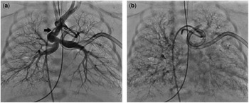
A full-term newborn male presented with tachypnoea and hypoxia immediately after birth. Hypoxia persisted even after relief of a spontaneous pneumothorax and maximum respiratory support. Transthoracic echocardiography suggested visceroatrial situs inversus, an unbalanced complete atrioventricular septal defect with a hypoplastic left ventricle, double-outlet right ventricle, and pulmonary atresia. A patent ductus arteriosus gave rise to two normally developed branch pulmonary arteries. The diagnosis of total anomalous pulmonary venous connection was suspected. Cardiac catheterisation confirmed the other cardiac findings, but demonstrated hypoplasia of the individual pulmonary veins, and a diminutive pulmonary venous confluence with several decompressing veins from the pulmonary capillary bed to the hepatic, innominate, and jugular venous systems (Fig 2). In light of the complex single ventricle anatomy and the absence of a pulmonary venous confluence, the patient was deemed inoperable. Care was withdrawn and the patient succumbed.
Cardiac catheterisation angiograms of Case 2 with absent pulmonary venous return to the heart. ( a ) Frontal projection with the catheter course retrograde via the descending aorta with the tip lying within the ductus arteriosus. The patent ductus arteriosus supplies the confluent branch pulmonary arteries in the absence of a main pulmonary artery connection. ( b ) Levophase in the same projection demonstrates retention of contrast in the pulmonary vascular bed with absent pulmonary venous connections to the heart or a major systemic vein. Decompressing vessels are not clearly demonstrated in this projection, but did return to the splanchnic veins.

Case 3
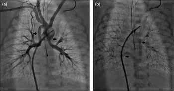
A female baby born at 34 weeks of gestation developed respiratory failure shortly after birth with severe cyanosis and persistent severe hypoxaemia, despite maximal respiratory support. Echocardiography demonstrated dextrocardia, complete atrioventricular septal defect, and apparently obstructed total anomalous pulmonary venous connection. Cardiac catheterisation performed within hours of birth confirmed dextrocardia, atrial situs inversus, complete atrioventricular septal defect, pulmonary valve atresia, and non-confluent pulmonary arteries. Both the branch pulmonary arteries were supplied by separate arterial ducts. There was apparent absence of the common pulmonary vein and a diminutive confluence behind the left atrium (Fig 3). The baby was considered inoperable and care was withdrawn.
Cardiac catheterisation angiograms of Case 3 demonstrating absent pulmonary venous return to the heart. ( a ) Frontal projection with a Berman catheter that courses retrograde via the descending aorta with balloon occlusion of the descending aorta to force contrast through the ducts and pulmonary arteries. The aorta gives rise to two patent ductus arteriosus, one arises from the innominate artery and supplies a non-confluent right pulmonary artery and the other arises from the undersurface of the transverse aortic arch and supplies the left pulmonary artery. No main pulmonary artery or pulmonary valve was evident during this study. ( b ) Levophase of the same projection demonstrates the pulmonary veins from both left and right sides, which join a large decompressing vein on each side with arrows depicting both these vessels. There is absence of the common pulmonary vein and a diminutive confluence behind the left atrium not seen in this projection. The right and left decompressing veins then course inferiorly to the portal venous system. There are several vessels arising from this system not seen here that course cephalad along the paraspinal plexus and eventually join the jugular veins on further prolongation of this angiogram.

Materials and methods
We conducted an English literature search via Pubmed and identified all the reported cases of common pulmonary vein atresia using the following search words: pulmonary veins, pulmonary vein atresia, common pulmonary vein, and common pulmonary vein atresia.Reference Lucas, Woolfrey and Anderson 1 , Reference DeLise, Schneider and Blackman 3 – Reference Vaideeswar, Tullu and Sathe 18 Each case was reviewed and data were collected on clinical presentation, laboratory findings, diagnostic study results, and interventions including extracorporeal membrane oxygenation and surgical interventions. The criterion used to define common pulmonary vein atresia was absence of pulmonary venous return to the heart or any major systemic veins as defined by Lucas et al.Reference Lucas, Woolfrey and Anderson 1 We identified several other cases of “common pulmonary vein atresia”, but upon review these cases did not meet the criterion for diagnosis, and thus were excluded.Reference Vaideeswar, Tullu and Sathe 18 In addition, four cases were not reviewed as they were not available in the English language for verification; two additional articles included in this review were available only as abstracts but provided sufficient information to warrant inclusion.Reference Sadou, Ciuffreda and Crupi 14 , Reference Sharda, Kurlandsky, Lacina and Radecki 15
Results
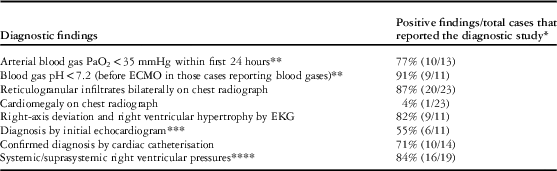
We identified a total of 35 cases of common pulmonary vein atresia.Reference Lucas, Woolfrey and Anderson 1 , Reference DeLise, Schneider and Blackman 3 – Reference Vaideeswar, Tullu and Sathe 18 The majority of patients presented with tachypnoea, cyanosis, and acidosis within 24 hours of birth, refractory to maximum medical support measures. Arterial blood gases typically demonstrated severe acidosis and hypoxia. Chest radiographs generally revealed bilateral reticular opacities in the lungs without cardiomegaly. On electrocardiography, right-axis deviation and right ventricular hypertrophy were found in most patients. Echocardiographic diagnosis was accurate in about half of the cases. Cardiac catheterisation confirmed the diagnosis of common pulmonary vein atresia in about 70% of the patients (Table 1).
Diagnostic findings in common pulmonary vein atresia.

ECMO=extracorporeal membrane oxygenation; EKG=electrocardiogram
* Many cases did not report any information on the data listed below. Furthermore, four case reports were limited to abstract information only, based on unavailability of the complete articleReference Sadou, Ciuffreda and Crupi 14 , Reference Sharda, Kurlandsky, Lacina and Radecki 15
** Only 13 cases reported any blood gas results with 11 of those reporting pH in addition to PaO2 measurements
*** Distinguishing atresia of the common pulmonary vein from total anomalous pulmonary venous return based on echocardiogram alone seemed difficult and beyond standard expectations for this study
**** Of the 19 cases reporting echocardiogram and/or cardiac catheterisation data, right ventricular pressures were not mentioned at all in the three cases and may have very well been elevated as well as would be expected
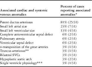
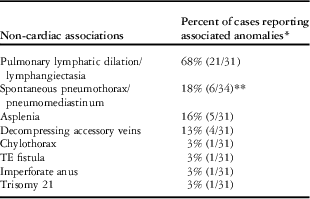
Common pulmonary vein atresia was usually an isolated cardiac anomaly (Table 2). The most common non-cardiac association was pulmonary lymphatic dilation or lymphangiectasia (64%). Trisomy 21 was noted in only one case.Reference Vaideeswar, Tullu and Sathe 18 Spontaneous pneumothorax and/or pneumomediastinum have been described in several cases as wellReference DeLise, Schneider and Blackman 3 , Reference Garcia, Ruiz and Sanchez-Redondo 6 , Reference Mas, Cochrane and Menahem 11 , Reference Sharda, Kurlandsky, Lacina and Radecki 15 (Table 3). It is unclear whether this was the result of aggressive resuscitative measures or primary lung disease; four cases were found to have accessory decompressing veins from either the pulmonary venous confluence or one of the pulmonary veins (Table 3). Most of these vessels joined the bronchial, paratracheal, or paraoesophageal venous systems.Reference Hawker, Celermajer, Gengos, Cartmill and Bowdler 7
Cardiovascular associated findings.

SVCs=superior caval veins
* Many reports may have not reported additional associated findings, such as the presence of a patent ductus arteriosus, when in fact they may have been present. Furthermore, two case reports (four cases) were limited to abstract information only, based on unavailability of complete article.Reference Mas, Cochrane and Menahem 11 , Reference Sadou, Ciuffreda and Crupi 14 Therefore, these four cases were excluded from this data set
** Type or classification of truncus arteriosus not specified
*** No further description as to which ventricle was hypoplastic or atrioventricular valve, semilunar valve, or great vessel findings
Non-cardiovascular associated findings.

TE=tracheoesophageal
* In all, 31 cases were included for most of this data set as four cases were limited to abstract information only as previously notedReference Sadou, Ciuffreda and Crupi 14 , Reference Sharda, Kurlandsky, Lacina and Radecki 15
** Only one case was excluded for this particular data pointReference Sadou, Ciuffreda and Crupi 14 as the second case report limited to abstract information did contain this information within the abstractReference Sharda, Kurlandsky, Lacina and Radecki 15
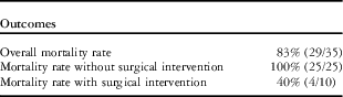
Without surgical intervention, common pulmonary vein atresia was universally fatal (Table 4). The first use of extracorporeal membrane oxygenation for common pulmonary vein atresia was described by Dudell et al, in which four patients showed improvement after venous-arterial extracorporeal membrane oxygenation. Extracorporeal membrane oxygenation was used for pre-operative stabilisation in four patients and post-operatively for pulmonary hypertension in three of them. The first successful surgical repair was reported in 1982 by Khonsari et al.Reference Khonsari, Saunders, Lees and Starr 8 Sporadic other successful repairs have been reported.Reference Dudell, Evans and Krous 5 , Reference Mas, Cochrane and Menahem 11 , Reference Shimazaki, Yagihara, Nakada, Sawa, Hirose and Sugimoto 16 , Reference Suzuki, Sato, Murai and Fukuda 17 Common features of such outcomes have been early surgical intervention and a sizeable pulmonary venous confluence; however, mortality remained high at 40% in those cases where surgical intervention was attempted (Table 4).
Outcomes in common pulmonary vein aresia.

Discussion
Embryology
Before formation of the lung buds, the mediastinum forms connections with the primitive atrium through the dorsal mesocardium. As the lungs develop, a pulmonary venous channel is formed through canalisation of the developing mediastinum.Reference Anderson, Hlavacek and Shirali 2 This primary pulmonary vein forms connections with a network of intrapulmonary veins. This fusion forms the pulmonary veins that connect with the atrial mass through the dorsal mesocardium. The pulmonary veins are subsequently engulfed by the atrium to form the pulmonary venous components of the morphological left atrium.Reference Anderson, Hlavacek and Shirali 2 A study from 2011 has elucidated some of the regulatory transcription factors – ISL-1, NKX2-5, and TBX2 – that may be involved in regulating this process 20–25 days into human development.Reference Sizarov, Ya, de Boer, Lamers, Christoffels and Moorman 19
Total anomalous pulmonary venous connection results from the failure of canalisation of the pulmonary venous channel in the mediastinum, which takes place early during the development of the pulmonary venous system; however, if canalisation occurs, but the pulmonary venous channel later becomes atretic in its course between the atrium and the intrapulmonary venous network, it results in common pulmonary vein atresia. This is a distinct diagnosis from total anomalous pulmonary venous connection as per the International Pediatric and Congenital Cardiac Code.Reference Walsh, Ungerleider, Aiello, Spicer and Giroud 20 In common pulmonary vein atresia, there is always a pulmonary venous confluence of varying size, and diminutive individual pulmonary veins decompress into the splanchnic system via small collaterals.Reference Anderson, Hlavacek and Shirali 2 , Reference Mavroudis and Backer 21 If there is absolutely no confluence at all, then the condition may represent atresia of the individual pulmonary veins and not atresia of the common pulmonary vein.
Presentation
Newborns with pulmonary vein atresia are extremely ill and present with respiratory distress, metabolic acidosis, and severe hypoxia. These findings are present within the first few hours of life and patients generally deteriorate rapidly. The natural history of the disease is characterised by respiratory failure and decreased systemic perfusion. Hepatomegaly is often present. The praecordium is usually silent except for a ductal murmur. Pallor and systemic hypoperfusion ensue despite aggressive supportive therapy, and death occurs usually within 48 hours of life.
Diagnostic findings
Cyanosis associated with profound metabolic acidosis is a universal finding among these neonates. A chest X-ray typically reveals bilateral reticular opacities without cardiomegaly. These diagnostic findings were consistent in our three patients as well.
Echocardiography may suggest common pulmonary vein atresia, but typically is not very reliable in making the diagnosis. In the report from Dudell et alReference Dudell, Evans and Krous 5 in 1993, the initial echocardiogram missed the diagnosis in four out of five cases, and repeat echocardiograms in two out of four of their patients. Despite this, echocardiography remains a useful diagnostic tool and is recommended as part of the initial evaluation. In our experience, the echocardiogram showed an absence of a pulmonary venous confluence and no demonstrable connections to the heart, suggestive of obstructed total anomalous pulmonary venous connection in all three cases. Nonetheless, one should suspect common pulmonary vein atresia when the echocardiogram shows no discernible pulmonary venous confluence.
Cardiac catheterisation is reliable in making the diagnosis of common pulmonary vein atresia in about 70% of cases. The definitive diagnosis of common pulmonary vein atresia can usually only be made during autopsy; however, cardiac catheterisation should be implemented when echocardiography is inconclusive. In our cases, cardiac catheterisation demonstrated decompressing veins from the pulmonary capillary bed to the splanchnic veins on prolonged levophase.
Associated findings
Common pulmonary vein atresia is usually an isolated cardiac anomaly, except for the presence of a patent ductus arteriosus (Table 2). There was only one reported case of visceroatrial situs inversus and two cases with pulmonary atresia, one of which had non-confluent pulmonary arteries. On the other hand, two of the cases at our institution were associated with complete atrioventricular septal defects, one of which was a double-outlet right ventricle with a moderately hypoplastic left ventricle. There was only one case with single ventricle physiology reported in the literature.
Pulmonary lymphangiectasia was the most common non-cardiac-associated finding. This is the result of pulmonary congestion and excess pulmonary venous blood flow being diverted into the lymph channels without the presence of significant alternative decompressing pathways.Reference Lucas, Woolfrey and Anderson 1 , Reference DeLise, Schneider and Blackman 3 , Reference Ledbetter, Wells and Connors 9 Pulmonary lymphangiectasia is typically noted at the time of autopsy, but dilated lymphatics were also described in some cases during surgical intervention. Spontaneous pneumothorax and/or pneumomediastinum have been described in several cases as well.Reference DeLise, Schneider and Blackman 3 , Reference Garcia, Ruiz and Sanchez-Redondo 6 , Reference Mas, Cochrane and Menahem 11 , Reference Sharda, Kurlandsky, Lacina and Radecki 15 This is a major associated complication that is likely secondary to barotrauma from aggressive ventilatory efforts. This complication was noted in only one of our cases.
Trisomy 21 was the only genetic defect reported, and was reported in only one case;Reference Vaideeswar, Tullu and Sathe 18 however, genetic testing may have not been performed in the majority of cases in which the patients died shortly after birth.
Management
Infants with common pulmonary vein atresia are extremely ill. Medical management consists of maximising ventilator support, usually requiring escalation to high-frequency oscillator ventilation. Nitric oxide therapy is typically implemented, but has little benefit.
The use of prostaglandin-infusion therapy is controversial, as it can potentially lead to lower oxygen saturations.Reference Dudell, Evans and Krous 5 Extracorporeal membrane oxygenation has become more routinely used over the last two decades as a means of stabilising the patient.Reference Dudell, Evans and Krous 5
Surgical intervention offers the only chance for survival. Before surgical intervention, one needs to determine whether there is a pulmonary venous confluence in close proximity to the left atrium. Successful surgical treatment has been contingent on there being sizeable pulmonary venous confluence. A few groups have reported successful outcome following surgical intervention;Reference Dudell, Evans and Krous 5 , Reference Khonsari, Saunders, Lees and Starr 8 , Reference Mas, Cochrane and Menahem 11 , Reference Shimazaki, Yagihara, Nakada, Sawa, Hirose and Sugimoto 16 , Reference Suzuki, Sato, Murai and Fukuda 17 however, in most cases, as also in ours, the common pulmonary venous confluence is hypoplastic, making surgical intervention difficult if not impossible. Also concerning is the long-term prognosis following a surgical intervention. There are scant follow-up data on surgical survivors, other than the fact that they commonly experience significant morbidity from persistent and recurrent chylous effusions and lymphangiectasia.Reference Dudell, Evans and Krous 5 If there is a sizeable pulmonary venous confluence, then surgery may be feasible and associated with survival; if, however, there is no significant confluence and the individual pulmonary veins are hypoplastic, then surgery is not appropriate and palliative care should be considered.
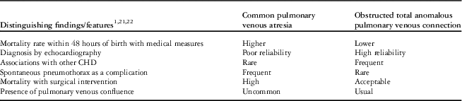
There are important prognostic distinctions between common pulmonary venous atresia and obstructed total anomalous pulmonary venous connection (Table 5). In 2005, the Boston Children’s Hospital reported early mortality rate of 22% for obstructed total anomalous pulmonary venous connection.Reference Hancock-Friesen, Zurakowski and Thiagarajan 22 There is much higher, almost prohibitive, mortality associated with common pulmonary vein atresia. Other differences simply reflect the natural history of each disease process, whereas some may be useful in guiding clinicians towards the correct diagnosis in the future. In the absence of a recognisable pulmonary venous confluence, palliative care may be the most humane management strategy.
Distinguishing common pulmonary vein atresia from total anomalous pulmonary venous connection.

Conclusions
Common pulmonary vein atresia is a rare form of CHD. The diagnosis must be entertained in any newborn that presents with cyanosis, refractory acidosis, and decreased systemic perfusion within the first 48 hours of life. A chest X-ray typically demonstrates bilateral reticular opacities. Echocardiography is a useful screening tool, although not diagnostic. Cardiac catheterisation may be helpful in establishing the diagnosis, but is not completely reliable. Extracorporeal membrane oxygenation is a valuable tool for stabilisation and may allow for further diagnostic testing to be performed. This disease is uniformly fatal without surgical intervention; however, survival after surgery continues to be poor with scant data available on post-operative prognosis.
Acknowledgements
None.
Financial Support
This research received no specific grant from any funding agency, commercial, or not-for-profit sectors.
Conflicts of Interest
None.