INTRODUCTION
The protozoan parasite Toxoplasma gondii is capable of infecting all mammals, including humans and livestock, and is one of the most successful parasites worldwide with one third of the human population predicted to have been exposed to the parasite (Tenter et al. Reference Tenter, Heckeroth and Weiss2000). Domestic and wild felids are thought to be the only definitive host and following primary infection can shed millions of T. gondii oocysts into the environment (Hutchison et al. Reference Hutchison, Dunachie, Siim and Work1969, Reference Hutchison, Dunachie, Work and Siim1971). Infected wildlife and livestock are known intermediate hosts, whereby parasitic cysts form in the tissues of these animals (tissue cysts), containing hundreds of infective bradyzoites (Dubey et al. Reference Dubey, Lindsay and Speer1998), which persist for the lifetime of the host. Wild mammalian carnivores can become infected by both consumption of T. gondii tissue cysts and oocysts present in the environment via contaminated water and soil. It is this factor which makes wild mammalian carnivores useful as potential sentinel species, providing an indication of which strains are present within the environment, however host behaviour and susceptibility to infection can also result in selective pressures on the parasite, and subsequently the genotypes found within a particular species.
In Europe and North America three main clonal lineages (types I, II and III) are predominantly found (Howe and Sibley, Reference Howe and Sibley1995), with type II being most frequently observed in Europe. Recently, a fourth clonal type (haplogroup 12) has been identified in North America and includes isolates from wildlife (Khan et al. Reference Khan, Dubey, Su, Ajioka, Rosenthal and Sibley2011). Moreover, molecular characterization using multilocus PCR–RFLP has identified recombinant or mixed infections containing combinations of alleles from types I, II and III (Lindstrom et al. Reference Lindstrom, Sundar, Lindh, Kironde, Kabasa, Kwok, Dubey and Smith2008; Berger-Schoch et al. Reference Berger-Schoch, Herrmann, Schares, Müller, Bernet, Gottstein and Frey2011) and atypical strains comprised of unique genotypes (Dubey et al. Reference Dubey, Sundar, Hill, Velmurugan, Bandini, Kwok, Majumdar and Su2008), which have otherwise predominantly been identified in South America. Although multilocus genotyping highlights a greater diversity of the parasite in Europe and North America than originally presumed, information on T. gondii strains circulating within the UK is lacking.
Recent figures regarding the prevalence of T. gondii in British wildlife is limited and is mostly based on serology rather than molecular detection of the parasite. In wild mammalian carnivores in the UK seroprevalence has been found to vary between 39·0–77·0% in Eurasian badgers (Meles meles) (Anwar et al. Reference Anwar, Knaggs, Service, McLaren, Riordan, Newman, Delahay, Cheesman and Macdonald2006), and 20·0% in red foxes (Vulpes vulpes) (Hamilton et al. Reference Hamilton, Gray, Wright, Gangadharan, Laurenson and Innes2005). A study in wild brown rats (Rattus norvegicus) detected a mean seroprevalence of 35·0% from an area designated as free of cats (Webster, Reference Webster1994). A recent study examining the seroprevalence of T. gondii in 125 Scottish sheep (Ovis aries) flocks found an overall prevalence of 56·6%, and interestingly every flock tested had at least one positive animal (Katzer et al. Reference Katzer, Brulisauer, Collantes-Fernandez, Bartley, Burrells, Gunn, Maley, Cousens and Innes2011), indicating a high level of environmental contamination with T. gondii oocysts in Scotland.
Studying the genotype of the parasite may have epidemiological significance, especially when comparing T. gondii genotypes found in livestock and human infections. Very few data are available of T. gondii genotypes within British wildlife, although a study carried out in woodmice (Apodemus sylvaticus), within North Yorkshire, UK, highlighted the presence of alleles for types I, II and III when genotyped using a single PCR–RFLP marker (Thomasson et al. Reference Thomasson, Wright, Hughes, Dodd, Cox, Boyce, Gerwash, Abushahma, Lun, Murphy, Rogan and Hide2011).
This aim of the current study was to determine the prevalence of T. gondii within wild British carnivores by PCR, and to genetically characterize the genotypes present using five PCR–RFLP markers and sequence analysis.
MATERIALS AND METHODS
Wildlife samples
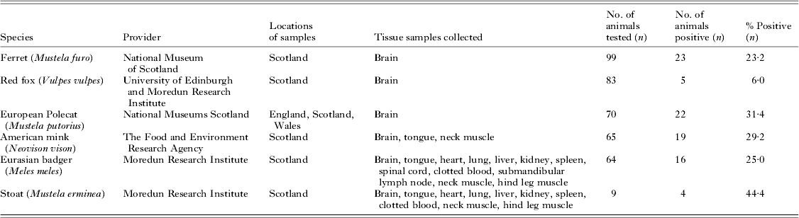
Tissue samples were collected from six different wildlife species, Eurasian badger (M. meles), ferret (Mustela furo), Red fox (V. vulpes), American mink (Neovison vison), European polecat (Mustela putorius) and stoat (Mustela erminea), from various locations in the UK, as further described in Table 1. Badger and stoat tissue samples were collected following fatal impacts with motor vehicles (badgers were collected with the permission of Scottish Natural Heritage). Where possible full necropsy was performed on the carcasses with tissue samples collected under sterile conditions. All other samples were kindly provided by The University of Edinburgh, The National Museums Scotland and The Food and Environment Research Agency (Table 1). All tissues processed showed no visible signs of decay or putrefaction. Samples were stored at −20 °C prior to DNA extraction and PCR amplification. Sample collection and the tissues derived for this study were identical to those described in a recent Neospora caninum publication (Bartley et al. Reference Bartley, Wright, Zimmer, Roy, Kitchener, Meredith, Innes and Katzer2013).
Samples collected for detection of Toxoplasma gondii and parasite prevalence by diagnostic nested ITS1 PCR

DNA extraction
DNA was extracted from approximately 1 g of homogenized tissue (tissue samples used are described in Table 1). Briefly, thawed tissue was transferred into a CK22 Precellys tissue homogenizer tube (Cepheid, Stretton Derbyshire, UK), containing 1 mL Nuclei Lysis Solution (Promega, Madison, WI, USA). Samples were homogenized for 2×50 s at 380 g using a Precellys 24 tissue homogeniser (Cepheid). The homogenate was processed using the Wizard® genomic DNA purification protocol (Promega), and volumes were scaled up to allow for 1 g of starting material. The final DNA pellet was re-suspended in 200 μL of DNase/RNase free water and stored at −20 °C prior to PCR analysis. To monitor potential cross-contamination between samples, extraction controls (sterile dH2O) were included within each batch of DNA extractions, and processed identically to homogenized tissue.
Detection of T. gondii DNA by ITS1 PCR
This nested PCR targets the multicopy 18S-5·8S rRNA internal transcribed spacer (ITS1) region of the parasite. Primers and methodology were adapted from Buxton et al. (Reference Buxton, Wright, Maley, Rae, Lunden and Innes2001) and Hurtado et al. (Reference Hurtado, Aduriz, Moreno, Barandika and Garcia-Perez2001). Briefly, each 20 μL reaction contained 2 μL 10× custom PCR mix – SM0005 (45 mm Tris-HCl 11 mm (NH4)2SO4, 4·5 mm MgCl2, 0·113 mg mL−1 BSA, 4·4 μ m EDTA and 1·0 mm dATP, dATC, dGTC, dTTP – ABgene, Epsom, Surrey, UK), 0·75 units BioTaq (Bioline, London, UK), and 2 μL DNA. First round reactions contained 0·05 μ m of forward primer and reverse primers NN1 5′-TCAACCTTTGAATCCAAA-3′, NN2 5′-CGAGCCAAGACATCCATT-3′ (Buxton et al. Reference Buxton, Wright, Maley, Rae, Lunden and Innes2001), the reaction was made to a final volume of 20 μL with DNase/RNase free water. To improve the sensitivity of the technique, each reaction was carried out in triplicate. Cycling conditions were 5 min at 95 °C, followed by 35 cycles of 1 min at 95 °C, 1 min at 55 °C, 1 min at 72 °C, and a final extension period of 5 min at 72 °C. To reduce unused primers from the primary PCR, first round PCR products were diluted 1:5 with dH2O, 2 μL of diluted product was used in the second round reaction. The conditions for each second round were identical to the first, however contained 0·05 μ m of internal forward and reverse primers Tg-NP1 5′-GTGATAGTATCGAAAGGTAT-3′, Tg-NP2 5′-ACTCTCTCTCAAATGTTCCT-3′ (Hurtado et al. Reference Hurtado, Aduriz, Moreno, Barandika and Garcia-Perez2001). The nested PCR products (10 μL) were electrophoresed on a 2% (w/v) agarose gel incorporating Gel Red™ (Biotum, Hayward, CA, USA), and visualized under UV light. A sample was regarded positive if an amplicon of 227 bp was present in any triplicate reaction. Multiple negative controls (dH2O) were included in each PCR run as well as DNA extraction controls, results were only accepted if these reactions were negative.
Actin PCR for detection of host DNA
To ensure the integrity of extracted DNA (and that it was free from PCR inhibitors), a reaction targeting host actin was performed using 2 μL of first round PCR reaction generated from the ITS1 PCR, previously used for the detection of T. gondii DNA. Each 20 μL reaction contained 2 μL 10× custom PCR mix – SM0005 (45 mm Tris-HCl, 11 mm (NH4)2SO4, 4·5 mm MgCl2, 0·113 mg mL−1 BSA, 4·4 μ m EDTA and 1·0 mm dATP, dATC, dGTP, dTTP – ABgene), 0·05 μ m of forward and reverse primers (Uni-Actin-F 5′-CATCCTGCGTCTGGACCT-3′, Uni-Actin-R 5′-TAGCAGAGCTTCTCCTTGATGTC-3′). Cycling conditions were identical to those previously described for the ITS1 PCR. Negative controls (dH2O) and an actin positive sample were included in each PCR run.
Genetic characterization of T. gondii by multiplex nested PCR–RFLP
Genetic characterization was performed using five markers spread over four different loci of the T. gondii genome. The first round of each 20 μL multiplex PCR reaction contained 2 μL 10× custom PCR mix – SM0005 (45 mm Tris-HCl, 11 mm (NH4)2SO4, 4·5 mm MgCl2, 0·113 mg mL−1 BSA, 4·4 μ m EDTA and 1·0 mm dATP, dATC, dGTP, dTTP – ABgene), 0·1 μ m of forward and reverse external primers for the following five markers – 5′SAG2 (Howe et al. Reference Howe, Honore, Derouin and Sibley1997; Su et al. 2006), 3′SAG2 (Howe et al. Reference Howe, Honore, Derouin and Sibley1997; Grigg et al. Reference Grigg, Ganatra, Boothroyd and Margolis2001), SAG3, GRA6 and BTUB (Prestrud et al. Reference Prestrud, Åsbakk, Mørk, Fuglei, Tryland and Su2008), 0·75 units BioTaq (Bioline), 5·85 μL dH2O and 2 μL DNA. To improve the sensitivity of the technique each multiplex reaction was carried out in quadruplicate. First round cycling conditions were 4 min at 95 °C, followed by 25 cycles of 30 s at 94 °C, 1 min at 53·7 °C and 2 min at 72 °C. First round PCR products were diluted 1 : 1 with dH2O. The nested second round reaction used separate internal primers for each marker and used the diluted first round PCR products instead of DNA. Each individual 20 μL second round reaction contained 2 μL 10× custom PCR mix (SM0005, ABgene), 0·3 μ m primers (individual forward and reverse internal markers for 5′SAG2, 3′SAG2, SAG3, GRA6 and BTUB – for details refer to references cited for first round PCR), 0·5 units BioTaq (Bioline), 13·85 μL DNase/RNase free water, and 2 μL of diluted first round PCR product. Cycling conditions were 4 min at 95 °C, followed by 40 cycles of 30 s at 94 °C, 1 min at 60 °C, and 1·5 min at 72 °C. A positive control, multiple negative controls and extraction controls were incorporated amongst samples to be genotyped. RFLP typing of positive samples was achieved by preparing an enzyme digest which contained 2 μL of the second round product, 2 μL of 10× NEB Buffer 4 (New England Biolabs, Herts, UK), 4 units of the appropriate restriction enzyme (New England Biolabs), made to a final volume of 20 μL in dH2O (for details of restriction enzymes used refer to references cited for first round PCR). Each digest was incubated for 1 h at the appropriate temperature depending on the restriction enzyme used. Digested products were visualized following electrophoresis using a 3% Metasieve Agarose gel incorporating Gel Red™ (Biotum). PCR-RFLP patterns were compared with those of control reference strains B1 (Type I), M4 (Type II) and NED (Type III) (B1 and NED obtained from BRC Toxoplasma, Limoges, France).
DNA sequencing
Twenty-five SAG3 PCR products were directly sequenced using both forward and reverse SAG3 internal primers. Products were purified (PCR and Gel Purification Kit, Bioline) and sequencing completed at GATC Biotech (Cologne, Germany). Results were analysed using BioEdit sequence analysis software (http://www.mbio.ncsu.edu/bioedit/bioedit.html), and consensus sequences generated. To enable identification of polymorphisms, a 166 bp fragment of the published sequences representing the three clonal archetypal T. gondii lineages were used as reference sequences, which included RH – type I, Pru – type II and CEP – type III (GenBank accession nos. AF340227, AF340228 and AF340229 respectively). Direct sequencing using additional forward and reverse internal primers was also completed to obtain representative sequences for 5′SAG2, 3′SAG2, GRA6 and BTUB.
RESULTS
Prevalence of T. gondii by ITS1 PCR
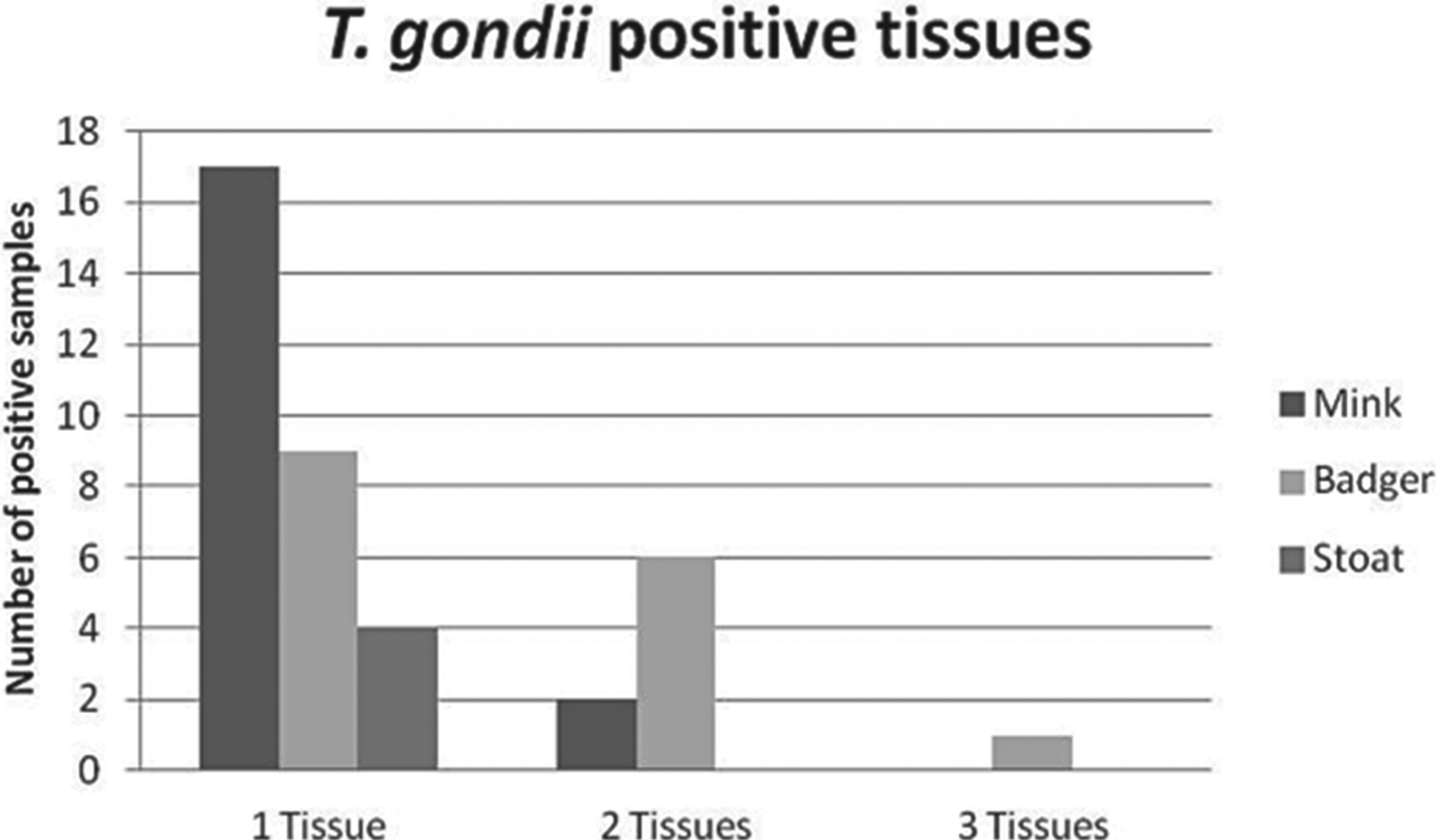
Overall 22·8% (89/390) of the animals tested were positive for T. gondii. The prevalence varied depending on the host species (Table 1), 31·4% (22/70) polecats tested positive, whilst only 6·0% (5/83) of foxes were positive. Mink, badger and ferret had a prevalence of 29·2% (19/65), 25·0% (16/64) and 23·2% (23/99) respectively. Stoats showed a higher prevalence with 44·4% (4/9) of animals tested PCR positive, however it should be noted that this group had a lower representative sample size and was collected from a geographically smaller area than the other species tested. Multiple tissues were used to test for the presence of T. gondii in mink, badger and stoat. Mink brain tissue accounted for 20·0% (13/65) of positive samples within this species, followed by tongue and heart tissue (10·8 and 1·5% respectively), whilst in badgers, brain tissue accounted for 5·0% (3/60) of positive samples, while tongue and heart tissue had a positivity of 6·3% (2/32) and 7·7% (4/54) respectively (Table 2). Multiple tissues from the same animal were found to be positive for mink, badger and stoat (Fig. 1).
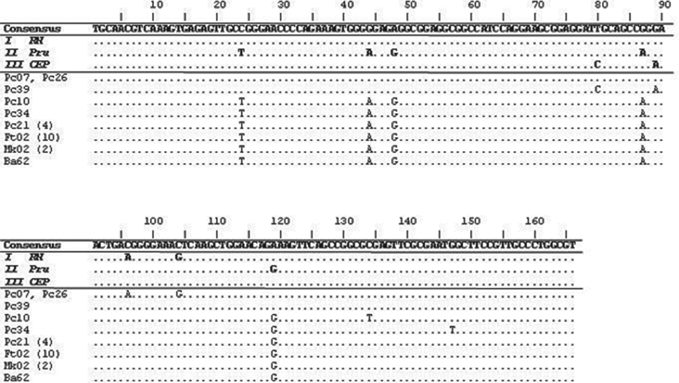
Sequence alignment at the SAG3 locus. Pc, Polecat; Ft, Ferret; Mk, Mink; Ba, Badger. Consensus sequence indicates the nucleotides shared by a minimum of two of the three archetypal clonal lineages RH (I), Pru (II) and CEP (III). The sequence alignment shown for Pc21 (4) also represents identical sequencing for four other polecat samples (Pc27, Pc30, Pc43 and Pc53), whilst Ft02 (10) represents identical sequencing for 10 other infected ferrets (Ft05, Ft15, Ft39, Ft42, Ft49, Ft50, Ft55, Ft64, Ft82 and Ft83), and Mk02 (2) represents identical sequencing for two other mink samples (Mk08 and Mk54).
Individual tissues collected and Toxoplasma gondii positivity
Br = Brain, Tn = Tongue, He = Heart, Ne = Neck Muscle, Lu = Lung, Kid = Kidney, Sp = Spleen, Bl = Clotted Blood, Hlm = Hind leg muscle, Sln = Submandibular lymph node, Sc = Spinal Cord.
Detection of host DNA
All wildlife DNA samples tested positive for the detection of host DNA, indicating that any negative result observed in either the ITS1 PCR or genotyping PCR–RFLP, was not due to degraded DNA or the presence of PCR inhibitors.
PCR–RFLP genotyping
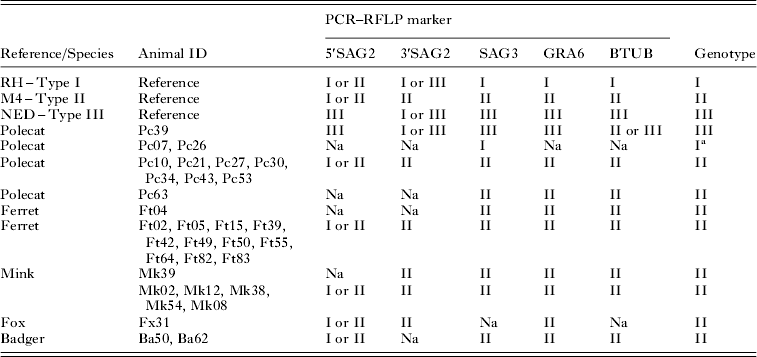
Of the 89 animals that tested positive for T. gondii by ITS1 PCR, 32 were successfully amplified with PCR–RFLP primers for three or more markers across three loci (36·0%). This included nine polecats, 12 ferrets, six mink, two badgers and one fox (Table 3). Two polecats (Pc07 and Pc26) only amplified at a single locus (SAG3), which identified a type I allele in both animals. Attempts to amplify products at additional loci in these animals failed.
Toxoplasma gondii PCR–RFLP genotyping from 32 infected animals across five markers
Na, no amplification.
a Based on a single allele identified at the SAG3 locus. I = type I; II = type II; III = type III; I or II = type I or II; I or III = type I or III; II or III = type II or III.
A type III lineage was identified across all five markers (four loci) in one polecat (Pc39), however the predominant lineage across all species genotyped was type II (90·6%). PCR–RFLP genotyping across all markers (four loci) for the type II lineage was achieved in 23 animals which included five mink, 11 ferrets, and seven polecats. Partial typing of the type II lineage was seen in six animals; two badgers, one fox, one mink, one ferret and one polecat.
Within this group of animals multilocus genotyping highlighted the presence of alleles for the three main clonal lineages of T. gondii.
Sequence analysis
Direct sequencing of 25 (11 ferrets, 10 polecats, three mink and one badger) SAG3 PCR products which had previously been genotyped by PCR–RFLP is highlighted in Fig. 1. The results confirm that the type I T. gondii alleles identified in two polecats by PCR–RFLP (Pc07 and Pc26) were identical to a 166 bp region of the archetypal type I lineage RH at the SAG3 locus (GenBank accession no. AF340227). The T. gondii strain identified in a polecat (Pc39), which was classified as type III by PCR–RFLP, showed a similar sequence identity to the archetypal type III lineage: CEP (GenBank accession no. AF340229). In 20 of the animals the T. gondii strain classified as type II by PCR–RFLP also had a sequence identity to the archetypal type II lineage: Pru (GenBank accession no. AF340228). Finally the T. gondii strain identified in two polecats (Pc10 and Pc34) showed slight variation compared with the type II archetypal lineage Pru, with sequences from both animals showing a single nucleotide substitution (Pc10 at 134 bp and Pc34 at 147 bp, GenBank accession numbers KC928252 and KC928253 respectively), both sequences were blasted against the NCBI and ToxoDB databases but did not show 100% identity to any other sequence. It should be noted that direct sequencing was also carried out for amplicons generated from 5′SAG2, 3′SAG2, GRA6 and BTUB. These sequences correlated with the results obtained from PCR-RFLP (data not shown). Sequencing using SAG3 proved to be more informative, providing additional information about the genotype than PCR-RFLP alone. Representative sequences were submitted to GenBank with the following accession numbers: KC928254 – representative of 20 isolates (SAG3 Type II allele), KC928250 – Pc07 (SAG3 Type I allele), KC928251 – Pc39 (SAG3 Type III allele), KC928252 – Pc10 (SAG3 Type II allele, single base pair substitution), KC928253 – Pc34 (SAG3 Type II allele, single base pair substitution), KC928255 (GRA6 Type II), KC928256 (GRA6 Type III), KC928257 (BTUB Type II), KC928258 (5′SAG2 Type II), KC928259 (3′SAG2 Type II).
DISCUSSION
Data describing the prevalence of T. gondi in wildlife within the UK are limited; even less information is known about the genotypes, which may be circulating within the host populations and subsequently the wider environment. However, research carried out in Europe has shown not only further information on the prevalence of the parasite, but also on the genotypes within these host species. The results from this current study help to complete the knowledge regarding the prevalence of T. gondii and the genotypes present in the environment within the UK. Our results have shown that the prevalence of T. gondii in British wild mammalian carnivores varies between 6·0–44·4% depending on the host species. Analysis of identical tissues for the detection of N. caninum was recently published (Bartley et al. Reference Bartley, Wright, Zimmer, Roy, Kitchener, Meredith, Innes and Katzer2013). A comparison between this research and the current study has identified seven animals (1·8%) that were infected with both T. gondii and N. caninum; this includes one badger, one fox and five polecats. The overall prevalence of T. gondii may be affected by the number and type of tissues available for testing within each species (Table 2). Stoats have the highest prevalence (44·4%), however, only nine animals were tested by PCR for the presence of T. gondii DNA, whereas for the other species examined the sample number was greater (n = 64–99), as described in Table 1. The availability of stoat samples was limited; with only nine animals being tested it can be concluded that this group does not constitute a representative sample for the species. To fully evaluate the prevalence of T. gondii within stoats a greater number of animals should be sampled. The lowest prevalence was observed in foxes (6·0%), however a previous serum-based study in red foxes from the UK found a T. gondii seroprevalence of 20·0% (Hamilton et al. Reference Hamilton, Gray, Wright, Gangadharan, Laurenson and Innes2005). Other studies within Europe have highlighted a higher seroprevalence, including 73·7% (14/19) in France (Aubert et al. Reference Aubert, Ajzenberg, Richomme, Gilot-Fromont, Terrier, de Gevigney, Game, Maillard, Gibert, Darde and Villena2010), 56·0% (115/206) in Ireland (Murphy et al. Reference Murphy, Walochnik, Hassl, Moriarty, Mooney, Toolan, Sanchez-Miguel, O'Loughlin and McAuliffe2007), and 74·5% (152/204) to 84·7% (149/176) in Germany depending on the region (Herrmann et al. Reference Herrmann, Maksimov, Maksimov, Sutor, Schwarz, Jaschke, Schliephake, Denzin, Conraths and Schares2012). It should be noted that lower figures are observed for the molecular detection of the parasite. In Belgium 18·8% (57/304) of foxes tested positive for T. gondii by real-time PCR (De Craeye et al. 2011) and in Germany 13·4% (20/149) to 18·4% (28/152) of foxes were identified as positive by PCR, which differed substantially from the serological results of 74·5–84·7% (Herrmann et al. Reference Herrmann, Maksimov, Maksimov, Sutor, Schwarz, Jaschke, Schliephake, Denzin, Conraths and Schares2012). The work carried out by Herrmann et al. (Reference Herrmann, Maksimov, Maksimov, Sutor, Schwarz, Jaschke, Schliephake, Denzin, Conraths and Schares2012) showed molecular detection resulted in a lower prevalence when compared with serological detection of the parasite. Unfortunately the tissues available for our study did not allow a seroprevalence study to be undertaken. Our prevalence rate is lower than many European seroprevalence studies, as serum-based detection appears to result in an increased T. gondii infection detection rate. Lower values for the molecular detection of the parasite are likely to be due to a combination of the inhomogeneous distribution of T. gondii tissue cysts in the host and the small sample size used for DNA isolation (approximately 1 g of tissue). Testing a greater number of tissue samples from each species by PCR could increase the overall detection rates, as it is possible that T. gondii infection was not detected in some animals as a particular tissue was not available for testing. An example of this is highlighted within the current study, where a selection of badger tissues was positive for T. gondii while corresponding brain samples tested negative (Table 2). It should also be noted that where multiple tissues were collected from individual animals (mink, badger and stoat), that more than one tissue could be positive for T. gondii by ITS1 PCR. This was particularly noted in a badger, where T. gondii was detected in three individual tissues from this animal (Fig. 2). Molecular detection of parasite DNA does enable strain genotyping by PCR–RFLP, which cannot be achieved by serology-based methods.
Samples which were positive for more than one tissue by ITS1 PCR.
Genotypic analysis of the parasite is important as it can identify mixed genotypes or atypical strains. Different strains of the parasite can be more virulent in different hosts, or when a species has not been previously exposed to the parasite (Innes, Reference Innes1997). Examples of this have been observed in Australian marsupials (Parameswaran et al. Reference Parameswaran, Thompson, Sundar, Pan, Johnson, Smith and Grigg2010), Californian sea otters (Miller et al. Reference Miller, Grigg, Kreuder, James, Melli, Crosbie, Jessup, Boothroyd, Brownstein and Conrad2004; Sundar et al. Reference Sundar, Cole, Thomas, Majumdar, Dubey and Su2008) and even humans (Carme et al. Reference Carme, Demar, Ajzenberg and Darde2009; Ferreira et al. Reference Ferreira, Vidal, de Mattos, de Mattos, Qu, Su and Pereira-Chioccola2011). Our research shows that the strain most prevalent throughout Europe and North America (type II) is the main lineage identified within the wildlife species tested (Table 3). However a T. gondii type III lineage was identified in one polecat and an allele for type I was identified within two different polecats (Table 3), indicating that alleles for the three main clonal lineages are present in the British environment. A recent publication, which incorporated genotyping for only one marker (SAG3) in woodmice (A. sylvaticus) from an area in North Yorkshire, UK, also described the presence of all three alleles within the study population; a total of 22 samples from T. gondii-infected mice were genotyped: 12 were identified to contain type I, nine as type II and one as type III (Thomasson et al. Reference Thomasson, Wright, Hughes, Dodd, Cox, Boyce, Gerwash, Abushahma, Lun, Murphy, Rogan and Hide2011). Although information relating to strains found in wildlife in Britain is still lacking there are reports of parasite genotypes belonging to type III and I lineages in European wildlife and livestock (de Sousa et al. Reference de Sousa, Ajzenberg, Canada, Freire, da Costa, Dardé, Thulliez and Dubey2006; Waap et al. Reference Waap, Vilares, Rebelo, Gomes and Angelo2008; Berger-Schoch et al. Reference Berger-Schoch, Herrmann, Schares, Müller, Bernet, Gottstein and Frey2011; De Craeye et al. Reference De Craeye, Speybroeck, Ajzenberg, Darde, Collinet, Tavernier, Van Gucht, Dorny and Dierick2011), and also evidence of atypical strains (Berger-Schoch et al. Reference Berger-Schoch, Herrmann, Schares, Müller, Bernet, Gottstein and Frey2011). Our current study using five RFLP markers did not identify any atypical strains or mixed genotypes within the species tested, however, it should be noted that application of additional markers at other loci may identify more novel T. gondii genotypes from these samples. Although we identified type II as the predominant lineage by PCR–RFLP (Table 3), sequence analysis at the SAG3 locus provided additional information across the amplicon. Two isolates identified as type II by PCR–RFLP differed by single base pair substitutions (Fig. 1), which was confirmed by repeating PCR and sequencing of the original samples. The two sequences are unique as they did not show 100% identity to any other sequence within the ToxoDB or NCBI databases. Additional sequencing of these PCR products at the GRA6 locus showed no further base pair polymorphisms (data not shown). Additional nucleotide changes, within the type II lineage, have also been reported from T. gondii-infected sheep in the Netherlands (Opsteegh et al. Reference Opsteegh, Langelaar, Sprong, den Hartog, De Craeye, Bokken, Ajzenberg, Kijlstra and van der Giessen2010), indicating a degree of variability within this lineage, which is not identifiable by PCR–RFLP analysis alone.
Not all DNA samples, which tested positive by ITS1 PCR, were successfully amplified for genotyping; only 35·6% of positive samples were successfully genotyped at three or more loci. Primers used for the detection of parasite DNA were designed around the multicopy ITS1 region (Hurtado et al. Reference Hurtado, Aduriz, Moreno, Barandika and Garcia-Perez2001); this PCR is therefore more sensitive than the genotyping PCR–RFLP. This is merely the result of multicopy vs single copy detection. Similarly, previous studies found only 22 out of 84 DNA samples from T. gondii positive mice could be genotyped at the SAG3 locus (Thomasson et al. Reference Thomasson, Wright, Hughes, Dodd, Cox, Boyce, Gerwash, Abushahma, Lun, Murphy, Rogan and Hide2011), 12 out of 20 T. gondii infected fox samples from the Saxony-Anhalt federal state in Germany could be genotyped across nine markers (Herrmann et al. Reference Herrmann, Maksimov, Maksimov, Sutor, Schwarz, Jaschke, Schliephake, Denzin, Conraths and Schares2012), and 31 out of 167 fox samples, which were seropositive, could be genotyped across 10 markers (Prestrud et al. Reference Prestrud, Åsbakk, Mørk, Fuglei, Tryland and Su2008). Interestingly, amplification of T. gondii DNA from Pc07 and Pc26 using markers 5′SAG2, 3′SAG2, GRA6 and BTUB was unsuccessful despite numerous attempts which included increased concentrations of target DNA. For these samples genotyping was only possible at the SAG3 locus, which identified an allele for type I by both PCR-RFLP (Table 3) and direct sequencing (Fig. 1). One possible explanation for this is that SAG3 has been reported to be the most sensitive genotyping marker (Khan et al. Reference Khan, Su, German, Storch, Clifford and Sibley2005), which suggests that parasite DNA was not present at a high enough concentration in the sample to enable detection by the less-sensitive PCR–RFLP markers.
This study highlights not only the prevalence of T. gondii across a variety of wild British mammalian carnivorous, but also provides novel data regarding the strains present within the environment that could potentially be transmitted to cats, livestock and even humans within the UK.
ACKNOWLEDGEMENTS
The authors would like to thank and acknowledge The Scottish Government, Rural and Environment Sciences and Analytical Services (RESAS) for funding this study and BRC Toxoplasma, Limoges, France for supplying T. gondii reference strains B1 and NED.
FINANCIAL SUPPORT
Fox samples were collected by A. Meredith with support by Defra grant SE01526.