Introduction
Hydrogels, crosslinked polymeric networks capable of containing large amount of water,[Reference Buhus, Peptu, Popa and Desbrieres1–Reference Williams, Wang, MacGregor, Siahaan, Stehno-Bittel and Berkland4] can be chemically or physically prepared from natural and/or synthetic polymers and have been widely used in the medical and pharmaceutical fields due to their biocompatibility and biodegradability;[Reference Tabata and Ikada5, Reference Vandelli, Rivasi, Guerra, Forni and Arletti6] for instance, synthetic hydrogels [e.g., PVA [poly(ethylene oxide) and poly(vinyl alcohol)]] have been used for tissue engineering due to the availability of synthesis methods to reproducibly manipulate molecular weights, block structures, degradable linkages, and other parameters that dictate their mechanical and chemical properties.[Reference Drury and Mooney7] In contrast, natural polymers typically exhibit poor mechanical properties but are popular for present biomedical applications since they are typically derived from living organisms, non-toxic, biocompatible,[Reference Coviello, Matricardi, Marianecci and Alhaique8] and cause no inflammatory response from the host organism.[Reference Buhus, Peptu, Popa and Desbrieres1] For these reasons, present studies extensively focus on natural polymers. In particular, gelatin (i.e., a protein obtained from the hydrolysis of collagen) has been an attractive candidate for preparing hydrogels used in long-term biomedical applications because it consists of a large number of functional groups and is easily crosslinked.[Reference Buhus, Peptu, Popa and Desbrieres1, Reference Bigi, Cojazzi, Panzavolta, Roveri and Rubini9]
Gelatin is easily soluble in water at 37 °C, non-immunogenic,[Reference Pierce, Pittermann, Ma, Gebauer, Neffe, Holscher, Jung and Lendlein10] and exhibits amphoteric behavior.[Reference Buhus, Peptu, Popa and Desbrieres1] Due to these properties, gelatin-based hydrogels are used in the manufacture of contact lenses, matrices for tissue engineering, and drug delivery systems. Developing new uses for gelatin-based hydrogels is another important area of academic research.[Reference Van Den Bulcke, Bogdanov, Rooze, Schacht, Cornelissen and Berghmans11] Additionally, the mechanical and chemical properties of gelatin can be modified using various kinds of crosslinking agents[Reference Neumann, Zur and Ehrenreich12–Reference Chang, Chang, Lai and Sung16] (e.g., glutaraldehyde, genipin, and dextran dialdehyde). For example, Crescenzi et al.[Reference Crescenzi, Francescangeli and Taglienti17] developed new gelatin-based hydrogels from high bloom purified gelatin A with/without hyaluronan by using transglutaminase-catalyzed crosslinking to form more densely connected networks that support cell regeneration.[Reference Peppas, Bures, Leobandung and Ichikawa18] This type of gelling system could be a promising candidate for nucleus pulposus replacement, which is one available treatment to restore function to the intervertebral discs.[Reference Crescenzi, Francescangeli and Taglienti17]
Chemically modified gelatin can exhibit tunable mechanical properties. For example, Bulcke et al.[Reference Van Den Bulcke, Bogdanov, Rooze, Schacht, Cornelissen and Berghmans11] studied the rheological properties of methacrylamide-modified gelatin-based hydrogels and found that the rheological properties of gelatin-based hydrogels can be regulated by the degree of modification, polymer concentration, photoinitiator concentration, and ultraviolet (UV) exposure time; gelatin-based hydrogels with a higher degree of modification can form more acrylate bonds, generate denser networks, and exhibit higher elastic modulus values. Photoinitiated chemical crosslinking of the methacrylamide-modified gelatin generates a controllable chemical network in which the strength of the hydrogels can be predictably modulated. This review will describe synthesis and fabrication techniques for preparing the gelatin-based hydrogels and illustrate the promising impact gelatin-based hydrogels have in various biomedical applications.
Synthesis and fabrication techniques of photosensitive gelatin-based hydrogels
The ability to process gelatin hydrogels using additive manufacturing would be an attractive prospect for fabrication of medical structures that are needed in limited amounts, including individualized medical devices. Conferring photocrosslinking abilities to gelatin would combine the ability to precisely define the geometry of the structure, while retaining some of the native cytocompatibility associated with gelatin. Gelatin-methacryloyl (GelMA) has well-described synthesis procedures; the biocompatibility of this materials has been demonstrated in multiple studies involving materials that were processed using UV stereolithography and two-photon polymerization (2PP).
Synthesis of gelatin-methacryloyl
GelMA is commercially available from multiple vendors [e.g., as a bio-ink for three-dimensional (3D) printing]; multiple facile methods are available for synthesizing GelMA. Amino acids with amine-containing side chains in gelatin readily react with methacrylic anhydride (MAA) to form GelMA. The synthesis method involves adding MAA to 10% w/v gelatin in phosphate-buffered saline (PBS) at a temperature of 50 °C over 1 h.[Reference Van Den Bulcke, Bogdanov, Rooze, Schacht, Cornelissen and Berghmans11] However, the pH is a critical process parameter especially since methacrylic acid is given off during the reaction; the methacrylic acid can reduce the reaction mixture below gelatin's isoelectric point where the amine-containing residues are not easily available for reaction. Another process iterates methacrylic acid addition and pH adjustment to keep the pH at the isoelectric point,[Reference Hoch, Schuh, Hirth, Tovar and Borchers19] which is able to achieve a high degree of substitution (DS), the proportion of amines that are methacrylated. Shirahama et al. performed a parametric study investigating the effects of buffer concentration, initial pH, gelatin concentration, MAA molar ratio and reaction temperature on the DS.[Reference Shirahama, Lee, Tan and Cho20] In their methods, a single pH adjustment and MAA addition is performed similar to Van den Bulke's methods but results in DS approaching the pH adjustment method. Briefly, Shirahama et al. found that a 0.25 M carbonate–bicarbonate buffer at an initial pH of 9 with a gelatin concentration at or above 10% w/v resulted in the highest DS. No effect was observed for reaction temperatures ranging between 35 and 50 °C and the reaction ran to completion within 60 min. The DS can be tuned very precisely by varying the MAA: gelatin ratio with most of the MAA consumed in the reaction. DS was investigated after adding between 0.012 and 0.2 mL/g MAA, which correspond to a 0.264–4.4 molar ratio of MAA to amine-containing residues. DS responded linearly between 0.012 and 0.05 g/mL, which corresponds to 0.264 and 1.1 molar ratios; DS for these values are near 25% and 100%.[Reference Shirahama, Lee, Tan and Cho20] Also, glycidylmethacrylate is an alternative methacrylating agent that reacts with amine groups using a similar procedure.[Reference Pierce, Tronci, Roble, Neffe, Jung and Lendlein21] For the above methods, the GelMA can be purified by filtering, dialysis, and lyophilization.
Ultraviolet stereolithography
GelMA has been reported to be photosensitive with 308 nm light;[Reference Pierce, Tronci, Roble, Neffe, Jung and Lendlein21] photoinitiators are generally added to improve photosensitivity. Common photoinitiators include 2-hydroxy-1-[4-(2-hydroxyethoxy)phenyl]-2-methyl-1-propanone (Irgacure 2959), lithium phenyl-2,4,6-trimethylbenzoylphosphinate (LAP), and an Eosin Y-based photoinitiator. Irgacure 2959 is a common photoinitiator with a peak absorbance around 275 nm, which can be excited by UV-C fluorescent bulbs or light emitting diodes (LEDs). Since this wavelength is extremely damaging to DNA (peak absorbance 260 nm), UV-A light at 365 nm can also be used at reduced efficiency. Irgacure 2959 at a concentration at 0.05–0.5 wt% has been used with no effect on cell viability on human OV-MZ-6 ovarian serous cystadenocarcinoma cells, chondrocytes, endothelial colony forming cells, and bone marrow-derived mesenchymal stem cells (MSCs).[Reference Loessner, Meinert, Kaemmerer, Martine, Yue, Levett, Klein, Melchels, Khademhosseini and Hutmacher22] LAP, on the other hand, has an absorbance peak around 380 nm, with a tail that reaches well into the visible spectrum. This photoinitiator allows for the photopolymerization with commonly available UV-A LEDs and is thus a popular choice for extrusion-based bioprinters. One interesting application taking advantage of the visible light absorption tail is to use LAP as the free-radical photoinitiator for negative photoresist functionality and incorporating an o-nitrobenzylether modified polyethyleneglycol UV-A photodegradable crosslinker for positive photoresist functionality; illumination with >400 nm light would polymerize GelMA, while exposure to UV-A would cleave the crosslinker. This hydrogel system was used to culture a cardiomyocyte–cardiac fibroblast co-culture, which exhibited spontaneous beating in vitro.[Reference Tsang, Annabi, Ercole, Zhou, Karst, Li, Haynes, Evans, Thissen, Khademhosseini and Forsythe23] Finally, Eosin Y absorbs well in the visible light spectrum with a peak absorbance at 517 nm. This fluorescent molecule was used as the chromophore of a type-II photoinitiator with triethanolamine as the co-initiator. A GelMA–polyethyleneglycol diacrylate (M n 700) co-polymer of various ratios was polymerized with this photoinitiator using a stereolithography-like method; this material showed >80% viability in a study involving NIH 3T3 fibroblasts. Furthermore, a grid structure and a university logo cell construct were printed in eight separate manually applied layers.[Reference Wang, Abdulla, Parker, Samanipour, Ghosh and Kim24]
Cross-linking by two-photon polymerization
Rapid prototyping approaches[Reference Hutmacher, Sittinger and Risbud25, Reference Yeong, Chua, Leong and Chandrasekaran26] (e.g., stereolithography, electrospinning, and 3D fiber deposition modeling) have garnered attention for fabricating materials for tissue engineering and other biomedical applications. However, those techniques have lower resolution and are not effectively used to mimic features of the architecture of the natural cells and/or tissues. Recently, 2PP has been employed to create the 3D materials because it can generate the structures with features from sub-100 nm to hundreds of micrometers.[Reference Nichol, Koshy, Bae, Hwang, Yamanlar and Khademhosseini27] In addition, it is a straightforward laser writing technique for fabricating 3D structures, including sophisticated and complex structures, from CAD files. The high resolution of 2PP stems from the nearly simultaneous absorption of two photons in a small volume of material, which excites the same energy transition as a single UV photon. Two photon absorption takes place in volumes with high laser intensities (i.e., at the focus of a femtosecond laser). As shown in Fig. 1, the 1030 nm pulses from the femtosecond laser are converted to 515 nm light by a SHG (second harmonic generator); this wavelength is more readily absorbed by photoinitiators that were designed for use around 250 nm (e.g., Irgacure 2959). Alternatively, titanium:sapphire lasers are commonly used for structuring further into the near-IR spectrum.
Schematic representation of two-photon polymerization (2PP) setup; reprinted with permission from Ref. Reference Ovsianikov, Deiwick, Vlierberghe, Dubruel, Moller, Drager and Chichkov28. Copyright (2011) American Chemical Society.

There are benefits and drawbacks to the use of two photon polymerization. One benefit of this technique stems from the localized absorption at the focal point; not only is the two-photon absorption a non-diffraction limited phenomenon that allows features to be produced that can be smaller than the wavelength of the laser source, but also polymerization can occur at any location within the bulk of the polymer. A mechanism to apply additional photopolymer to the surface of the structure during processing, as in stereolithography, is not required; processing of an additional layer involves the simple movement of the z-stage. In fact, 2PP breaks out of the “layer-by-layer” paradigm and could do away with layer-based fabrication altogether. For example, a 2PP apparatus containing a spatial light modulator demonstrates the fabrication of a tetrahedron using only the spatial light modulator to move the focus in all three dimensions; no mechanical movement of the sample was required.[Reference Zalesskii, Danil'chenko, Barbashov, Zapadinskii and Sarkisov29] On the other hand, 2PP is a slow process due to its resolution and is often equated to painting with a fine-tip pen; it should be noted that the aforementioned spatial light modulators[Reference Gittard, Nguyen, Obata, Koroleva, Narayan and Chichkov30] or galvanoscanners[Reference Koroleva, Deiwick, Nguyen, Wolter, Narayan, Timashev, Popov, Bagratashvili and Chichkov31] can be utilized to reduce processing times. While some stereolithography approaches (e.g., digital micromirror device-based microstereolithography) can project an entire 2D image per layer using well-established technologies, 2PP processing only takes place at the focal point. As such, 2PP processing of wide structures requires long periods of time.
Cross-linking by gamma irradiation
At present, radiation-induced co-polymerization has been employed for crosslinking the polymeric structures instead of using chemical methods. Since it does not require additives, the generated structures are capable of sterilization and free of carcinogenic materials.[Reference Guven, Sen, Karadag and Saraydin32–Reference Sen, Uzun and Guven34] Because of these advantages, the radiation-induced crosslinking can be one of the most attractive fabrication methods. Gamma rays have been employed to crosslink the gelatin-based hydrogels. For instance, Eid et al.[Reference Eid, Abdel-Ghaffar and Dessouki35] performed the assessment on the impacts of gamma rays irradiation dose rate on the properties of poly (acrylamide/maleic acid/gelatin) hydrogels (e.g., swelling and thermal stability). As the gamma rays irradiates P(AAM/MA/G), it induces polymerization and crosslinking reactions simultaneously; the radiolysis products of water (e.g., hydroxyl free radicals) cause the monomer to form P(AAM/MA/G) hydrogels. At moderate gamma ray dose rates, the swelling ratio of gelatin-based hydrogels increases with increasing maleic acid and gelatin concentrations. Hydrogels generated with low irradiation dose rate exhibit higher thermal stability than those generated with moderate dose rate. This thermal stability increases with increasing dose until 20 kGy after which the stability declines. Since the strength of the covalent bond between the atoms forming the polymer determines the thermal stability of any polymer materials; thus, lower irradiation doses elevate the strength of the covalent bond.
Scaffolds fabricated via three-dimensional plotting
The composition and structure of tissue engineering scaffold materials define parameters such as cell responses, degradation, and mechanical properties.[Reference Karageorgiou and Kaplan36, Reference Bose, Roy and Bandyopadhyay37] An ideal multi-functional platform in tissue engineering and regenerative medicine should be composed of: (a) a material providing sufficient mechanical support, cell attachment, and stimulation of the new tissue growth as well as (b) a method to fabricate precise, user-defined geometries with this material. Fabrication of 3D porous scaffolds can be achieved with multiple methods with a variety of different materials.
Alginate is a biocompatible and biodegradable hydrogel that is commonly used to prepare tissue engineering scaffolds.[Reference Augst, Kong and Mooney38, Reference Schlossmacher, Schroder, Wang, Feng, Diehl-Seifert, Neumann, Trautwein and Muller39] Due to the dearth of efficient sites for cell adhesion, alginate scaffolds do not support significant cell attachment and proliferation.[Reference Suarez-Gonzalez, Barnhart, Saito, Vanderby, Hollister and Murphy40] As a consequence, the inclusion of other biomaterials in alginate-based scaffolds is commonly performed to improve cell attachment. Gelatin has been shown to be capable of promoting cell adhesion, proliferation, and differentiation[Reference Ito, Mase, Takizawa, Shinkai, Honda, Hata, Ueda and Kobayashi41–Reference Xia, Liu, Cui, Liu, Zhong, Liu, Wu, Chua and Cao43] which makes it an appropriate candidate for inclusion in scaffolds.
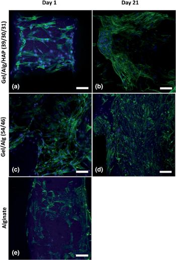
Multiple methods exist for the production of pores within the scaffold. For instance, freeze-drying is a common approach for preparing gelatin scaffolds;[Reference Eslaminejad, Mirzadeh, Mohamadi and Nickmahzar44–Reference Liu and Ma46] however, this technique does not allow for control over the pore dimensions. An electrospinning approach[Reference Meng, Wang, Ma, Zheng, Li and Zheng47, Reference Panzavolta, Gioffre, Focarete, Gualandi, Foroni and Bigi48] is also widely used for preparing gelatin-scaffolds; however, it has limited capabilities for fabricating 3D porous scaffolds with controllable macroscopic pores, which are needed for cell penetration and new tissue growth. The 3D plotting, an extrusion-based rapid prototyping method, show great advantages for fabricating predesigned scaffold architectures.[Reference Hutmacher and Cool49, Reference Derby50] In particular, Luo et al. employed a 3D-plotting method for making gelatin/alginate and gelatin/alginate/hydroxyapatite composites (Fig. 2); the 3D-plotted composite scaffolds showed significantly greater strength and modulus values than most gelatin scaffolds that were prepared by conventional methods.[Reference Luo, Lode, Akkineni and Gelinsky51] Moreover, the gelatin/alginate composite scaffolds were shown to promote cell adhesion and proliferation of human-bone marrow-derived mesenchymal stem cells (hBMSCs); the cells exhibited a homogeneous distribution within the inner regions of the 3D-plotted composite scaffolds and remained viable over 21 days (Fig. 3). The 3D-plotted gelatin/alginate composite porous scaffolds that exhibit a shape that matches a patient-specific defect could have superior performance over non-porous implants due to the cell networks within the porous scaffolds.
The structure of 3D plotted composite scaffolds of pure alginate (a, d, and g); the structure of 3D plotted composite scaffolds of gelatin/alginate with a mass ratio of 54/46 (b, e, and h); and the structure of 3D-plotted scaffolds of gelatin/alginate/hydroxyapatite with a mass ratio of 39/30/31 (c, f, and i). Note that A–C are microscopic images with scale bar = 1 mm; (d–f) are SEM images of the surface with scale bar = 20 µm; and (g–i) are SEM images of rods cross-sections with scale bar = 20 µm; reproduced from, Ref. Reference Luo, Lode, Akkineni and Gelinsky51. Copyright (2015), with permission of The Royal Society of Chemistry.

Confocal laser scanning microscopy images of human bone marrow-derived mesenchymal stem cells seeded on (a) and (b) 3D-plotted gelatin/alginate/hydroxyapatite scaffold with a mass ratio of 39/30/31; (c) and (d) gelatin/alginate with a mass ratio of (54/46); (e) alginate scaffolds; (a, c, e) scaffolds on day 1 and (b and d) scaffolds on day 21 with scale bar = 100 µm; reproduced from, Ref. Reference Luo, Lode, Akkineni and Gelinsky51. Copyright (2015), with permission of The Royal Society of Chemistry.

Biomedical applications of gelatin-based hydrogels
Gelatin-based hydrogels for drug delivery
Hydrogels are insoluble but can absorb a significant amount of water compared to hydrophobic polymeric networks [e.g., poly(lactic acid), or poly(lactic acid-co-glycolic acid)].[Reference Peppas, Bures, Leobandung and Ichikawa18, Reference Kashyap, Kumar and Kumar52] The lack of hydrophobic interactions allows for the encapsulation of biomacromolecules that are generally water soluble.[Reference Young, Wong, Tabata and Mikos53] Hydrogels can also absorb a significant amount of aqueous solutions.[Reference Einerson, Stevens and Kao54] Due to the low levels of immunogenicity and cytotoxicity associated with gelatin,[Reference Rathna, Mohan Rao and Chatterji55] hydrogels are attractive materials for drug delivery devices.
Drug release systems and antibacterial activities
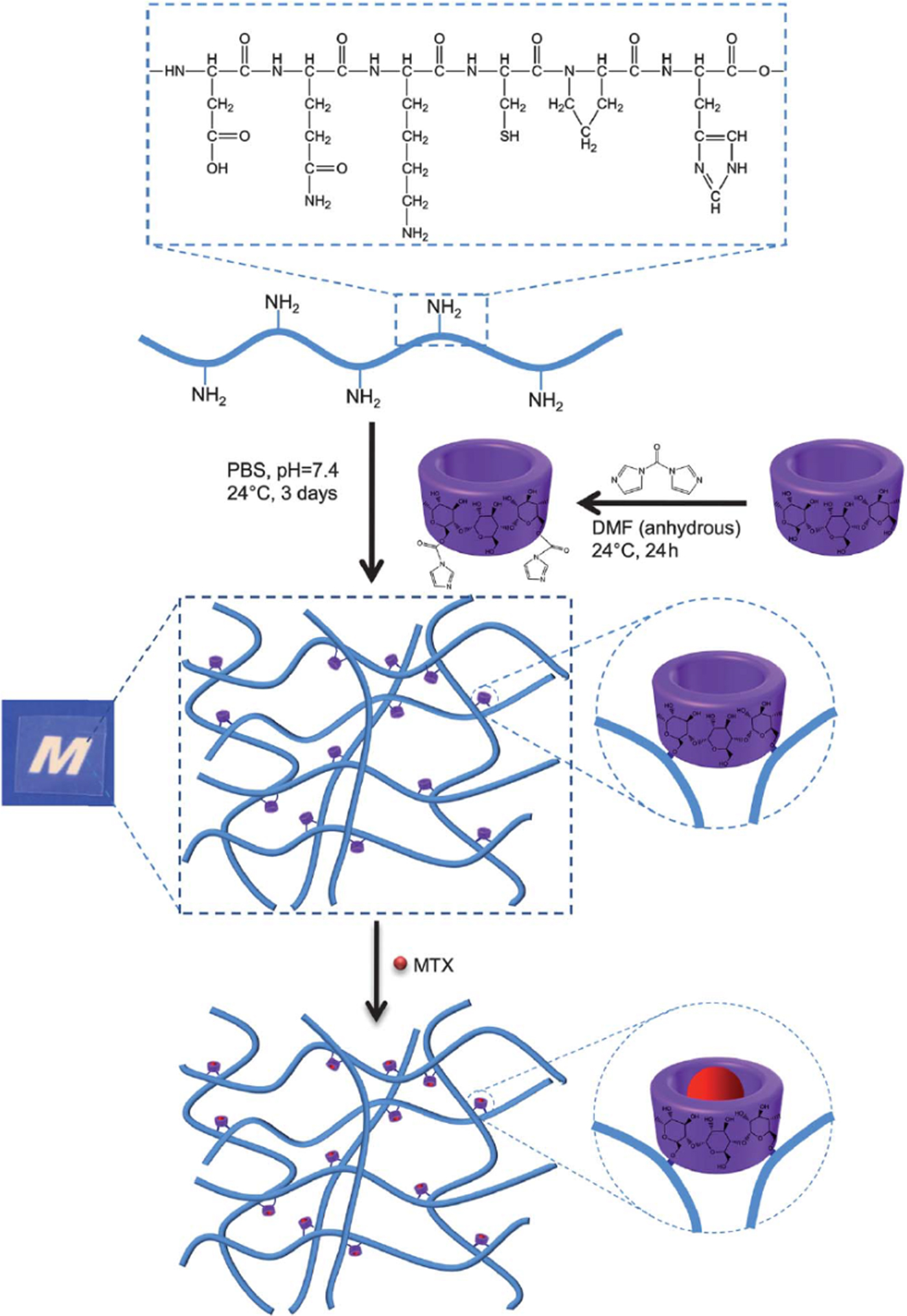
Particularly, Einerson et al.[Reference Einerson, Stevens and Kao54] studied the effect of gelatin modifications on in vitro drug release; the results indicated that the modification of gelatin backbone with polyethylene glycol-dialdehyde and/or ethylenediaminetetraacetic dianhydride decreased the maximum mass ratio of drug release and the time to reach the maximum mass ratio of the drug release. Liu et al.[Reference Liu, Zhang, Liu, Ni and Li56] made gelatin-based hydrogels with β-cyclodextrin (β-CD) as a crosslinking agent for improved drug carrier capacity and sustained release of anticancer drug methotrexate (MTX). A β-CD-Gel-3 formulation with a β-CD content in the hydrogel of 15.2% by weight exhibited the highest MTX loading level with 16.4 MTX mg per gram of hydrogel. MTX loading levels of hydrogels containing 11.1% or 13.5% by weight of β-CD contained 12.2 mg and 14.9 mg MTX per gram of hydrogel, respectively. A dextran-crosslinked gelatin-based hydrogel control contained 7.8 mg of MTX per gram. β-CD crosslinkers played an important role as binding sites for incorporating and complexing MTX. Formation of the complex elevated the MTX solubility within the hydrogels and caused MTX molecules to diffuse into the hydrogels (Fig. 4). Moreover, their MTX release study showed that an increasing β-CD content slowed elution of drug. β-CD-Gel-3 released loaded MTX much slower than the other three study groups (i.e., Dex-Gel, β-CD-Gel-1, and β-CD-Gel-2). Dex-Gel was the fastest, followed by β-CD-Gel-1, β-CD-Gel-2; complexation of MTX with β-CD decelerated diffusion of MTX and consequently lengthened the duration of drug release from the hydrogels.
Schematic representation for synthesis of the (β-CD)-crosslinked gelatin-based hydrogel for anticancer drug MTX loading; reproduced from Ref. Reference Liu, Zhang, Liu, Ni and Li56. Copyright (2013), with permission of The Royal Society of Chemistry.

Buhus et al.[Reference Buhus, Peptu, Popa and Desbrieres1] fabricated carboxymethylcellulose- and gelatin-based hydrogels crosslinked with epichlorohydrin as the controlled release drug system. Carboxymethylcellulose- and gelatin-based hydrogels with the highest swelling degree can contain high levels of water-soluble drugs such as chloramphenicol (ClPh); up to 198.7 mg of CIPh was loaded per gram of hydrogel. Zero-order release kinetics was also achieved with this system, which is associated with a constant, predictable elution of drug over time. This hydrogel system demonstrated antimicrobial activity against Staphylococcus aureus.
Gelatin-based hydrogels for tissue engineering
Matrix materials for tissue engineering scaffolds should mechanically support cells and should be able to mimic natural cell components, such as the extracellular matrix.[Reference Ovsianikov, Deiwick, Vlierberghe, Dubruel, Moller, Drager and Chichkov28] Gelatin-based hydrogels are capable of providing sites for cell adhesion and proliferation. Gelatin is biodegradable and is one of the main extracellular matrix components in many tissues.[Reference Van Den Bulcke, Bogdanov, Rooze, Schacht, Cornelissen and Berghmans11] Due to these characteristics, Gelform, a commercial gelatin-based biomaterial approved by the FDA, has been successfully employed in wound healing[Reference Ovsianikov, Deiwick, Vlierberghe, Dubruel, Moller, Drager and Chichkov28] and tissue engineering.[Reference Van Den Bulcke, Bogdanov, Rooze, Schacht, Cornelissen and Berghmans11, Reference Liu and Ma46, Reference Rohanizadeh, Swain and Mason57] Alongside the biologic benefits, the ability to tune the stiffness of the tissue engineering material is essential to recreate the stem cell niche. Methacrylamide-modified gelatin possesses both properties natural and synthetic gelatins having cell adhesion sites and tunable mechanical properties.[Reference Van Den Bulcke, Bogdanov, Rooze, Schacht, Cornelissen and Berghmans11, Reference Van Vlierberghe, Cnudde, Dubruel, Masschaele, Cosijns, De Paepe, Jacobs, Van Hoorebeke, Remon and Schacht58, Reference Van Vlierberghe, Dubruel, Lippens, Cornelissen and Schacht59]
Ovsianikov et al.[Reference Ovsianikov, Deiwick, Vlierberghe, Dubruel, Moller, Drager and Chichkov28] fabricated 3D CAD scaffolds for tissue engineering applications from methacrylamide-modified gelatin by using 2PP. The biocompatible photoinitiator Irgacure 2959 was employed for crosslinking the scaffolds. The photo-polymerized methacrylamide-modified gelatin preserves its enzymatic degradation capability. Additionally, the produced scaffolds support porcine MSC adhesion and proliferation. Calcium deposition was observed on the scaffold, which demonstrates their suitability for bone tissue engineering (Fig. 5).
Gelatin-based hydrogel scaffold seeded with porcine mesenchymal stem cells (MSCs) after 11 days: (a) phase contrast image; (b–e) blue and green stain fluorescence images indicating cell distributions; (f, g) magnified view locating the deposition of calcium phosphate; and (h) the energy dispersive x-ray spectrum confirming the presence of calcium and phosphate on the 2PP-generated scaffold; reprinted with permission from Ref. Reference Ovsianikov, Deiwick, Vlierberghe, Dubruel, Moller, Drager and Chichkov28. Copyright (2011) American Chemical Society.

In vitro human mesenchymal stem cells proliferation
MSCs have been used for studies of biomaterial-supported regenerative therapies;[Reference Shastri and Lendlein60, Reference Shastri and Lendlein61] however, controlling the MSC viability on the substrates remains a significant technical challenge. Bulk polymer materials (e.g., polycarbonate and polystyrene) have commonly been used as substrates for MSC attachment, differentiation, and proliferation. However, the degradability and high water uptake abilities of conventional scaffold materials are deficient.[Reference Pierce, Pittermann, Ma, Gebauer, Neffe, Holscher, Jung and Lendlein10, Reference Engler, Sen, Sweeney and Discher62] As a consequence, gelatin has been developed as an alternative material for MSC substrates due to its degradability, improved MSC attachment, and proliferation over traditional substrates.
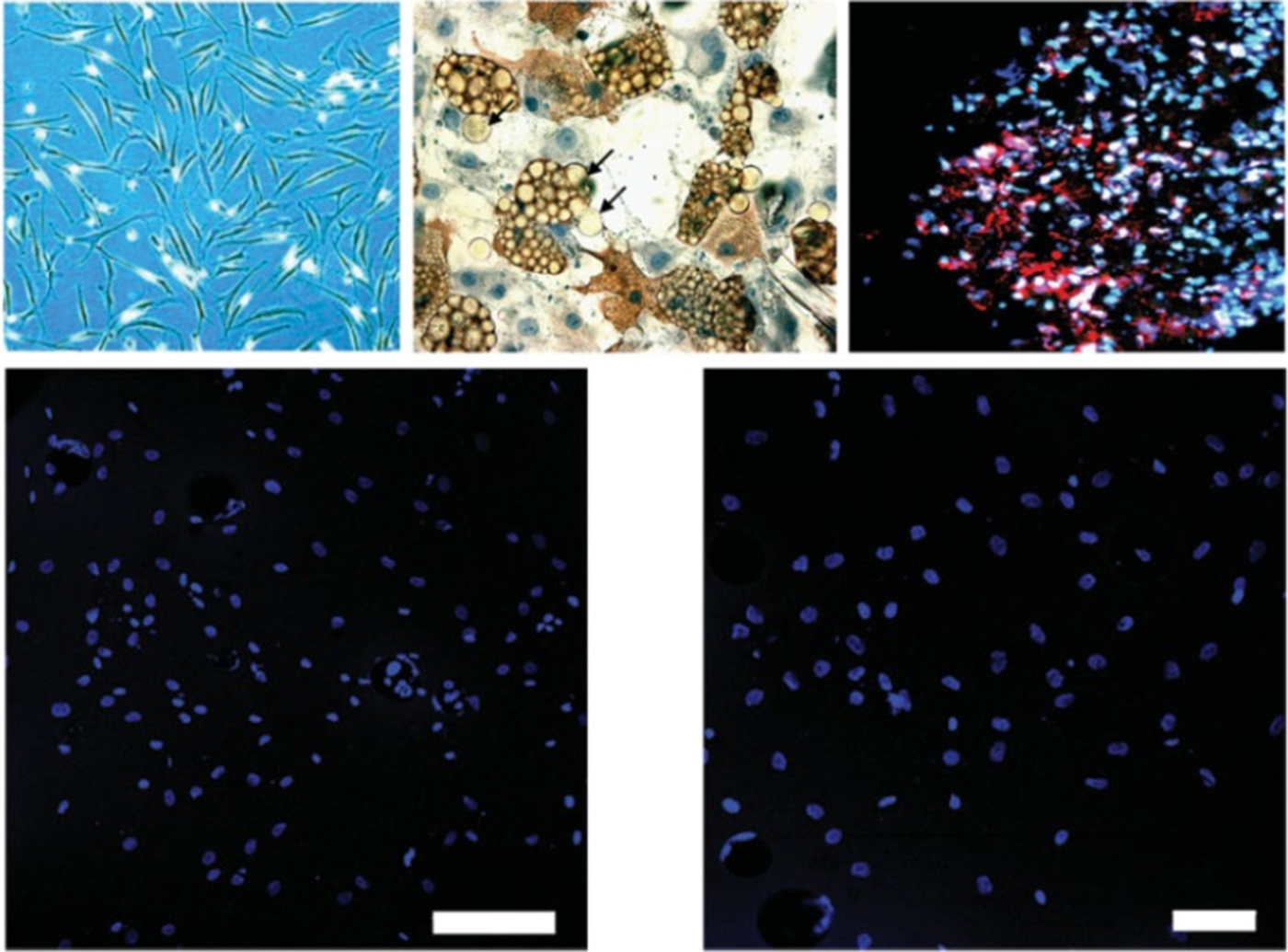
Pierce et al.[Reference Pierce, Pittermann, Ma, Gebauer, Neffe, Holscher, Jung and Lendlein10] fabricated a gelatin-based hydrogel with tunable mechanical properties by crosslinking different concentrations of gelatin using various amounts of ethyl lysine diisocyanate in PBS solution; they investigated the viability of the human MSCs seeded on these hydrogels. These gelatin-based materials show promising results for in vitro MSC proliferation. Preliminary cytotoxicity testing with L929 murine fibroblasts showed low cytotoxicity as measured by examining cell morphology (Fig. 6), release of extracellular lactate dehydrogenase, and mitochondrial activity after 48 h of culture time; the eluates from all samples showed a non-toxic response. Long-term cell culture was possible with viable bone marrow-derived MSCs cultures for nine days. Additionally, preadsorption with fibronectin enhanced MSC attachment, leading to an increase in the number of viable MSCs; the mean cell number of the highest proliferation of MSCs for fibronectin-adsorbed materials was 10,260 ± 312 cells/film and the lowest cell count on fibronectin-adsorbed materials was 2895 ± 293 cells/film.
The top images illustrating morphological appearance of MSCs and the bottom representing the viable MSCs after 9 days culture seeded on G13_LNO5, and Hoechst 33258 (blue) has been used for clarifying viable MSC nuclei; reprinted from Ref. Reference Pierce, Pittermann, Ma, Gebauer, Neffe, Holscher, Jung and Lendlein10. Copyright (2012), with permission from John Wiley & Sons, Inc.

Synthetic stem cell niches with mesenchymal stem cells differentiation
Understanding the effect of the chemical, mechanical, and geometric properties of stem cell niches is essential to stem-cell-based therapies, which include the regeneration of injured cell, tissue, and/or organ function. Engineering synthetic niches, artificial microenvironments mimicking particular interactions between stem cells and the extracellular surroundings in three dimensions, is a promising solution to regulate stem cells fate.[Reference Nava, Raimondi, Credi, Marco, Turri, Cerullo and Osellame63, Reference Joddar and Ito64] Varying the geometry and mechanical properties can be achieved using additive manufacturing techniques and previously discussed chemical modifications. Additional manipulation of the chemical microenvironment can be obtained by introduction of additives or by functionalization with various peptides. Angele et al. showed that composite scaffolds containing hyaluronan and gelatin promoted chondrogenic differentiation of human MSCs. Gelatin-containing composite scaffolds exhibited stronger promotion of collagen type II than pure hyaluronan scaffolds.[Reference Angele, Muller, Schumann, Englert, Zellner, Johnstone, Yoo, Hammer, Fierlbeck, Anglle, Nerlich and Kujat65] Bian et al. showed that functionalizing HA hydrogels with N-cadherin mimetic peptides can promote early chondrogenesis of human bone marrow-derived MSCs and cartilage-specific matrix production.[Reference Bian, Guvendiren, Mauck and Burdick66] Nava et al. fabricated 3D structural niches in an organic–inorganic photoresist (SZ2080) with 1% concentration of Irgacure 369 photoinitiator [2-benzyl-2-dimethylamino-1-(-4-morpholinophenyl)-butanone-1] via 2PP and investigated tuning the mechanical properties of the niches through coating with thin layers of biomimetic hyaluronan- and gelatin-based hydrogels. A higher metabolic activity of MG63 cells, indicative of differentiation toward the osteochondral lineage, was observed in gelatin-coated niches relative to hyaluronan-coated niches because cells had a tendency to adhere to and proliferate on flat surfaces with relatively high stiffness substrates (e.g., hyaluronan-coated samples). In contrast, cells on softer gelatin-coated niches did not move toward nor proliferate on the flat coated niches.[Reference Nava, Raimondi, Credi, Marco, Turri, Cerullo and Osellame63] These studies illustrated the role of matrix microenvironment in chondrogenic differentiation of MSCs.
Encapsulating growth factors and live human endothelial cells by three-dimensional plotting
An approach for combining more than one material by using extrusion-based additive manufacturing[Reference Mandrycky, Wang, Kim and Kim67] (e.g., multi-channel printers) has been attractive for years. The 3D extrusion of two different biomaterials in a core/shell (c/s) platform has been of great interest for fabricating novel biomedical materials. For instance, composite c/s biomaterials with different mechanical properties that consist of a stiffer shell that supports a softer core can be used to load drugs or growth factors; these structures are capable of performing dual release with controllable release kinetics.[Reference Akkineni, Ahlfeld, Lode and Gelinsky68] Moroni et al.[Reference Moroni, Hendriks, Schotel, de Wijn and van Blitterswijk69] successfully fabricated c/s scaffolds via extrusion-based 3D printing of molten polymers for cartilage tissue engineering. It should be noted that 3D printing at high temperatures is incompatible with incorporation of drugs, incorporation of living cells, or incorporation of biologic components (e.g., growth factors) due to the destruction of these substances at these high temperatures. The 3D plotting,[Reference Akkineni, Ahlfeld, Lode and Gelinsky68] extrusion-based additive manufacturing at physiological or room-temperature conditions, has been used for these purposes with the added benefit of being a simple, precise, and highly controllable process for fabricating biomaterials. Akkineni et al.[Reference Akkineni, Ahlfeld, Lode and Gelinsky68] successfully plotted robust and mechanically stable c/s 3D scaffolds with: (a) highly concentrated (16.7 wt%) alginate hydrogels as a shell material and (b) soft biopolymer hydrogels, including alginate, chitosan, gellan gum, gelatin, and collagen hydrogels, as core materials. The shell materials act as a physical barrier and protect the cells/growth factors encapsulated in the core. In addition, the release kinetics of the drug or growth factors can be regulated by tuning the thickness and degradability of the shell. Their c/s scaffolds demonstrated dual release of vascular endothelial growth factor and bone morphogenetic protein 2 loaded in the core and shell, respectively. In addition, human endothelial cells were encapsulated in the core material of the scaffolds, illustrating that living cells can be included in the 3D-plotting process.
Scaffolds for wound dressings
Wound care materials should provide a warm and moist environment for a rapid healing process; in addition, they should prevent the proliferation of bacteria around the wound area.[Reference Winter70–Reference Choi, Hong, Lee, Song, Park and Nam73]. Consequently, wound dressing hydrogels with biodegradability, good fluid absorbance, transparency, and optimal water vapor permeability are preferred over the preformed dressings (e.g., commercial dressings in the forms of membranes and sheets) for the wound healing process.[Reference Balakrishnan, Mohanty, Umashankar and Jayakrishnan74]
Balakrishnan et al. investigated an oxidized alginate- and gelatin-based hydrogel for wound dressing application via in vivo study in a rat model. Their hydrogel dressing shows promising results with relatively low water vapor transmission rate compared with commercially available wound dressing products and good water absorptivity. The improved water retention facilitated the development of a moist environment that is conducive to wound healing; the alginate- and gelatin-based hydrogel was shown to enhance cell migration and re-epithelialization. At 15 days, the wound defects in the rat model filled up to 95.3%[Reference Balakrishnan, Mohanty, Umashankar and Jayakrishnan74] (as shown in Fig. 7).
Images illustrating the appearance of (a) the wound excised on the rat model; (b) gel applied on wound; (c) control wounds at 5, (e) 10, (g) 15 days, respectively; (d) test wounds at 5 days, (f) 10 days, and (h) 15 days; reprinted from. Ref. Reference Balakrishnan, Mohanty, Umashankar and Jayakrishnan74. Copyright (2005), with permission from Elsevier.

Conclusions
Gelatin-based hydrogels have exhibited many attractive aspects for uses and improvements in biomedical applications, including drug delivery devices, tissue engineering scaffolds, and wound dressings. The hydrogels can also be patterned via novel fabrication approaches (e.g., UV stereolithography, 2PP, and gamma irradiation) and their mechanical properties tuned using multiple established procedures. Because of its fascinating features, gelatin hydrogels are one of the most promising materials for use in new types of medical devices.
Acknowledgments
The authors would like to acknowledge support from the US National Institutes of Health (5 R21 AI117748 02).