
Introduction
Electrospinning has quickly grown as an attractive method to generate polymeric nanofibrous mats to be used for applications in biomedical engineering and tissue regeneration.[ Reference Agarwal, Wendorff and Greiner 1 ] Electrospinning offers the capacity of replicating the morphological characteristics of tissue matrix in chemistry, mechanical properties, orientation, geometry, and scale.[ Reference Walters and Stegemann 2 , Reference Almodovar, Castilla Casadiego, Ramos Avilez and Vrana 3 ] The versatility of this technique allows for the fine-tuning of the final properties of the scaffold by controlling the different operational variables.[ Reference Schiffman and Schauer 4 ] Biopolymers—including proteins and polysaccharides—can be electrospun in order to create structures that replicate native soft tissues.[ Reference Schiffman and Schauer 4 ] Moreover, polymeric solutions containing inorganic particles may be electrospun in order to obtain composite nanofibrous scaffolds that are mimetic to mineralized bone tissue. However, electrospinning of biopolymers is challenging due to their limited solubility in ideal electrospinning toxic solvents.[ Reference Schiffman and Schauer 4 ]
Collagen, which is the most abundant protein in the extracellular matrix (ECM) of human tissues, has been previously electrospun from toxic solvents such as trifluoroacetic acid,[ Reference Huang, Shanmugasundaram, Masih, Pandya, Amara, Collins and Arinzeh 5 ] and 1,1,1,3,3,3,-hexafluoro-2-propanol.[ Reference Rho, Jeong, Lee, Seo, Park, Hong, Roh, Cho, Park and Min 6 ] Although nanofibers are produced with these solvents, they tend to denature the collagen, yielding a gelatinous scaffold.[ Reference Zeugolis, Khew, Yew, Ekaputra, Tong, Yung, Hutmacher, Sheppard and Raghunath 7 ] Moreover, the use of these solvents poses a risk to the manufacturer as toxic fumes are produced, requiring extra care and proper facilities when performing the electrospinning process. Lastly, extensive post-processing procedures are required to remove solvent traces prior to contact with any biologic agent.
To avoid problems associated with using harsh solvents, investigators have focused on fabricating electrospun scaffolds using mild “green” solvents.[ Reference Liverani and Boccaccini 8 , Reference Agarwal and Greiner 9 ] Recently, several researchers[ Reference Kazanci 10 – Reference Liu, Teng, Chan and Chew 14 ] and our previous work[ Reference Castilla Casadiego, Ramos Avilez, Herrera-Posada, Calcagno, Loyo, Shipmon, Acevedo, Quintana and Almodovar 15 ] have demonstrated the formation of electrospun collagen nanofibers using acetic acid as a mild solvent. The advantage of using acetic acid as an electrospinning solvent for collagen is that it preserves the secondary structure of collagen.[ Reference Castilla Casadiego, Ramos Avilez, Herrera-Posada, Calcagno, Loyo, Shipmon, Acevedo, Quintana and Almodovar 15 ] Moreover, we have previously demonstrated that by adjusting the voltage or the flow rate, the diameter and orientation of the electrospun collagen nanofibers can be adequately controlled. We obtained a collagen nanofiber diameter ranging from 150 to 400 nm, by operating the electrospinning equipment at conditions involving an applied voltage between 25 and 45 kV and a solution flow rate of 0.5–3 mL/h.[ Reference Castilla Casadiego, Ramos Avilez, Herrera-Posada, Calcagno, Loyo, Shipmon, Acevedo, Quintana and Almodovar 15 ] We also demonstrated that it is possible to modulate the elastic modulus of the nanofibers in the range of 2.7–4.1 MPa, by the selection of the crosslinking method.[ Reference Castilla Casadiego, Ramos Avilez, Herrera-Posada, Calcagno, Loyo, Shipmon, Acevedo, Quintana and Almodovar 15 ]
Electrospinning of composite inorganic/polymeric scaffolds for biomedical applications also suffers from the same limitations of the requirement of harsh solvents. A collagen/hydroxyapatite (COL/HA) electrospun scaffold would be ideal for bone tissue engineering applications, since the ECM of bone is composed of an organic phase (elastic proteins that provide fracture resistance) and a mineral phase (inorganic mineral that contributes to the rigidity in bones). The organic matrix of bone is composed of structural proteins, such as collagen, which corresponds to 90% of the total protein content in bone ECM.[ Reference Young 16 ] Of the different types of collagen, the most abundant in bone tissue is type I collagen.[ Reference Young 16 ] Its mineral phase, which represents 70%–90% of the bone, is primarily composed of hydroxyapatite.[ Reference Young 16 ]
Initial efforts to produce electrospun COL/HA nanofibrous scaffolds used highly toxic fluorinated solvents.[ Reference Teng, Lee, Wang and Kim 17 – Reference Ji, Bar-On and Wagner 19 ] The use of these solvents limits the potential application of these scaffolds in tissue engineering. Another approach to prepare COL/HA nanofibers is to include a carrier polymer to aid during the electrospinning process. COL/HA nanofibers blended with chitosan[ Reference Zhang, Reddy, Wong, Li, Su, Ramakrishna and Lim 20 ] and polyvinyl alcohol[ Reference Asran, Henning and Michler 21 , Reference Song, Markel, Wang, Shi, Mao and Ren 22 ] have been successfully electrospun as previously reported. As a way to overcome the limitations of fabricating electrospun, COL/HA nanofibers using toxic solvents, Li and co-workers recently fabricated a nanofibrous mat using a phosphate buffered saline/ethanol mixture as the electrospinning solvent.[ Reference Zhou, Yao, Wang, Wang, Liu and Li 23 ] They obtained a collagen nanofibrous scaffold with preserved secondary structure and a fiber diameter range of 500–820 nm.[ Reference Zhou, Yao, Wang, Wang, Liu and Li 23 ]
In this work, we demonstrate a simpler and more efficient way of producing COL/HA nanofibers with a diameter of approximately 342 ± 67 nm using acetic acid and water as a “green” electrospinning solvent. This formulation yields a nanofibrous structure, that maintains collagen's chemical structure, and that supports pre-osteoblast adhesion. This work highlights the potential of acetic acid as a “green” solvent for the fabrication of a collagen composite scaffold with possible biomedical applications.
Experimental section
Materials
sAcetic acid (Acros Organics) was prepared at 90% (v/v) diluted in ultra-pure water at 18 MΩ (NANOPURE unit, Thermo Scientific, Marietta, OH, USA). The electrospinning solution consisted of 10% (w/v) of hydroxyapatite nanopowder with a particle size of <200 nm (Sigma Aldrich, St. Louis, MO, USA) and 20% (w/v) of lyophilized type I collagen from cow tendons (generously donated by Integra LifeSciences Holdings Corporation, Añasco, PR) dissolved in 90% (v/v) acetic acid.
Solution preparation
A solution containing 10% (w/v) hydroxyapatite nanopowder in 90% (v/v) acetic acid was first heated to 90 °C for 5 min with constant agitation at 125 rpm. The collagen was then added slowly in small pieces to the heated solution and allowed to stand under constant agitation for 90 min for a final collagen concentration of 20% (w/v).
Electrospinning collagen/hydroxyapatite
The prepared electrospinning solution was loaded into a 3 mL luerlock syringe (BD™, VWR International) with a blunt end nozzle, controlled by a syringe pump (Model No. ne-300, Farmingdale, NY, USA). The solution was pumped through a capillary blunt steel 23-gauge needle (CML Supply). The needle was connected to the positive output of a high-voltage power supply (GAMMA HIGH, Ormond Beach, FL, USA). To collect the nanofibers, a rotating drum was wrapped with aluminum foil and located at a fixed distance of 10 cm from the needle. The flow rate of the solution, relative humidity, applied voltage, and spinning time were fixed at 3 mL/h, 55%, 47 kV, and 3 h, respectively, at a temperature of 20 °C. The resulting membrane was crosslinked for 24 h using glutaraldehyde vapor from a 25% (v/v) glutaraldehyde solution as described in our earlier work.[ Reference Castilla Casadiego, Ramos Avilez, Herrera-Posada, Calcagno, Loyo, Shipmon, Acevedo, Quintana and Almodovar 15 ]
Scanning electron microscopy (SEM) and energy-dispersive X-ray spectroscopy (EDAX)
A JEOL JSM-6390 SEM operated at 10 keV was used to evaluate nanofiber morphology and pre-osteoblast adhesion. The nanofibers were coated with gold using a Denton Vacuum Desk IV sputter coater to improve conductivity. The instrument was placed in the EDAX mode to analyze the chemistry and the distribution of HA in the collagen nanofiber.
Fourier transform infrared (FTIR) spectroscopy and Particle Size Analyzer
FTIR was used to confirm the presence of the functional groups of type I collagen and HA. The infrared spectra of the samples were collected using an ALPHA Platinum Bruker with a Diamond ATR holder with a wavenumber range of 500–1800 cm−1 using 300 scans at 4 cm−1 resolution. The diameter of the as-received HA particles was evaluated using a Particle Size Analyzer (NanoBrook 90Plus, Brookhaven Instruments).
In vitro study
An in vitro study was conducted to observe cellular adhesion over the COL/HA crosslinked nanofibrous scaffold using rat pre-osteoblasts MC3T3-E1 (ATCC, Manassas, VA). The MC3T3 subline of cells was used in this work because it has been categorized as a model system in the biology of bone tissue, and because of its potential to differentiate towards the osteogenic lineage.[ Reference Towler, Arnaud, Bilezikian, Raisz and Rodan 24 ] COL/HA scaffolds, crosslinked with 25% glutaraldehyde vapor, were cut with a punch hole to produce thin disks which fit into the wells of a 96-well plate. The membranes were soaked in phosphate buffered saline overnight to remove any possible solvent or crosslinker residue. Prior to cell seeding, the membranes were sterilized by exposure to UV light while being submerged in sterile PBS for 30 min. MC3T3-E1 cells dispersed in growth medium (αMEM containing 10% calf serum from Gibco®) were seeded on the sterilized samples at 5000 cells/well and incubated for 24 h at 37 °C and 5% CO2. Once the incubation period was completed, the cells were fixed by depositing 200 µL of formaldehyde at a concentration of 3.7% in each well and their nuclei were stained with the Hoechst (Sigma) fluorescent stain. Fluorescence images were captured using an Olympus IX71 confocal microscope (Olympus America; PA, USA). For SEM evaluation, the samples were fixed with 3.7% formaldehyde, dehydrated using ethanol solutions of sequentially increasing concentration (30%, 50%, 70%, and 100%), and further dried in a desiccator overnight. The samples were observed in the SEM as mentioned above.
Results and discussion
Nanofiber morphology
SEM micrographs presented in Fig. 1 reveals the as-spun collagen nanofibrous mat without HA [Fig. 1(a) and 1(b)] and that we can successfully electrospin the COL/HA solution to yield a nanofibrous composite mat [Fig. 1(c) and 1(d)]. On comparing the images, it is evident that the surface of the COL/HA nanofiber mat is drastically altered by inclusion of hydroxyapatite [Fig. 1(c)]. An increase in roughness for the COL/HA scaffolds is observed as compared with the smooth collagen fibers [Fig. 1(a)]. At higher magnifications, we can observe the distribution of the HA nanoparticles within the nanofibrous scaffold [Fig. 1(d)], in which larger particles are embedded within the fibers, while smaller particles decorate the surface of the fiber. In Fig. 1(d), the different diameters of the hydroxyapatite particles (acquired from Sigma Aldrich, manufacturer reports sizes less than 200 nm) can be appreciated. We evaluated the diameter of the as-received HA particles using a particle size analyzer, and we observed two distinct populations with respect to particle diameter [Fig. 1(f)]. We observe a population with an average particle diameter of about 149 nm, and a smaller population with an average diameter of about 448 nm. This distribution is in agreement with the size of the HA particles in the COL/HA fibers [Fig. 1(d)] and in accord with the particles dispersed in an SEM stub and imaged separately [Fig. 1(e)]. Using ImageJ®, we compared changes in fiber diameter as a result of the addition of HA. We observed that, under the operating conditions described in the methodology, as-spun collagen fibers without HA yields an average diameter of 142 ± 73 nm, while the addition of HA causes an increase in the average diameter to 342 ± 67 nm. However, the average diameter of the collagen nanofibers may be modulated by varying the electrospinning operating conditions as we have previously demonstrated by adjusting the injection flow or the voltage.[ Reference Castilla Casadiego, Ramos Avilez, Herrera-Posada, Calcagno, Loyo, Shipmon, Acevedo, Quintana and Almodovar 15 ]
SEM micrographs of (a) collagen nanofibers (5500× magnification), (b) collagen nanofibers (11000× magnification), (c) collagen/hydroxyapatite nanofibers (5500× magnification), (d) collagen/hydroxyapatite nanofibers (11000× magnification), (e) hydroxyapatite dispersed on a SEM stub, inset represents higher magnification (16000× magnification), and (f) size distribution of the hydroxyapatite nanoparticles in solution as received.

Nanofibers composition
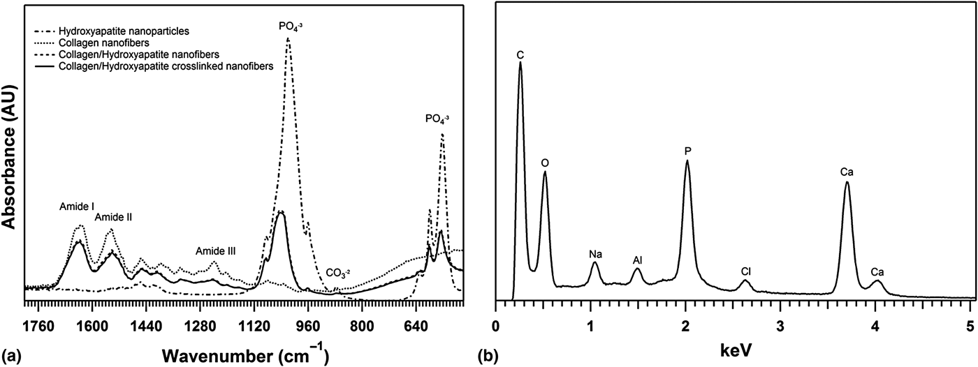
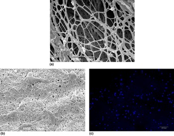
FTIR studies confirmed that the characteristic bands of type I collagen and HA were clearly present, after the nanofiber fabrication process. In Fig. 2(a), the characteristic absorption peaks of collagen can be observed including the amide I (1700–1600 cm−1), amide II (1600–1500 cm−1), and amide III (1300–1180 cm−1) peaks for the electrospun collagen without HA.[ Reference Castilla Casadiego, Ramos Avilez, Herrera-Posada, Calcagno, Loyo, Shipmon, Acevedo, Quintana and Almodovar 15 , Reference Camacho, West, Torzilli and Mendelsohn 25 ] The HA particles exhibit absorptions bands corresponding to the phosphate PO4 −3 (900–1100 cm−1 and 500–650 cm−1)[ Reference Trommer, dos Santos and Bergmann 26 ] and carbonate CO3 −2 (877 cm−1 and 1400–1500 cm−1[ Reference Venugopal, Low, Choon, Sampath Kumar and Ramakrishna 18 , Reference Trommer, dos Santos and Bergmann 26 ]) groups. These characteristic peaks of HA were present in the COL/HA nanofibers spectra, thus confirming the inclusion of the HA particles within the collagen nanofibrous scaffold. In order to use the nanofibrous scaffold for cell culture, it must be crosslinked to a small degree to avoid instantaneous dissolution. We have previously demonstrated a methodology for crosslinking collagen nanofibers by exposure to glutaraldehyde vapor from a 25% (w/v) solution.[ Reference Castilla Casadiego, Ramos Avilez, Herrera-Posada, Calcagno, Loyo, Shipmon, Acevedo, Quintana and Almodovar 15 ] In this work, we crosslinked the COL/HA composite nanofibers by exposing the sample to glutaraldehyde vapor from a 25% (w/v) solution. FTIR confirms [Fig. 2(a)] that the crosslinking procedure did not affect the chemical structure of the native collagen or the HA. SEM reveals that the fibrillar structure of the COL/HA fibers is maintained after crosslinking [Fig. 3(a)].
(a) Infrared spectra of hydroxyapatite nanoparticles, collagen nanofibers, and COL/HA nanofibers with and without crosslinking. (b) EDAX spectrum of COL/HA nanofibers.

(a) SEM micrograph of COL/HA nanofibers after glutaraldehyde crosslinking. (b) SEM micrograph of MC3T3-E1 pre-osteoblast cultured on a COL/HA nanofibrous scaffold. (c) Fluorescent image of MC3T3-E1, cultured on a COL/HA, cell nucleus staining (scale 200 µm).

EDAX was used to observe the distribution of HA in the collagen nanofiber. The results reveal that calcium and phosphorus elements were present on the nanofiber surface. Spectra in Fig. 2(b) reveal that the peaks at 2 keV refer to P and the peak at 3.7 keV to Ca.[ Reference Venugopal, Low, Choon, Sampath Kumar and Ramakrishna 18 , Reference Apella, Venegas and Rodenas 27 ] Elemental analysis yields a Ca:P ratio of 1.74, which is close to that expected for HA.[ Reference Varma, Yokogawa, Espinosa, Kawamoto, Nishizawa, Nagata and Kameyama 28 ]
In vitro cellular evaluation
To demonstrate that the fabricated COL/HA scaffold supports cell adhesion, MC3T3-E1 cells were cultured on the scaffold. For these experiments, 5000 cells were seeded in wells containing the crosslinked COL/HA scaffold. In Fig. 3(b), the SEM micrograph demonstrates that cells adhere well to the COL/HA scaffold after 24 h of culture. Figure 3(c) shows a fluorescent image of MC3T3-E1 cell nuclei stained with Hoescht. Counting the cells adhered per area, we estimate that 50% of the initial amount seeded successfully adhered on the COL/HA scaffold. These results are in agreement with our previous results that demonstrate that electrospun collagen scaffolds supports fibroblast cell culture.[ Reference Castilla Casadiego, Ramos Avilez, Herrera-Posada, Calcagno, Loyo, Shipmon, Acevedo, Quintana and Almodovar 15 ] The inclusion of HA on collagen scaffolds is ideal given that HA promotes osteoblastic matrix formation as well as pre-osteoblast and mesenchymal stem cell differentiation.[ Reference Manjubala, Ponomarev, Wilke and Jandt 29 , Reference Shah, Hong, Hyder and Hammond 30 ] Incorporating HA into electrospun collagen nanofibers using a “green” solvent may lead to solutions in bone regeneration as an ECM bone mimetic structure containing both an organic and a mineral phase with similar chemistry, morphology, and mechanical cues as that of native ECM.
Conclusions
Our results demonstrate a new and simple methodology to prepare a composite COL/HA nanofibrous scaffold using an acetic acid/water solution as a “green” solvent. SEM revealed the stability of the developed nanofibers, with an average diameter of 342 ± 67 nm. We were able to demonstrate that these nanofibers preserve the chemical composition in the organic phase as well as in the mineral phase. HA particles either decorated the surface of the fibers, or appear to be embedded within the fibers. This phenomenon was observed to be dependent on the diameter of the particle. In vitro studies with pre-osteoblasts showed the ability of the cells to adhere to the nanofibrous scaffold, which indicates that this non-toxic ECM mimetic structure of bone tissue could be useful in tissue regeneration.
Acknowledgements
This work was financially supported by the NSF's Puerto Rico Institute for Functional Nanomaterials (EPS-1002410), by the Puerto Rico Research, Science, and Technology Trust (Agreement Number 2016–00067), and by the “Programa de Apoyo Institucional Para la Formación en Estudios de Posgrados en Maestrías y Doctorados de La Universidad del Atlántico, Colombia” by providing DCC a scholarship. The authors thank Dr. Ricky Valentín for access to the electrospinning equipment; Dr. David Suleiman for access to the FTIR apparatus; Christian Rivera for assistance using the EDAX equipment; Beatriz Quiñones for her support in the use of the FTIR; Ana M. Reyes for her support with the Particle Size Apparatus; Edwin Burgos, Coral Irizarry, & the UPRM Center for Biomedical Engineering and Nanomedicine for support of the in vitro experiments; and José Almodóvar for providing assistance with the confocal microscope. The authors thank Dr. Anibal Quintana from Integra Lifesciences for generously donating collagen.