Introduction
Considering the time-honoured principle of non-maleficence (‘Primum non nocere’)Reference Aggarwal, Mytton, Derbrew, Hananel, Heydenburg and Issenberg1 and the changes made to the Brazilian national medical school curriculumReference Flores, Bez and Bruno2 in recent years, the search for alternatives to conventional medical education has become imperative. In this scenario, the use of simulators has proved to be an efficient, feasible alternative for several Brazilian medical schools, offering an important tool at a time of broad transition between passive and active teaching methodsReference Luna and Spight3 that can help to improve care quality and patient satisfaction.Reference Nogueira Júnior and Cruz4
The motor learning process consists of three distinct stages.Reference de Sousa, Okada and Suzuki5 The cognitive stage is when the student acquires the necessary technical knowledge to perform a certain task. The associative stage consists of supervised training on live human patients. Finally, during the autonomous stage, the student has consolidated both knowledge and technique and gains progressive independence to act. Simulators allow enhancement of the cognitive stage and task-based teaching,Reference Pazin Filho and Scarpelini6 which can optimise the learning process.
Otology has stood out as the foremost user of virtual and physical simulators over the years.Reference Musbahi, Aydin, Omran, Skilbeck and Ahmed7 Recently, several simulators, for different purposes, have been fabricated with the aid of three-dimensional (3D) printing technology,Reference Gadaleta, Huang, Rankin, Hsue, Sakkal and Bovenzi8,Reference Barber, Kozin, Dedmon, Lin, Lee and Sinha9 demonstrating their varied applications.
Tympanostomy is one of the most prevalent surgical procedures in the paediatric age groupReference Testa, Dimatos, Greggio and Duarte10 and is the most common procedure performed by otolaryngologists in the USA.11 At over 600 000 procedures per year, it accounts for 20 per cent of all out-patient surgical procedures.Reference Cullen, Hall and Golosinskiy12 It is therefore imperative that tympanostomy technique is mastered by otological and general surgeons. Tympanostomy consists of the placement of ventilation tubes in the tympanic membrane in order to treat conditions such as otitis media with effusion.Reference da Costa, Francesco, Lessa, Abdo, Felix and Moraes e Silva13
It is not a complication-free procedure. Early extrusion of the tympanostomy tube, improper tube placement into the middle ear and tympanic granuloma have all been reported.Reference Kay, Nelson and Rosenfeld14 Therefore, theoretical and practical expertise is required from the surgeon, who can only acquire such experience in a good medical residency programme.
Within this context, the present article proposes a simulation model for training of myringotomy, tympanostomy and ventilation tube placement, assisted by 3D printing. The simulator was designed to have the lowest possible cost, be easily reproducible and pose no danger to patients or surgeons in training.
Materials and methods
Image acquisition and simulator dimensions
Three coronal computed tomography sections of the temporal bone (0.3 mm thick bone window) of an adult, an eight-year-old child and a five-year-old child were used to obtain internal measurements between the midpoint of the opening of the external auditory canal to the midpoint of the tympanic membrane and from there via the shortest distance to the promontory. These dimensions, captured through RadiAnt™ Dicom® Viewer 2020.1 medical image viewing software, were used to generate a 3D model from Dicom files.
Tympanic membrane
A clear plastic bag interposed between the detachable portion and base of the simulator was used to simulate the tympanic membrane. This was used because the plastic bag was free of charge and has similar properties to the tympanic membrane.
Digital fabrication of simulator
The 3D modelling was done in Rhinoceros® (version 5.0) graphics and design software, followed by Simplify 3D® (version 3.0) 3D-printing software to generate files in the 3D-printable stereolithographic (.stl) format.
Three-dimensional printing
Fused deposition modelling technology was used to print the design. Fabrication was performed in a GTMAX3D Graber I3 3D (GTMax3D Equipamentos Eletrônicos e Materiais Plásticos LTDa, Americana, Brazil) printer running Repetier Version 1.0 control firmware (Hot-World GmbH & Co. KG, Willich, Germany). The selected material was acrylonitrile butadiene styrene polymer in natural white.
Proposed simulator design
The simulator was designed with several principles in mind: affordable cost; replicability; portability; intuitiveness and ease of assembly, use and reuse; and fidelity to the actual structures. Taking into account the aforementioned factors, a small, polygonal simulator was designed in which surgery could be performed both under an operating microscope and through an endoscope.
The simulator was designed in a rectangular shape and divided into a base and three upper detachable portions, which connected to the base by magnets. These detachable portions each have a cylindrical canal with an internal aspect that simulates the external auditory canal and tympanic cavity, separated by the ‘tympanic membrane’, a piece of plastic bag interposed at the interface where the base and detachable portions connect. The magnetised interface holds the tympanic membrane and allows it to be changed easily. The simulator is 12 cm wide, 4 cm tall and 4 cm long.
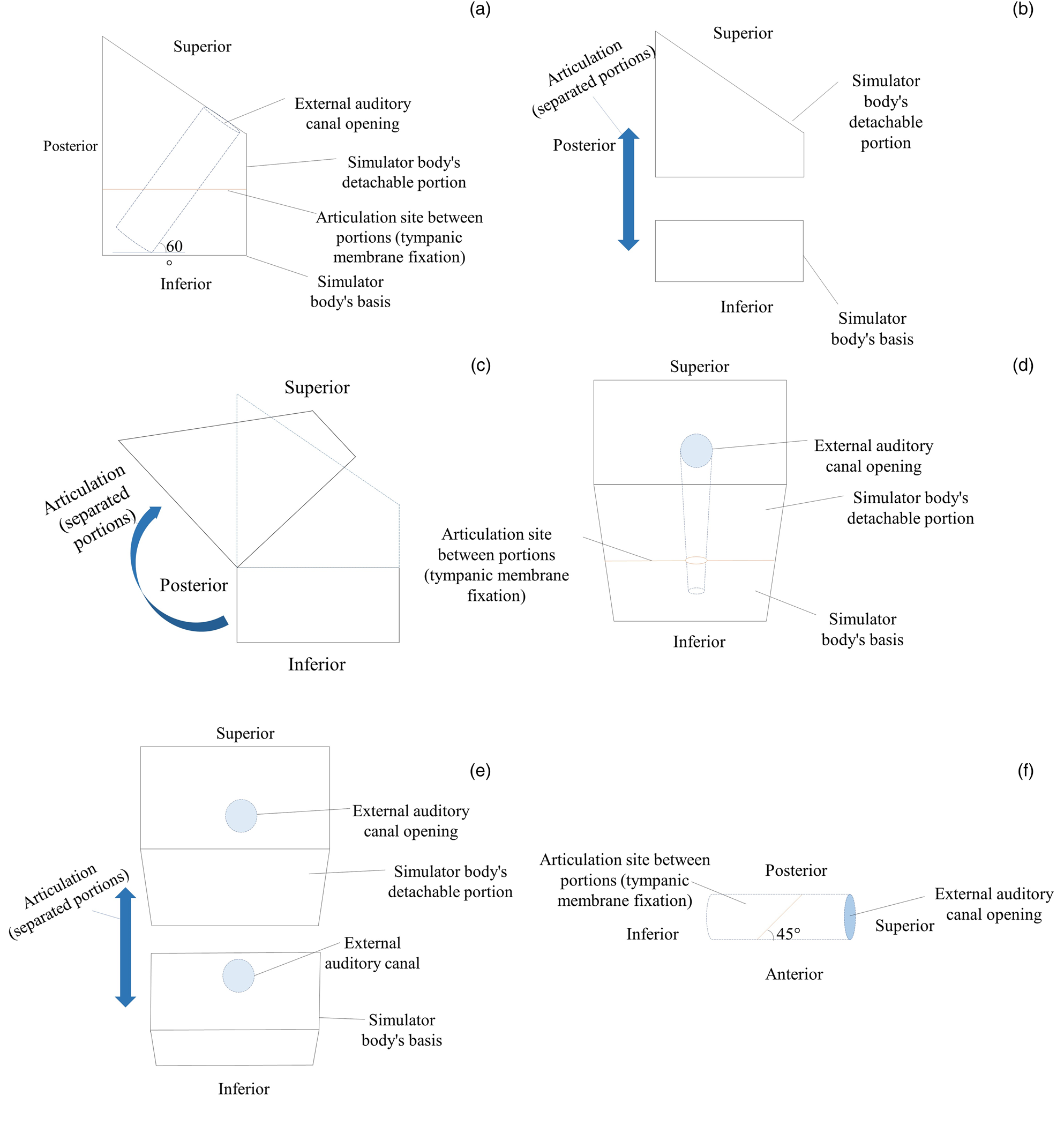
Considering the surgery position for tympanostomy, the external auditory canal of the simulator was angled at 60° in relation to the horizontal surface (Figure 1). The tympanic membrane was angled at 45° in relation to the external auditory canal as seen on the Dicom images.
Initial (general) proposed model sketch, with no dimensions specified for the Dicom image samples. (a) Side view; (b) side view with exploded parts, showing a distance between the detachable portions and the base; (c) side view showing the articulation between the detachable portions and the base; (d) anterior view; (e) anterior view with exploded parts, showing a distance between the detachable portions and the base; (f) anatomical view of the cylindrical canal (simulated external auditory canal).

To enhance the realism of the model, the simulated external auditory canal and surface of the upper aspect of the detachable portions were coloured with orange and beige acrylic paint, respectively.
Surgery simulation
The simulated procedure consisted of myringotomy, tympanostomy and placement of a Teflon-coated Shepard 1011 (Medicone Projetos E Soluções Para A Indústria E A Saúde, Cachoeirinha, Brazil) short-term ventilation tube (grommet type). The equipment used was as follows: a 13-cm length, 3-mm thickness, zero-degree Xion fibre-optic otoscope (Berlin, Germany), an Olympus Exera II CLV-180 light source (Hamburg, Germany), a Ferrari Medical MFX 10 G camera (Sao Paulo, Brazil), a Radiance NDS HD display (San Jose, USA), a Carl Zeiss Opmi Visu 150 S7 surgical microscope (with 200f lens and 20 cm focal length; Jena, Germany), a bayonet-handled spear-tip myringotomy knife, a bayonet-handled straight ear pick and a 9 cm Hartmann ear forceps.
The first step was visualisation of the simulated external auditory canal and the tympanic membrane. Myringotomy was performed with the myringotomy knife in the anteroinferior quadrant, and the tympanostomy tube was inserted with the aid of the straight ear pick.
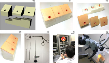
The authors considered three external auditory canals from an adult, an eight-year-old child and a five-year-old child to challenge the surgeons with different dimensions. Therefore, the final version of the simulator contained three types of external auditory canal and tympanic cavity in parallel (Figure 2a–d).
Final planned and printed model. (a) Model of the simulator showing the base and detachable portions. (b) Anterior view, with the detachable portions joined to the base via magnets. (c) Anterior view showing the detachable portions and shared base. Note that magnets are glued to the base. (d) Right lateral view, with the detachable portions joined to the base via magnets. (e) Instruments used in the simulated procedure: top left: Shepard 1011 short-term ventilation tube, white; top right, Hartmann ear forceps; bottom left, bayonet-handled myringotomy knife; bottom middle, bayonet-handled straight ear pick; and bottom right, rigid fibreoptic endoscope. (f) Endoscopic procedure. (g) Microscopic procedure with the plastic bag in the simulator.

Project cost
The cost of the 3D model was budgeted at R$300.00 with printing at R$150.00. The two tubes of acrylic paint cost R$8.00 each. The plastic bag was obtained free of charge. The 36 magnets cost R$0.60 each. The total was therefore R$487.60 and yielded a sturdy model that did not require reprinting or finishing. Because of the small area of the simulated tympanic membrane, the single plastic bag could be reused several times.
Data collection and registration
Project data were recorded in Microsoft Office Word® (2007) word processing software. A photographic record was obtained using an iPhone® 6S smartphone camera and duly captioned.
Results
The planning stage was followed by designing, fabricating and finishing the simulator, as illustrated in Figure 2.
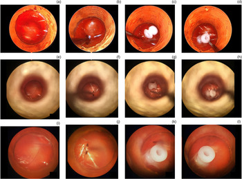
Consecutive tests with several inorganic materials were conducted to find out which was best for simulation of the surgical procedure. Paper (different types and weights) and clear plastics from hospital supply packaging, plastic bags and polyvinyl chloride film were trialled. Of these, the material closest to the tympanic membrane in texture, elasticity, compliance, thickness and transparency was the clear plastic bag (Figure 3).
View of the adult module. (a–d) Operator's view under the 0° rigid endoscope, showing: (a) initial view, (b) myringotomy with the bayonet-handled myringotomy knife, (c) ventilation tube placement with a bayonet-handled straight ear pick, and (d) final view. (e–h) Operator's view under an optical microscope, ×100 magnification, showing: (e) initial view, (f) myringotomy, (g) ventilation tube placement, and (h) final view. (i–l) Operator's view under an optical microscope, ×250 magnification, showing: (i) initial view, (j) myringotomy, (k) ventilation tube placement, and (l) final view.

Discussion
When it comes to surgical education, objective practices must be adopted to improve operative technique and patient safety.Reference Agha and Fowler15 In this study, it was shown that the use of synthetic materials allowed us to achieve these goals and ensure reproducibility through the use of readily accessible and affordable supplies, provided that the necessary surgical instruments and magnifying optics are available. In simulation settings, synthetic material, albeit less tactile, elastic and compliant than organic tissue, enables repetitive use because of its characteristic low cost. Furthermore, it eliminates the risk of contamination, ethical conflicts and difficulty of acquisition.Reference Tan and Sarker16
The cost of model fabrication was low and could be made even lower if the service had a 3D printer and a qualified designer on its staff, which would reduce expenditures to the cost of the filament, paint and magnets used. The cost of the simulator is thus limited to the cost of printing and finishing. On comparison with simulators used in other areas of otolaryngology, as reported in a systematic review,Reference Nogueira Júnior and Cruz4 the production cost of this model is compatible with its fabrication at scale. Compared with existing models of the same procedure,Reference Sparks, Kavanagh, Vargas and Valdez17 the 3D-printed simulator presented here is less expensive and offers the possibility for local production, showing that high-cost devices are unnecessary to achieving an educational objective. Chan et al.Reference Chan, Carrillo and Ong18 also published a low-cost model assembled from pre-existing materials. Therefore, simulators for this procedure can be designed to be affordable to interested institutions and reproducible across different otolaryngology teaching environments. There are several examples in the literature of authors pursuing such goals, including in otological surgery.Reference Barber, Kozin, Dedmon, Lin, Lee and Sinha9,Reference Sparks, Kavanagh, Vargas and Valdez17,Reference Ganguli, Pagan-Diaz, Grant, Cvetkovic, Bramlet and Vozenilek19,Reference Vaitaitis, Dunham, Kwon, Mayer, Evans and Baker20 As of 27 July 2020, the Brazilian real exchange rate was approximately R$5.55 to the dollar (US$ 1.00 = R$0.18); the total cost of the project was therefore US$88.99.
Anschuetz et al. reported better adaptation to use of the endoscope as opposed to the microscope by otologic surgeons, especially in students and residents with little experience in the field.Reference Anschuetz, Stricker, Yacoub, Wimmer, Caversaccio and Huwendiek21 In cadaver training, endoscopy was the method that best avoided accidents, in addition to providing the advantage of wide visualisation in a restricted space.
Nevertheless, both endoscopic and microscopic surgery could be reproduced in the model (Figure 3), although ease of manipulation was greatest with the microscope because there was no technical difficulty due to an excess of instruments in the restricted space of the external auditory canal. Barber et al. Reference Barber, Kozin, Dedmon, Lin, Lee and Sinha9 designed a polylactic acid model for simulation of endoscopic ear surgery in order to improve dominant and non-dominant hand performance through simple transcanal activity; they also noted the difficulty of operating through an endoscope in an inflexible cavity and the challenge inherent to endoscopic surgical technique of being single-handed.
Otological surgery techniques are essentially dependent on operator experience and on the armamentarium available at each service, as evaluated in a comparative pilot study specifically regarding insertion of ventilation tube.Reference Martellucci, Pagliuca, de Vincentiis, De Virgilio, Fusconi and Gallipoli22 Therefore, apprentice surgeons have the possibility of acquiring two distinct techniques, depending on how well they adapt to each. It also bears stressing that it is important to invest in the more recent modality of endoscopic ear surgery, as its popularity is on the rise and it allows better visualisation of structures than the operating microscope, which is limited by lighting and angulation constraints.Reference Cohen, Landegger, Kozin and Lee23 The model proposed in this article allows training in both modalities.
Another advantage of the model is its portability. Its reduced dimensions mean it is easily transported, as are previous models reported in the literature,Reference Sparks, Kavanagh, Vargas and Valdez17,Reference Vaitaitis, Dunham, Kwon, Mayer, Evans and Baker20,Reference Anschuetz, Stricker, Yacoub, Wimmer, Caversaccio and Huwendiek21 and may even allow its later sale and wide distribution.
As for the tympanic membrane simulation, the authors were satisfied with the attributes of a clear plastic bag, which is readily acquired anywhere at little to no cost and can be used countless times. Sparks et al. used tympanic membranes made from rubber at a cost of a few cents of a dollar each; however, these still had to be made to order.Reference Sparks, Kavanagh, Vargas and Valdez17
The use of magnets at the interface between the base and detachable portions of the simulator provided some degree of seal. However, considering that the presence of fluid effusion in the middle ear is common in diseases which indicate tympanostomy, an improved watertight design would be required to simulate this; infusion of water into our simulator caused rapid extravasation. We are planning a new version of the design with this capability, in addition to the possibility of increasing volume in the tympanic cavity, to better simulate the volume of effusion aspirated during the procedure and to avoid leakage during model assembly. In another study, the authors assembled their model from individually printed parts which join together and allow simulation of surgery under the operating microscope.Reference Vaitaitis, Dunham, Kwon, Mayer, Evans and Baker20
Future research might test improvements of the model described herein, which would allow its use for simulation of other invasive techniques, such as transtympanic injection of medication and type I (inlay) tympanoplasty with tragus cartilage graft, for example. Adjustments will be planned for a new and enhanced version of the simulator.
As for the limitations of the model, its light weight was found to cause some difficulty during manipulation, as it was easily displaced during both endoscopy and microscopy, increasing training time during tests performed by the authors.
Additionally, dealing with synthetic materials reduces the technical difficulty inherent to the anatomy and physiology of each individual ear. Such materials do not simulate complicating elements such as bleeding, tortuous or stenotic external auditory canals, manipulation of the ossicular chain, facial nerve canal dehiscence, or adhesive otitis, among others.Reference Ragab, Mohammed, Abdel-Fattah and Afifi24 These are not necessarily disadvantages, but the resulting ease of the simulated procedure is beyond the realm of probability in human patients. Therefore, the aim of the simulator is to provide the surgeon-in-training with the ability to handle instruments in restricted surgical fields, such as the external auditory canal and the tympanic cavity, improving fine motor dexterity in a near-optimal setting before applying these skills to real cases.
It is also worth noting that this is the only description of a model capable of simulating myringotomy and tympanostomy both by microscopy and by endoscopy. The proposed model fulfilled the authors’ expectations and criteria as a safe, inexpensive, reproducible, user-friendly and accessible surgical education tool, which can be improved and reassessed for further research.
• This study proposes the first model for simulating both microscopic and endoscopic surgery
• It is simple to produce a simulation model for otological surgery using three-dimensional (3D) printing or even handmade simulators
• Models from 3D printing are low cost and should be continuously promoted to the medical community
• Medical residency and fellowship services should invest in these technologies for training purposes
• Models can be easily adapted and modified to allow acquisition of new and diverse skills
Competing interests
None declared