




No CrossRef data available.
Hypoplasia of the left heart
Published online by Cambridge University Press: 21 September 2005
Extract
The lesion that, nowadays, is most usually described as “hypoplastic left heart syndrome”, was initially described in terms of “hypoplasia of the aortic outflow tract complex”.1 Sporadic cases with aortic atresia, an intact ventricular septum, and gross hypoplasia of the left ventricle, had been described long before, but it was Noonan and Nadas, in a landmark study, who coined the term “hypoplastic left heart syndrome”.2 The paediatric cardiac community has now accepted this term uniformly, although as we will see, problems remain with regard to precisely which malformations should be included within the “syndrome”. The term itself, nonetheless, is not beyond criticism. This is because, for those working in the genetic community, a “syndrome”, by definition, is a constellation of anomalies afflicting multiple systems of organs. The so-called “hypoplastic left heart syndrome”, however, almost always involves only the heart and the great arteries. Thus, according to the geneticists, it should not strictly be described as a “syndrome”, although the Nomenclature committee of the International Coding Project have marshalled arguments in favour of the term. In this review, nonetheless, we will skirt these problems with the use of “syndrome”, and simply describe the morphology as seen in patients unified because they have hypoplasia of the left heart.
Information
- Type
- Anatomical Considerations
- Information
- Copyright
- © 2004 Cambridge University Press
There is then little difficulty in describing the structure of the “classical” examples having such hypoplastic left-sided structures. The ventricular septum is intact, and there is aortic atresia (Fig. 1), combined either with mitral stenosis and fibroelastosis of the left ventricular cavity (Fig. 2), or mitral atresia and a virtual left ventricle (Fig. 3).
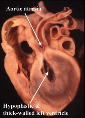
Figure 1. A section replicating the “four-chamber” echocardiographic cut showing the essence of hypoplasia of the left heart as seen with aortic atresia and an intact ventricular septum. There is usual atrial arrangement, the atrioventricular connections are concordant (see Fig. 2), and the ventriculo-arterial connections are potentially concordant, the aorta being in potential continuity with the left ventricle.

Figure 2. This section replicating the echocardiographic four-chamber plane is through a similar heart to that shown in Figure 1, demonstrating the patent mitral valve, and showing gross fibroelastosis of the hypoplastic left ventricle. In this heart, the atrioventricular connections are concordant.

Figure 3. In the section, again replicating the echocardiographic four-chamber plane, and again from a patient with aortic atresia and intact ventricular septum, there is absence of the left atrioventricular connection. The left ventricle is no more than a slit in the posterior wall of the ventricular mass, but lacks any fibroelastotic lining. The heart has a univentricular atrioventricular connection.
In the majority of the cases described by Noonan and Nadas,2 the aortic valve was patent, albeit critically stenotic, and many of the cases had severe coarctation as the leading morphologic feature. Hearts of this type have recently created further problems in terminology. This is because, when there is aortic atresia and an intact ventricular septum, then the only surgical option for treatment is to create a functionally univentricular circulation. In some cases with critical aortic stenosis, nonetheless, it has been shown that it is possible to open the diseased aortic valve and restore a biventricular circulation. In still others, with hypoplasia of the aortic arch and left ventricle, but in the absence of overt stenosis of the aortic and mitral valves, it has been argued that the hearts should be described as the “hypoplastic left heart complex”, thus distinguishing them from those falling at the severe end of the syndrome because they are suitable for biventricular repair.3 As morphologists, we see little to recommend in this suggestion. In our opinion, if a heart was seen by the morphologist with severe hypoplasia of the aortic arch, but without obvious stenosis of the aortic and mitral valves, and in the absence of fibroelastosis within the left ventricle, it is unlikely that it would be diagnosed as falling within the hypoplastic left heart syndrome. In this regard, there is no question but that certain patients with critical aortic stenosis can undergo biventricular repair. Diagnosticians, however, are still seeking for rigid criterions to distinguish these patients during life,4, 5 and a significant proportion fall into the group requiring functionally univentricular repair. The key morphologic feature that permits the morphologist to diagnose these cases as unequivocal examples of hypoplastic left heart syndrome is the presence of fibroelastosis within the left ventricle (Fig. 4). It is not surprising, therefore, that patients with severe aortic coarctation, but without overtly stenotic aortic and mitral valves, and in the absence of any left ventricular fibroelastosis, can be repaired in biventricular fashion. This fact, in our opinion, does not in itself justify creating a new category for their description. Indeed, if this criterion was adopted for definition of the “hypoplastic left heart complex”, then many patients with totally anomalous pulmonary venous connection would also be candidates for inclusion, since it is well known that the left ventricle can be remarkably hypoplastic in this setting, yet still capable of sustaining the systemic circulation subsequent to anastomosis of the anomalous pulmonary veins to the body of the left atrium. As morphologists, we consider it better simply to describe the structural variation seen in the setting of hypoplasia of the left heart, with some of these combinations, but not all, then falling within the grouping of the hypoplastic left heart syndrome.

Figure 4. This heart comes from a patient with critical aortic stenosis and an intact ventricular septum. The right ventricle is apex-forming, and there is gross fibroelastosis of the left ventricle. This patient would unequivocally have required a functionally univentricular repair had surgery been attempted during life.
Segmental combinations
Almost always, hypoplasia of the left heart is found with usual atrial arrangement and potentially concordant atrioventricular and ventriculo-arterial connections (Figs 1 and 2). In the setting of mitral atresia, however, the left atrioventricular connection can be absent, and then the heart shows univentricular connection to a dominant right ventricle with a rudimentary and incomplete left ventricle (Fig. 3). Indeed, in some hearts with mitral atresia, the left ventricle may be no more than an endothelial lined crevice in the posterior wall of the dominant right ventricle, its position being discovered morphologically only when incisions are made between the delimiting coronary arteries. Very rarely, nonetheless, hearts can be found when the ventricular septum is intact, but the atretic pulmonary trunk is in potential connection with the cavity of the hypoplastic left ventricle. These anomalies have been described with either concordant6 or discordant7 atrioventricular connections. Hearts with gross hypoplasia of the left ventricle are also rarely encountered with isomerism of the left atrial appendages. But, in most instances, the segmental combinations are potentially normal, and these are the hearts on which we will concentrate in this review.
The pattern of the circulation
The essence of the hearts having hypoplasia of the left heart is that the circulations are dependent on a functionally single right ventricle. The left ventricle is present, but is either a virtual slit in the ventricular wall, or else a small cavity lined by gross fibroelastosis. The pulmonary venous return, therefore, needs to cross the atrial septum so as to be distributed effectively to the body, whilst the circulation to the upper body and coronary arteries is duct-dependent (Fig. 5). Two key morphological areas, therefore, are the atrial septum and the junction of the arterial duct with the isthmus and the descending aorta, since obstructions at these points can seriously compromise the overall circulatory patterns. There are then, of course, further variations to be found, which can afflict the atrial chambers, the ventricular mass, and the arterial pathways. We will review all these anatomic variations.

Figure 5. The circulation in hypoplasia of the left heart is dependant on the dominant right ventricle, which pumps blood through the pulmonary trunk and via the arterial duct to the descending aorta. The upper body is fed retrogradely through the isthmus, with the retrograde circulation then feeding the coronary arteries through the ascending aorta. The critical areas in the circulation are the isthmus and the atrial septum.
The atrial chambers
As already explained, almost without exception the atrial chambers are arranged in usual fashion, although rarely there may be isomerism of the left atrial appendages. The systemic venous connections are normal, although on occasion there may be fenestrations present between the cavity of the left atrium and the coronary sinus (Fig. 6) that can provide a route for mixing of the venous returns should the atrial septum be intact, which occurs in about one-tenth of cases (Fig. 7). In most instances, the atrial septum is either deficient, or the oval fossa is probe-patent. Indeed, the flap valve of the oval fossa is often aneurysmal, while the group from Children's Hospital of Philadelphia8 described a frequent variation in which the flap valve is deviated to the left relative to the superior interatrial fold, diminishing the size of the left atrium in the process (Fig. 8). In the minority of cases in which the atrial septum is intact, further changes are to be anticipated. In some instances,9, 10 the pulmonary veins become thickened and arterialised (Figs 9 and 10), and both lungs show severe lymphangiectasia (Figs 11 and 12). Fenestrations to the coronary sinus provide the “overflow” in some of these cases, but in others, an alternative route for pulmonary venous return is provided by the so-called “levoatrial cardinal vein”,11 a collateral venous channel running from the roof of the left atrium to a systemic vein, usually the superior caval vein (Fig. 13). The left atrium, as already emphasised, is often small, and in some cases the left atrial appendage can be grossly dilated and abnormally positioned. In most instances, the pulmonary veins are normally connected, but cases have been described with totally or partially anomalous pulmonary venous connection, and rare cases can have division of the left atrium. The latter malformation may be of the “classical” variety, or produced by a supravalvar ridge.

Figure 6. The coronary sinus has been opened from behind, showing fenestrations between its cavity and that of the left atrium. They provided an “overflow” from the atrium, since the patient had had an intact atrial septum in the setting of hypolasia of the left heart.

Figure 7. The right atrium has been opened in a heart from a patient with hypoplasia of the left heart. The oval fossa is intact.

Figure 8. This “four-chamber” section is viewed from behind. It shows leftward deviation of the flap valve of the oval fossa, diminishing the size of the left atrium.

Figure 9. This section is taken transversely across the thorax of a fetus with hypoplasia of the left heart and an intact atrial septum. There is gross thickening of the pulmonary veins, which are also arterialised (see Fig. 10).

Figure 10. This histological section of a pulmonary vein comes from the heart shown in Figure 9. Note the arterial nature of the thickened walls (brackets).

Figure 11. These lungs are removed from another fetus having hypoplasia of the left heart and an intact atrial septum. Note the “cobble stone” nature of the parenchymal surface.

Figure 12. This section from the lungs shown in Figure 11 illustrates the gross lypmangiectasia that ensues in the presence of hypoplasia of the left heart when the atrial septum is intact.

Figure 13. The cartoon shows the course of the so-called “levoatrial cardinal vein” that drains blood from the left atrial roof to the superior caval vein in some patients with an intact atrial septum in the setting of hypoplasia of the left heart.
The atrioventricular junctions
In the majority of cases, the atrioventricular connections are concordant, and the mitral valve, albeit normally formed, is miniaturised in keeping with the hypoplasia of the left ventricle (Fig. 2). When patent, then without exception the left ventricle itself is thick-walled and its lining is thickened and pearly white – showing the changes known as fibroelastosis (Figs 2 and 4). The tendinous cords of the miniaturised mitral valve can then be involved in the fibroelastotic process, producing additional stenosis. In a minority of cases, the valve can become imperforate, producing mitral atresia. More frequently, however, mitral atresia is the consequence of absence of the left atrioventricular connection, the left ventricle then being no more than a slit in the posterior wall of the dominant right ventricle (Fig. 3). Even in the presence of concordant atrioventricular connections with an imperforate left atrioventricular valve, it is unusual for the lining of the left ventricle to be fibroelastotic. Only in the presence of fibroelastosis and mitral stenosis are the coronary arteries abnormal, with microscopic fistulous connections then being demonstrable with the cavity of the ventricle.12, 13 The epicardial coronary arteries do not typically show the type of changes that are frequent with pulmonary atresia and intact ventricular septum.14, 15
The ventricular mass
As already emphasised, the ventricular mass is arranged in the form of a dominant right ventricle, with either a normally constituted left ventricle with a hypoplastic cavity but hypertrophied walls (Fig. 1), or else an incomplete and rudimentary chamber as seen when the left atrioventricular connection is absent (Fig. 3). Even when the left ventricle is merely hypoplastic with less thick walls, it will be incapable of supporting the systemic circulation because of the severe fibroelastosis (Fig. 4). Typically, the small chamber fails to extend to the ventricular apex, which is formed by the dominant right ventricle (Figs 2 and 4). The tricuspid valve can be dysplastic, but more usually it is normally formed, albeit supported by multiple accessory apical papillary muscles, often arising from a free-standing septomarginal trabeculation (Fig. 14). There is a similarity of subtle abnormalities of the trabeculations of the right ventricle that occurs across the spectrum of left-sided morphology in hypoplasia of the left heart. Care must be taken in this situation not to confuse this remodelling of the muscle bundles within the right ventricle as a ventricular septum.

Figure 14. In this heart with hypoplasia of the left ventricle, there is a free-standing septomarginal trabeculation supporting the tension apparatus of the tricuspid valve.
The ventriculo-arterial connections
In the classical cases of hypoplasia of the left heart, the aortic outflow tract is atretic, with fibroelastosis interposed between the roof of the small left ventricle and the atretic aortic root giving rise to the coronary arteries (Fig. 1). More rarely, the aortic valve can be imperforate. In a substantial number of cases, the aortic valve is critically stenotic (Fig. 4). In the example shown in Figure 4, the presence of the apex-forming right ventricle, coupled with the gross fibroelastosis, leaves no doubt that the specimen is justifiably included within the category of hypoplasia of the left heart. In other instances, however, the left ventricle in cases with critical aortic stenosis will be larger, and less afflicted by fibroelastosis. These are the cases that are amenable either to balloon dilation of the stenotic valve, or else biventricular surgical repair. The dimensions that distinguish these examples of critical aortic stenosis from the others with unequivocal hypoplasia of the left heart have still to be agreed. As already discussed, we see no justification for creating a separate category of “hypoplastic left heart complex” for those patients who are able to survive attempted biventricular repair in “borderline” situations.3
The outflow tract of the right ventricle supports the pulmonary trunk, which arises from the normal free-standing muscular infundibulum.
The arterial pathways
The essence of hypoplasia of the left heart is that the systemic circulation is duct-dependent. Thus, the major flow pathway is from the pulmonary trunk and through the duct to the descending aorta. The isthmus is a side-branch from this major pathway (Fig. 5), as are the right and left pulmonary arteries. The brachiocephalic arteries are fed in retrograde fashion, albeit that there can also be antegrade flow to these vessels when there is critical aortic stenosis co-existing with hypoplasia of the left heart (Fig. 4). Equally significantly, the coronary arteries are typically fed in retrograde fashion via the ascending aorta. The ascending aorta itself varies markedly in size, being smallest when there is aortic atresia. Even with aortic atresia, the calibre of the ascending aorta can vary markedly in size, this probably reflecting the time during fetal life at which the aortic root became atretic. In the severest cases, the ascending aorta can be no more than one to two millimetres in diameter (Fig. 15). There is also marked variability in the structure of the isthmus. In some cases, the subclavian artery can arise distal to the union of the duct with the descending aorta (Fig. 16). There is then a spectrum of origin of the subclavian artery relative to the junction of the duct with the descending aorta, with the isthmus itself varying in length according to the location of the subclavian artery (Fig. 17).

Figure 15. In this fetal example of hypoplasia of the left heart, as shown by the centimetre marker, the ascending aorta (arrow) is no more than 1 mm in diameter.

Figure 16. In this fetal specimen with hypoplasia of the left heart, the left subclavian artery arises from the descending aorta distally relative to the junction of the aortic arch and the duct.

Figure 17. The isthmus is the region of the aortic arch (bracket) between the union of the duct with the descending aorta and the origin of the left subclavian artery. Its length can vary according to the site of origin of the subclavian artery, with there being no isthmus in the heart shown in Figure 16.
More significantly, the ductal tissue itself extends typically around the mouth of the isthmus, sometimes incorporating the origin of the left subclavian artery (Fig. 18). This produces the potential for coarctation, sometimes in paraductal position (Fig. 19), but much more frequently, in our experience,16 in preductal location (Fig. 20).

Figure 18. In this heart, the ductal tissue forms a coarctation shelf around the origin of the transverse aortic arch, and also encircles separately the mouth of the left subclavian artery.
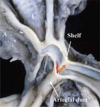
Figure 19. In this fetal heart, the shelf of ductal tissue produces paraductal coarctation. In our experience, coarctation in this location is much rarer than the preductal variant (see Fig. 20).

Figure 20. This specimen shows the typical form of preductal coarctation produced by a shelf of ductal tissue.
Hypoplasia of the left ventricle with deficient ventricular septation
In the “classical” forms of hypoplasia of the left heart, the ventricular septum is intact, giving the counterpart on the left side to “pulmonary atresia with intact septum”. In some instances, nonetheless, the left ventricle can be grossly hypoplastic, and incapable of supporting the circulation, when there is a ventricular septal defect. The most obvious example of this situation is found with double inlet right ventricle, when the left ventricle is not only hypoplastic, but also incomplete, lacking its inlet, and usually also its outlet component. Extreme forms of atrioventricular septal defect with common atrioventricular junction and right ventricular dominance also have unequivocal hypoplasia of the left ventricle (Fig. 21), and may do better with univentricular as opposed to biventricular surgical repair. In our experience, it is extremely rare to find severe left ventricular hypoplasia with an uncomplicated ventricular septal defect, but examples can be found with, for instance, absence of the left atrioventricular connection (Fig. 22). In all these instances, the decision as to whether the left ventricle is capable of supporting the circulation is likely to be based on clinical and physiologic rather than anatomic criterions, since echocardiographic measurements, as yet, are insufficiently robust prospectively to underpin therapeutic decision-making.

Figure 21. In this heart with atrioventricular septal defect and common atrioventricular junction, there is unequivocal hypoplasia of the left ventricle.

Figure 22. This cast from a fetal heart shows hypoplasia of the left ventricle in the setting of absent left atrioventricular connection, with the incomplete and rudimentary left ventricle fed through a ventricular septal defect.
Conclusions
Hypoplasia of the left heart is a well-recognised constellation of cardiac anomalies that, in only a minority of cases, is associated with associated abnormalities of other systems of organs. Because of this, geneticists would argue that it is inappropriate to describe the constellation of cardiac anomalies as a “syndrome”. In essence, the situation is the counterpart of pulmonary atresia or stenosis with intact ventricular septum, since although some cases of unequivocal left ventricular hypoplasia can be found with deficient ventricular septation, in the overwhelming majority of cases the ventricular septum will be intact. All cases with aortic atresia and intact ventricular septum will fit into this category, albeit that the mitral valve can be stenotic or atretic. Some cases of critical aortic stenosis will also appropriately be categorised as having hypoplasia of the left heart. These will be the examples with an apex-forming right ventricle, and with gross left ventricular fibroelastosis. Other cases of critical aortic stenosis will be amenable to treatment by interventional catheterisation, or biventricular surgical repair. These patients are unlikely to have significant hypoplasia of the left ventricle. Prospective distinction of these cases during life will depend on clinical rather than anatomic investigations.
References
Lev M. Pathological anatomy and interrelationship of hypoplasia of the aortic tract complexes. Lab Invest 1952; 1: 61–70.Google Scholar
Noonan JA, Nadas AS. The hypoplastic left heart: an analysis of 101 cases. Paediatr Clin North Am 1958; 5: 1029–1057.Google Scholar
Tchervenkov CI, Tahta SA, Jutras LC, Bjorkland MJ. Biventricular repair in neonatal hypoplastic left heart complex. Ann Thorac Surg 1998; 66: 1350–1357.Google Scholar
Rhodes LA, Colan S, Perry SB, Jonas RA, Sanders SP. Predictors of survival in neonates with critical aortic stenosis. Circulation 1991; 84: 2325–2335.Google Scholar
Kovalchin JP, Brook MM, Rosenthal GL, Suda K, Hoffman JI, Silverman NH. Echocardiographic hemodynamic and morphometric predictors of survival after two-ventricle repair in infants with critical aortic stenosis. J Am Coll Cardiol 1998; 32: 237–244.Google Scholar
Lang D, Hofstetter R, Kuperschmid C. Hypoplastic left heart with complete transposition of the great arteries. Br Heart J 1985; 53: 650–653.Google Scholar
Steeg CN, Ellis K, Bransilver B, Gersony W. Pulmonary atresia and intact ventricular septum complicating corrected transposition of the great vessels. Am Heart J 1971; 82: 382–386.Google Scholar
Weinberg PM, Chin AJ, Murphy JD, Pigott JD, Norwood WI. Postmortem echocardiography and tomographic anatomy of hyoplastic left heart syndrome after palliative surgery. Am J Cardiol 1986; 58: 1228–1232.Google Scholar
Rychik J, Rome JJ, Collins MH, DeCampli WM, Spray TL. The hypoplastic left heart syndrome with intact atrial septum: atrial morphology, pulmonary vascular histopathology and outcome. J Am Coll Cardiol 1999; 34: 554–560.Google Scholar
Brann S, Brawn WJ, Raafat F, Sreeram N. Neonatal pulmonary vascular disease in hypoplastic left heart syndrome. Ann Thorac Surg 1995; 60: 433–434.Google Scholar
Pinto CA, Ho SY, Redington A, Shinebourne EA, Anderson RH. Morphological features of the levoatriocardinal (or pulmonary-to-systemic collateral) vein. Pediatr Pathol 1993; 13: 751–761.Google Scholar
O'Connor WN, Cash JB, Cottril CM, Johnson GL, Noonan JA. Ventriculocoronary abnormalities in hypoplastic left hearts. An autopsy microscopic study. Circulation 1982; 66: 1078–1086.Google Scholar
Baffa JM, Chen SL, Guttenberg ME, Norwood WI, Weinberg PM. Coronary artery abnormalities and right ventricular histology in hypoplastic left heart syndrome. J Am Coll Cardiol 1992; 20: 350–358.Google Scholar
Calder AL, Co EE, Sage MD. Coronary arterial abnormalities in pulmonary atresia with intact ventricular septum. J Thorac Cardiovasc Surg 1986; 59: 436–442.Google Scholar
Anderson RH, Zuberbuhler JR, Anderson C. Further morphological studies on hearts with pulmonary atresia and intact ventricular septum. Cardiol Young 1991; 1: 105–114.Google Scholar
Aiello VD, Ho SY, Anderson RH, Thiene G. Morphologic features of the hypoplastic left heart syndrome. Pediatr Pathol 1990; 10: 931–943.Google Scholar

The circulation in hypoplasia of the left heart is dependant on the dominant right ventricle, which pumps blood through the pulmonary trunk and via the arterial duct to the descending aorta. The upper body is fed retrogradely through the isthmus, with the retrograde circulation then feeding the coronary arteries through the ascending aorta. The critical areas in the circulation are the isthmus and the atrial septum.

The coronary sinus has been opened from behind, showing fenestrations between its cavity and that of the left atrium. They provided an “overflow” from the atrium, since the patient had had an intact atrial septum in the setting of hypolasia of the left heart.

The right atrium has been opened in a heart from a patient with hypoplasia of the left heart. The oval fossa is intact.

This “four-chamber” section is viewed from behind. It shows leftward deviation of the flap valve of the oval fossa, diminishing the size of the left atrium.

This section is taken transversely across the thorax of a fetus with hypoplasia of the left heart and an intact atrial septum. There is gross thickening of the pulmonary veins, which are also arterialised (see Fig. 10).

This histological section of a pulmonary vein comes from the heart shown in Figure 9. Note the arterial nature of the thickened walls (brackets).

These lungs are removed from another fetus having hypoplasia of the left heart and an intact atrial septum. Note the “cobble stone” nature of the parenchymal surface.

This section from the lungs shown in Figure 11 illustrates the gross lypmangiectasia that ensues in the presence of hypoplasia of the left heart when the atrial septum is intact.

The cartoon shows the course of the so-called “levoatrial cardinal vein” that drains blood from the left atrial roof to the superior caval vein in some patients with an intact atrial septum in the setting of hypoplasia of the left heart.

In this heart with hypoplasia of the left ventricle, there is a free-standing septomarginal trabeculation supporting the tension apparatus of the tricuspid valve.

In this fetal example of hypoplasia of the left heart, as shown by the centimetre marker, the ascending aorta (arrow) is no more than 1 mm in diameter.

In this fetal specimen with hypoplasia of the left heart, the left subclavian artery arises from the descending aorta distally relative to the junction of the aortic arch and the duct.

The isthmus is the region of the aortic arch (bracket) between the union of the duct with the descending aorta and the origin of the left subclavian artery. Its length can vary according to the site of origin of the subclavian artery, with there being no isthmus in the heart shown in Figure 16.

In this heart, the ductal tissue forms a coarctation shelf around the origin of the transverse aortic arch, and also encircles separately the mouth of the left subclavian artery.

In this fetal heart, the shelf of ductal tissue produces paraductal coarctation. In our experience, coarctation in this location is much rarer than the preductal variant (see Fig. 20).

This specimen shows the typical form of preductal coarctation produced by a shelf of ductal tissue.

In this heart with atrioventricular septal defect and common atrioventricular junction, there is unequivocal hypoplasia of the left ventricle.

This cast from a fetal heart shows hypoplasia of the left ventricle in the setting of absent left atrioventricular connection, with the incomplete and rudimentary left ventricle fed through a ventricular septal defect.
 You have Access
- 13
- Cited by