Introduction
Dirofilariasis is an infection caused by filarial nematodes of the genus dirofilaria. These roundworms are natural parasites of dogs, but humans are aberrant hosts for dirofilaria species.Reference Marty1 In humans, the worms usually die before maturing, provoking a focal granulomatous reaction in subcutaneous tissue.
The distribution of sites of Dirofilaria repens infection in the human is of great importance to clinicians, as patients with dirofilariasis may present to dermatologists, ophthalmologists and even otolaryngologists.Reference Pampiglione and Rivasi2 Using the keywords ‘dirofilaria’ and ‘neck’, we searched PubMed Medical Subject Headings for all articles published up to February 2012 and found only one documented case report of D repens infection in the neck.Reference Permi, Veena, Prasad, Kumar, Mohan and Shetty3 Most of the documented cases of human dirofilariasis presented with ocular infections.
Case report
A 47-year-old Caucasian male presented to his general practitioner with an 8-week history of a right-sided, lower anterolateral neck swelling that was 1 cm in size, associated with weight loss of 2 kg. The lump was non-tender. He occasionally had loose stools (around once a fortnight). He was married to a Greek lady and had spent a lot of time in Greece due to his previous occupation as a marine biologist. He denied any close animal contact other than a visit to the petting zoo with his children. He had no known previous history of neck disease.
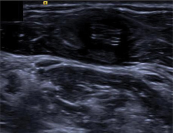
On examination, the patient looked pale with signs of recent weight loss. There was a 1 cm, non-tender, round, mobile mass at the right anterior cervical triangle. The lump was located precisely at the lower third of the anterior border of the sternocleidomastoid muscle. There were no other masses or evidence of lymphadenopathy. Oropharyngeal examination was normal. Flexible nasendoscopy was performed and the findings were unremarkable. An ultrasound scan of the neck was carried out (Figure 1), which showed a 1 cm, cystic mass within (inferiorly to) the right sternocleidomastoid muscle. The mass contained a coiled tubular structure which moved spontaneously. A cinematic video of the intramuscular cyst was taken, which showed images of a parasitic worm moving.
Ultrasound image of the neck showing a 1 cm, cystic mass containing a coiled tubular structure within (inferiorly to) the right sternocleidomastoid muscle.

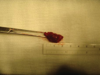
The patient was subsequently referred to infectious diseases and ENT for definite management. It was decided by both disciplines to remove the parasitic worm under local anaesthetic. During the operation, an elliptical skin incision was made to reveal the underlying cyst (Figure 2). Meticulous dissection enabled the cyst to be excised as a whole.
The excised cyst, measuring approximately 2 cm in size.

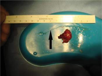
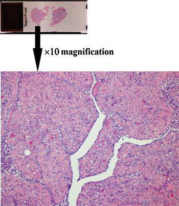
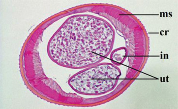
The sample was sent for pathological diagnosis. Macroscopic examination revealed a yellow, well circumscribed lesion containing a coiled, 10-cm long, white, thread-like worm (Figure 3). Representative blocks of the mass were submitted for histology. In addition, the worm was extracted and separately sampled transversely and longitudinally. Microscopic examination showed skeletal muscle containing a collapsed cavity surrounded by fibro-inflammatory stroma. The inflammatory infiltrate was composed of histiocytes, neutrophils, lymphocytes and a number of eosinophils (Figure 4). Cross-sections of the worm showed a gut and two uteri surrounded by a thick cuticle and a muscle. In addition, external longitudinal cuticular ridges were present (Figure 5). These are features of D repens. The diagnosis was confirmed by an infectious disease specialist and pathologist.
The 10 cm parasite (arrow), removed from the cyst after incision of the sac.

Microscopic examination of the excised cyst: the photomicrograph shows the collapsed cavity which previously contained Dirofilaria repens, and surrounding granulation tissue with lymphocytes, plasma cells, eosinophils and histiocytes. (H&E; ×10)

Transverse section through the midbody of a non-gravid female Dirofilaria repens (H&E; ×20). Ms = somatic musculature; cr = cuticular ridges; in = intestine; ut = paired uteri

The patient was discharged on the same day as surgery. At follow up, there was no recurrence of the swelling and the patient had regained his weight. No anti-parasitic treatment was recommended.
Discussion
Human nematode infection is endemic in certain regions. A 2005 report by the World Health Organization estimates that filariasis affects more than 90 million people worldwide.4Dirofilaria repens is a subcutaneous parasite of dogs and cats in Europe, Africa and Asia, which accounts for incidental infections in humans in affected areas. Almost all human infections by D repens are localised in the upper parts of the body (75.8 per cent). Dirofilariasis may also affect the male genital organs (6.5 per cent), the female breast (5.4 per cent), the lungs (2.6 per cent), and the abdominal viscera comprising the mesentery and omentum (1.3 per cent).Reference Pampiglione and Rivasi2
Dirofilaria repens infections in the upper body occur mostly at the ocular region, accounting for 30.5 per cent of the total. However, the parasites have the ability to migrate up to 30 cm in 2 days. There have been reports of subcutaneous migrations of the parasite over considerable distances, such as from the lower limbs to the head or from one side of the body to the other.
All species of dirofilaria have insects such as mosquitoes as vectors. Aedes caspius is the species that is most aggressive towards humans; it is capable of travelling 20 km without wind assistance and bites all day long. Generally speaking, most patients do not recall being bitten by mosquitoes. There were two cases of patients who consistently complained of ‘a worm under the skin’, which led physicians to diagnose delusory parasitosis and have them admitted to a psychiatric ward, until the appearance of a nodule and its subsequent histological analysis cleared up the matter.Reference Jelinek, Schulte-Hillen and Loscher5
• This paper is the first to report a case of anterolateral neck swelling secondary to Dirofilaria repens infection in Europe
• Neck swelling and weight loss in a patient with a travel history to dirofilaria endemic countries necessitates a detailed history and tailored investigation
• Neck swellings are often related to infections; due to the recent rise in human dirofilariasis, this may include Dirofilaria repens infection
The only treatment for dirofilariasis in humans is surgical removal of the lesion or extraction of the worm, as demonstrated in our case report. Routine protection against mosquito and fly bites helps to prevent infection. Deworming pets, especially dogs, protects against D repens infection in humans.