INTRODUCTION
Balamuthia mandrillaris is a free-living soil amoeba that is the causative agent of granulomatous amoebic encephalitis, a life-threatening brain infection (Schuster and Visvesvara, 2004). First isolated from brain tissue of a mandrill baboon (Rideout et al. 1997), B. mandrillaris is naturally found in soil (Dunnebacke et al. 2004), and occasionally can enter humans to cause an insidious infection, which can occur both in immunocompetent and immunosuppressed individuals. The incubation period may be long and usually results in drastic neurological damage that includes single and multiple space-occupying brain lesions. Portals of entry for amoebae may be the nasal mucosa or breaks in the skin resulting from trauma (Martinez and Visvesvara, 2001). Studies utilizing mouse models have shown that B. mandrillaris is able to migrate by the olfactory nerve pathway and access the brain (Janitschke et al. 1996; Kiderlen and Laube, 2004). The existence of skin lesions that lead to brain infections can be an indication also of haematogenous dissemination of amoebae in the mammalian host (Martinez and Visvesvara, 2001).
There have been few in vitro studies with B. mandrillaris. This amoeba has been shown to be cytotoxic to monkey kidney cells (Schuster and Visvesvara, 1996; Schuster et al. 2003) and human brain microvascular cells (Jayasekera et al. 2005), but cellular mechanisms of cytopathogenesis are unknown.
In the migratory process to the brain, whether by the olfactory nerve pathway or the haematogenous route, B. mandrillaris encounters a group of molecules that constitute the extracellular matrix (ECM) of the mammalian host. The ECM is an intricate meshwork of biomolecules, that fills intercellular spaces in tissues, such as connective and nerve tissue, and that also forms the basal lamina of epithelia (Alberts et al. 2002). ECM molecules play an integral role in diverse cellular physiological processes, including proliferation, differentiation and migration (Fuller and Shields, 1998). In addition, the ECM is involved in metastasis, especially in tumor cell invasion to other tissues (Mareel and Leroy, 2002). Similarly, a diverse array of microorganisms including parasitic protozoa can interact with the ECM (Silva-Filho et al. 1988; Gordon et al. 1993; Guillen, 1996; Wadstrom and Ljungh, 1999; Han et al. 2004), resulting in tissue or cell invasion (Somner et al. 2000; Meza, 2000).
Since tissue invasion is requisite for progression of B. mandrillaris infection, the goals of this study were (1) to determine whether B. mandrillaris recognizes 3 relevant constituents of the ECM: fibronectin, type I collagen and laminin-1, (2) to characterize the interaction between B. mandrillaris and these molecules, and (3) to obtain insight into factors that mediate B. mandrillaris-ECM binding.
MATERIALS AND METHODS
Amoeba
Balamuthia mandrillaris (ATCC 50209) isolated from brain tissue of a female mandrill baboon that died of amoebic encephalitis, was grown for 24 h at 37 °C in plastic tissue culture flasks prior to assay in BM-3 medium (Schuster, 2002), which is composed of Biosate peptone, yeast extract, Torula yeast RNA and liver extract, supplemented with calf serum, taurine, haemin, glucose, amino acids, vitamins, lipids and salts.
Extracellular matrix (ECM) glycoprotein surface coating
Twenty-four well plates (Costar, Cambridge, MA) or glass cover-slips were incubated at 4 °C overnight with solutions containing 10 μgċml−1 of fibronectin (isolated from human plasma), type I collagen (isolated from rat tails) (Sigma, St Louis, MO) or laminin-1 (isolated from Engelbreth-Holm-Swarm mouse sarcoma, Invitrogen, Grand Island, NY) diluted in 0·01 M phosphate-buffered saline (PBS), pH 7·2. After coating, the surfaces were washed gently once with PBS, pH 7·2, to remove unassociated glycoproteins.
Binding assay
Amoebae grown for 24 h at 37 °C in BM-3 medium were detached by bumping from the flasks, washed twice with PBS, counted by using a haemacytometer and suspended to a final concentration of 6×10⁴ amoebae ml−1. Amoebae were added to the ECM coated plates for 20 min at 37 °C. Following incubation the wells were washed once to remove unbound amoebae and fixed with 2·5% glutaraldehyde made in cacodylate buffer 0·1 M (pH 7·2). Digital photographs were taken using an Olympus CK2 phase-contrast microscope at a 100× magnification. The number of amoebae bound was counted in 10 frames (selected randomly) for each experimental condition. Alternatively, amoebae were pre-treated for 20 min at 37 °C in PBS with 100 mM mannose, galactose or sialic acid, 10 and 100 mM CaCl2, 10 or 100 mM MgCl2, 1 mM MnCl2 or 5 mM EDTA and added to coated wells. Also, amoebae were pre-treated with 0·25% trypsin or 1 mM periodate for 30 min at 37 °C prior to incubation with ECM glycoproteins. Trypsin was inactivated by adding fresh medium and the samples were washed once and resuspended in PBS. Binding was measured as the number of amoebae per cm2, where 100% of the amoebae bound represents approximately 1000 amoebae per cm2.
Scanning electron microscopy
Amoebae were incubated with cover-slips coated with 10 μgċml−1 of the ECM proteins under the conditions cited in the binding assay. After removing unbound amoebae, the cover-slips were treated for 1 h with 2·5% glutaraldehyde diluted in 0·1 M cacodylate buffer, washed 4 times with the same buffer and immersed for 40 min in the dark in 2% osmium tetroxide made up in 0·1 M cacodylate buffer. Following extensive washing with cacodylate buffer, cover-slips were subjected to a dehydration protocol using increasing amounts of ethanol and subjected to critical-point drying (CO2 as a transitional fluid). Cover-slips were attached to metal stubs, coated with a thin layer of gold in an ion-sputtering device, and observed in a scanning electron microscope (JEOL), operating at an accelerating voltage of 15 kV as described previously (Pettit et al. 1996).
Statistical analysis
Data were expressed as mean values and standard error of the mean. For statistical significance (P<0·05), a two-tailed, unpaired Student's t-test was used.
RESULTS
Balamuthia mandrillaris binding to extracellular matrix components
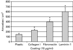
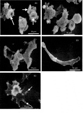
To determine whether B. mandrillaris trophozoites associate with 3 types of ECM components, we quantified the interaction between the amoebae and ECM-coated wells. A greater number of trophozoites bound to the ECM-coated wells as compared with uncoated plastic wells (Fig. 1). An ordered preference for binding was observed, with greatest binding noted for laminin, followed by fibronectin and collagen. Scanning electron microscopy demonstrated that binding to ECM was associated with shape modifications in the trophozoites dependent on the substratum. B. mandrillaris adhered to plastic in amoeboid form and exhibited a ‘rounded’ compact shape with surface projections known as food-cups (Fig. 2A) that are observed in diverse amoeba species such as Entamoeba histolytica (Rigothier et al. 1991), Naegleria fowleri (Marciano-Cabral and Fulford, 1986), and Acanthamoeba spp. (Pettit et al. 1996). Distinctive morphologies were noted for B. mandrillaris interacted with ECM components (Fig. 2B, C, D, E). Amoebae attached to ECM-coated wells were observed as spread out in various conformations. Fibronectin-associated amoebae (Fig. 2C) exhibited an elongated shape containing several lobose protrusions, observed in 69% of the fibronectin-adhered amoebae. Collagen-associated amoebae also possessed some cellular protrusions (Fig. 2B). However, these were not as extensive as those observed for amoebae maintained on fibronectin-coated plates, being slightly similar to control amoebae on plastic surfaces. In contrast, laminin-associated amoebae exhibited a distinctive morphological transformation to elongate (in 24% of the B. mandrillaris population adhered on laminin, Fig. 2D) and highly spread forms (51% of the population, Fig. 2E). Amoebae in the latter conformation contained adhesion structures that were morphologically similar to mammalian focal adhesions (Fig. 2E).

Fig. 1. Binding of Balamuthia mandrillaris to ECM components. Amoebae exihibited highest levels of binding to plates coated with laminin, followed by fibronectin and collagen when compared to untreated plastic surfaces (*P<0·05, Student t-test, as compared with amoebae maintained on plastic).

Fig. 2. Scanning electron micrographs of Balamuthia mandrillaris bound to ECM glycoproteins. Trophozoites interacting with (A) plastic, (B) collagen, (C) fibronectin and (D, E) laminin were fixed and processed for scanning electron microscopy, as described in the Materials and Methods section. The arrows designate surface projections known as ‘food-cups’. Lobose protrusions were observed for amoeba adhered on fibronectin or collagen. Amoebae placed on laminin exhibited either an elongated form (D) or a ‘flattened’ shape with focal adhesion (arrows, E).
Effect of sugars on binding of B. mandrillaris to ECM components
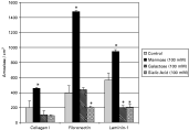
To determine the effect of sugars on B. mandrillaris binding to ECM molecules, trophozoites were incubated with 3 saccharides that are related to adhesion in other parasitic protozoa: mannose, galactose and sialic acid. Pre-treatment of amoebae with sugars exerted differential effects on binding by B. mandrillaris (Fig. 3). Binding to fibronectin was augmented significantly (P<0·05) by mannose and slightly diminished by sialic acid; however, galactose did not affect binding to fibronectin. Mannose also stimulated the binding of B. mandrillaris trophozoites to collagen or laminin-coated plates while galactose and sialic acid decreased binding to these substrata.

Fig. 3. Effect of sugars on binding of Balamuthia mandrillaris to ECM as compared with untreated amoebae interacted with ECM components. Mannose exerted a significant effect on the increase of B. mandrillaris binding to all ECM components, sialic acid was able to diminish amoebic association with fibronectin and laminin and galactose decreased B. mandrillaris association to laminin (*P<0·05, Student's t-test).
Effect of trypsin and periodate on B. mandrillaris binding to laminin-1 coated plates
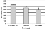
Laminin-1 was shown to elicit preferential binding of amoebae. Thus, in order to obtain insight as to the biochemical nature of this binding, amoebae were pre-treated with the broad-spectrum proteolytic enzyme trypsin or with the sugar oxidant agent periodate. Pre-treatment with either agent had a minor diminishing effect on B. mandrillaris binding to laminin-1 (Fig. 4).

Fig. 4. Effect of trypsin and periodate on binding of Balamuthia mandrillaris to laminin-1. Both agents exerted a minor but significant inhibitory effect on binding of amoebae on laminin-1 coated plates (*P<0·05, student's t-test).
Effect of EDTA and divalent cations (Ca+2, Mg+2 and Mn+2) on B. mandrillaris binding to laminin-1 coated plates
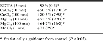
The effect of the divalent cation chelator EDTA and the free divalent cations Ca+2, Mg+2 and Mn+2 on B. mandrillaris binding to laminin-1 was examined. Interaction between amoebae and laminin was highly dependent on the presence of divalent cations, since EDTA treatment greatly inhibited B. mandrillaris association on laminin-1 coated plates (Table 1). All of the divalent cations tested elicited an increase in amoebae binding on laminin-1. Calcium at a concentration of 100 mM exerted the highest augmentation of binding.
Table 1. Effect of EDTA and divalent cations on Balamuthia mandrillaris binding on laminin-1-coated plates (Results expressed as % binding (standard error of the mean) from the control interactions with laminin-1.)

DISCUSSION
Granulomatous amoebic encephalitis caused by B. mandrillaris is an invasive infection, since amoebae have the ability to spread from skin abrasions or nasal epithelium to the central nervous system as well as disseminate to other organs (Schuster and Visvesvara, 2004). This biological characteristic of B. mandrillaris indicates that during the course of invasion, the amoebae encounter molecules of the host extracellular matrix (ECM), including components of the basal lamina as detected previously in the mouse model of Balamuthia infection (Kiderlen and Laube, 2004). Therefore, it is plausible that recognition of ECM constituents could be relevant to the process of tissue invasion by B. mandrillaris.
In this report, we demonstrate that B. mandrillaris trophozoites recognize 3 relevant ECM components: collagen type I, the main component of connective tissue ECM; fibronectin, one of the most abundant and ubiquitous ECM molecules that is critical in cell adhesion processes; and laminin-1, a key molecule in the formation of the basal lamina. Diverse species of pathogenic microorganisms are able to recognize ECM molecules, including bacteria (Westerlund and Korhonen, 1993; Schwarz-Linek et al. 2004), fungi (Klotz, 1994; Gonzalez et al. 2005) and protozoa, (Ouassi et al. 1984; Ghosh et al. 1996; Eda and Sherman, 2004). In the present study, amoebae were able to bind with the greatest intensity to laminin, followed by fibronectin and collagen, in that order, demonstrating that a free-living soil amoeba isolated from brain tissue is able to recognize typical mammalian glycoproteins. The apparent preferential association of B. mandrillaris with laminin has been observed also with another species of pathogenic free-living amoeba, Acanthamoeba polyphaga (Gordon et al. 1993) that bound more avidly to basal lamina components (laminin and collagen IV) than to fibronectin. Those investigations and the close phylogenetic relationship between Acanthamoeba spp. and B. mandrillaris (Smirnov et al. 2005) led us to suggest that the recognition of basal lamina molecules might be important for the establishment of pathogenic free-living amoebae in the mammalian host.
Major morphological transformations were observed in the ECM-adhered amoebae, indicating that the association between amoebae and ECM molecules triggers signalling pathways that lead to structural rearrangement. It is possible that ECM receptors on amoebae mediate these interactions, inducing cytoskeletal rearrangements and changes in shape, as has been observed in the pathogenic free-living amoeba Naegleria fowleri (Han et al. 2004) and in the gastrointestinal parasite Entamoeba histolytica (Talamas-Rohana and Meza, 1988), in which fibronectin binding induced the formation of actin-containing structures called adhesion plates. In our model, the most remarkable morphological differences were observed on laminin-adhered amoebae, where very elongated forms and spread forms were observed. The elongated forms, were similar to the elongated forms described by Kiderlen and Laube (2004), who used mouse models of intranasal B. mandrillaris infection to demonstrate that amoebae in the olfactory epithelium stall at the basement membrane and orientate parallel to it, presenting an elongated shape. These observations are consistent with those obtained for our laminin interactions. The amoebae spread on laminin exhibited a structural feature that was similar to the well-described mammalian ECM-adhesion structures known as focal adhesions. These adhesion structures link microfilaments to integrin ECM receptors resulting in activation of diverse signalling pathways that lead to cell survival, proliferation and migration (Burridge and Chrzanowska-Wodnicka, 1996). For instance, the urogenital parasite Trichomonas vaginalis (Silva-Filho, 1998) and E. histolytica (Vazquez-Prado and Meza, 1992) possess focal adhesion structures and molecules, which are activated when they adhere to fibronectin. Thus, B. mandrillaris interaction with laminin may trigger a similar complex cellular response.
It has been reported that extracellular matrix binding can be mediated by cell surface lectins, which recognize specific sugar residues of ECM glycoproteins (Chammas et al. 1994). Gordon and coworkers (1993) observed that binding to the ECM by the free-living amoeba Acanthamoeba is affected by mannose. Thus, to determine the influence of sugars on binding of B. mandrillaris to ECM molecules, we incubated amoebae with 3 sugar molecules related to microbial adhesion processes. Incubation with mannose resulted in an increase in amoeba association with all ECM substrata tested, an outcome distinctive from that for A. polyphaga, where mannose inhibited almost totally the binding of amoeba to laminin, collagen IV and fibronectin (Gordon et al. 1993). The increased adhesion observed for B. mandrillaris could be due to the presence of a mannose-binding element on the cell surface that serves as a co-receptor to ECM binding. Alternatively, mannose could stimulate the expression of ECM receptors. In contrast, addition of galactose had no effect on adhesion to collagen and fibronectin, but resulted in a decrease in laminin adhesion. Thus, for B. mandrillaris, the recognition of collagen I and fibronectin apparently is galactose-independent, while that for laminin appears to be dependent on a galactose-binding protein. Typical molecules of this type in eukaryotic cells are galectins that constitute a family of galactose lectins observed in a wide variety of animal cells (Leffler et al. 2004). These molecules are able to bind to laminin (Ramkumar and Podder, 2000), since this ECM glycoprotein possesses α-galactose terminations (Hughes, 1992). Galactoside-binding lectins also can be involved in central nervous system tumor metastasis, indicating a role in tissue invasion (Bresalier et al. 1997). Our results, taken in the context of those observations, suggest the presence of a similar cell surface molecule on B. mandrillaris, that is specific for association with laminin and that may be involved in host tissue invasion. Sialic acid pre-treatment was able to diminish amoeba adhesion on all ECM coverings studied, indicating that B. mandrillaris possesses a sialic acid binding molecule that is involved in ECM binding. Select species of pathogenic bacteria (Roberts et al. 1989) and fungi (Hamilton et al. 1999; Tronchin et al. 2002) also require sialic acid for recognition of ECM glycoproteins. In this context, Hughes (1992) reported that sialic acid is present at the end of sugar chains of fibronectin and laminin, indicating that the interaction of amoebae with ECM could be mediated, at least in part, by this component.
Since laminin binding by B. mandrillaris was greater than that by collagen I and fibronectin, and its interaction resulted in the greatest morphological transformation, we characterized further the biochemical nature of the interaction. Trypsin and periodate treatment have been shown to diminish the laminin binding of the extracellular parasite T. vaginalis (Crouch and Alderete, 1999). In our model, the interaction between amoebae and laminin seems to be dependent on both surface proteins and sugars, since both agents diminished laminin binding. However, the observed inhibition of binding was minimal, probably because of an insensitivity of the amoeba to the trypsin and periodate concentrations used or to a rapid protein/carbohydrate turnover at the amoeba cell surface during the period of assay.
Since the main class of extracellular matrix receptors in eukaryotic cells is the integrin family of glycoproteins (Giancotti and Ruoslathi, 1999), the binding of which is mediated by divalent cations in the extracellular milieu (Plow et al. 2000), we tested these components on B. mandrillaris binding to laminin. We observed, by using known protozoa effective divalent cation concentrations (Wang et al. 1994; Silva-Filho and Arroyo, 1998; Carbajal et al. 1996) that laminin binding was highly dependent on divalent cations and that this association was increased by divalent cation input. These results suggest that an integrin-like molecule mediates binding of B. mandrillaris to laminin. Divalent cation dependence on ECM binding has been described in diverse parasitic protozoa. For example, calcium has been reported to enhance A. polyphaga binding to collagen IV, laminin and fibronectin (Wang et al. 1994) and to increase binding to fibronectin by T. vaginalis (Crouch et al. 2001) and to collagen I by E. histolytica (Munoz et al. 2001).
In summary, we have demonstrated that a pathogenic protist, B. mandrillaris, recognizes typical molecules that compose the extracellular matrix of mammalian tissues. The amoeba binding was highest for laminin-1, followed by fibronectin and collagen-I and such binding was dependent on select sugars. Mannose was able to increase amoeba binding to all substrata tested; galactose was ineffective in altering B. mandrillaris association for collagen and fibronectin, but was able to inhibit binding on laminin-coated plates. Sialic acid inhibited binding in all conditions tested. Laminin binding may be more biologically relevant. The association that correlated with drastic morphological alterations was that for amoebae interacted with laminin. Such interaction was dependent on divalent cations and the glycoprotein nature of the amoeba surface. B. mandrillaris recognition of ECM may be relevant in the pathogenesis of infections caused by B. mandrillaris, since ECM recognition is important for tissue invasion and migration of pathogens in infected hosts (Lauwaet et al. 2000; Mareel and Leroy, 2003).
The following agencies have supported this study: CAPES-MEC, CNPq, MCT-PRONEX and FUJB-UFRJ (Brazil) and the National Institutes of Health (DA 05274 and DA 15608). B.R.A. was a PDEE (CAPES-MEC) research scholar for the period May 2005 to April 2006.