Neurofibromatosis is a genetically transmitted multi-system disease showing clinical manifestations of dysplasia of mesodermal and ectodermal tissues. Extra-cardiac vascular manifestations of neurofibromatosis have been previously described in many reports.Reference Hamilton and Friedman 1 However, coronary arterial involvements have been rarely described. We report a case of thrombosed coronary aneurysm causing acute myocardial infarction in association with neurofibromatosis, which was treated with coronary bypass graft surgery.
Case report
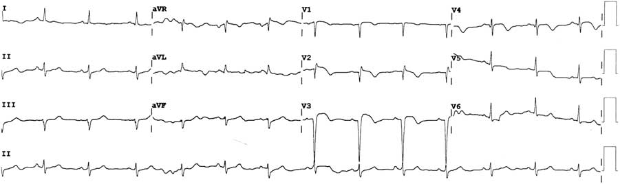
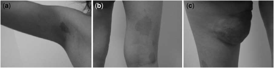
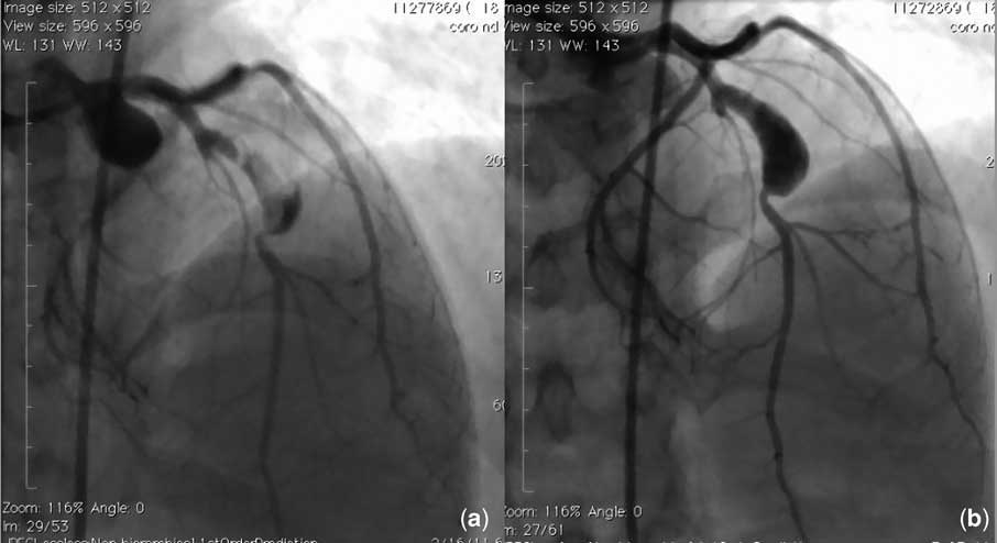
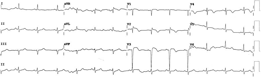
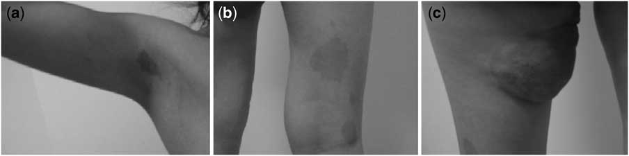
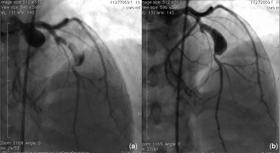
A 17-year-old girl presented to our clinic with subacute myocardial infarction. She had no cardiac risk factors or history of Kawasaki disease. She had been diagnosed with neurofibromatosis 3 years ago. Her blood pressure was normal and heart rate was 82 per minute. On cardiac auscultation, an S3 was heard, but no murmurs were noted. She had a 4 × 8 centimetres biopsy-proven neurofibroma on the medial side of her upper leg (Fig 1), and 12 café au lait spots larger than 10 millimetres over her body. Electrocardiogram on admission showed sinus rhythm and QS and ST elevation in leads V1–V3 (Fig 2). Transthoracic echocardiography revealed akinesis of the apex and hypokinesis of the anterior wall, with an estimated ejection fraction of 45%. No valvular pathologies were noted. Laboratory results were normal except for the cardiac enzymes. She underwent cardiac angiography for the ongoing post-myocardial infarction angina, which revealed a 7.3 × 14.1 millimetres aneurysm with thrombus in the left anterior descending artery (Fig 3a). Her angina was controlled with antiaggregant, anticoagulant, and antiischaemic therapy. She was discharged with aspirin, clopidogrel, beta-blocker, and nitrate. Her thrombophilia screen, including protein C and S, antithrombin, activated protein C resistance, homocysteine, fibrinogen, and factor V Leiden and prothrombin gene mutation analysis were negative. After 3 months, she was admitted to our hospital again with angina. A coronary angiogram was performed a second time, revealing an aneurysm with increased diameter (7.9 × 14.2 millimetres; Fig 3b). The patient was referred to the department of cardiothoracic surgery where a single-vessel coronary artery bypass graft surgery using the left internal mammarian artery was performed for the angina episodes occurring despite medical therapy. She was discharged with aspirin, clopidogrel, and beta-blocker therapy without any complications. She was symptom free on the 3-month follow-up.
Electrocardiogram showing ST elevations and V1–V3 QS pattern.
( a and b ) Examples of the café au lait spots over the body and ( c ) biopsy-proven neurofibroma on the thigh.
( a ) Thrombus image inside the aneurysm and ( b ) coronary aneurysm.
Discussion
Neurofibromatosis is an autosomal dominant multi-system genetic disorder with an incidence rate of 1 in 3000. There are two major subtypes: type 1, also known as von Recklinghausen disease, and type 2. Neurofibromatosis 1 results from a mutation in or deletion of the NF1 gene located in the long arm of the 17th chromosome. This gene encodes a protein named neurofibromin that appears to function in a tumour-suppressive role. This protein is expressed in a variety of tissues, and the function of neurofibromin is critical for regulation of the proliferation and differentiation of the hematopoietic, neural, and mesenchymal lineage progenitor cells.Reference Hamilton and Friedman 1 , Reference Kossler, Stricker and Rödelsperger 2 Thus, diminished levels can affect almost any system including bone, nervous system, soft tissue and skin, as well as blood vessels. Renal arteries are affected most commonly; however, neurofibromatosis 1 vasculopathy may involve any level from the aorta to the small arterioles and may cause vascular stenosis, occlusion, aneurysm, pseudoaneurysm, rupture, or fistula formation.Reference Friedman, Arbiser and Epstein 3 , Reference Bilsel, Akbulut, Terzi, Sayar and Yesilcimen 4 Nevertheless, coronary arterial involvements have been rarely described.
In this report, we present a 17-year-old girl with a coronary artery aneurysm in the left anterior descending artery associated with neurofibromatosis vasculopathy. To the best of our knowledge, this is the youngest patient treated with coronary artery bypass graft surgery because of a coronary lesion attributable to neurofibromatosis vasculopathy. Kandarpa et al were the first to report a case of myocardial infarction attributable to a coronary aneurysm in a 30-year-old patient with neurofibromatosis.Reference Kandarpa, Stoll, Reiss, Rutherford and Cohn 5 Multiple coronary aneurysms in neurofibromatosis have also been rarely reported.Reference Bilsel, Akbulut, Terzi, Sayar and Yesilcimen 4 , Reference Ruggieri, D'Arrigo, Abbate, Distefano and Upadhyaya 6 , Reference Trevelyan, Been and Patel 7 Fuchi et al reported coronary vasospasm reflecting abnormality of the cardiac sympathetic nerve in a case with neurofibromatosis and an organic coronary lesion, which was treated by percutaneous coronary angioplasty.Reference Fuchi, Ishimoto, Kajinami, Kajinami, Ohmichi and Kinoshita 8 Talay et al treated a 67-year-old man with coronary and peripheral artery occlusive disease and neurofibromatosis with simultaneous coronary artery and peripheral artery bypass graft surgery.Reference Talay, Dag, Ozmen and Erkut 9
The pathophysiology of vascular pathologies in neurofibromatosis is yet to be fully elucidated. However, some explanations have been suggested. Vasculopathy may result from failure to suppress excessive smooth muscle migration and proliferation, impaired vascular histogenesis, impaired vascular maintenance and healing due to abnormal neurofibromin function, or invasion and weakening of the arterial wall by neurofibromatous tissue.Reference Hamilton and Friedman 1 , Reference Talay, Dag, Ozmen and Erkut 9 , Reference Norton, Xu and Gutmann 10
Coronary artery aneurysm is a localised dilatation in the coronary artery segment more than 1.5-fold normal size compared with adjacent normal segments. It is most commonly observed in the right coronary artery. The most common cause of coronary artery aneurysms is atherosclerosis; however, other causes include trauma, infective endocarditis, Kawasaki disease, percutaneous transluminal angioplasty, fibromuscular dysplasia, and arteritis.Reference Trevelyan, Been and Patel 7 Coronary aneurysms may cause thrombosis and embolisation, formation of arteriovenous fistulae, vasospasm, and rupture. Treatments comprise surgical, percutaneous, or medical interventions. Despite limited data, medical management includes antiplatelet and/or antithrombotic agents, and if medical therapy agents fail coronary artery bypass graft surgery or intracoronary stent placement should be considered. In this patient, medical therapy was chosen first considering the young age of the patient. However, recurrence of symptoms led us to surgical therapy.
In conclusion, this report presents a young patient admitted with myocardial infarction due to coronary aneurysm attributed to neurofibromatosis and, despite the rarity, the clinical manifestations of neurofibromatosis vasculopathy include myocardial infarction and may have important clinical consequences.