1. Introduction
The double-helical structure of DNA has many benefits as a means for stably storing genetic information. Sequestering information-rich nucleotides away from solvent protects them from chemical damage, while complementarity between the sister strands allows mistakes, lesions, and discontinuities in the genome to be addressed and repaired. The intertwined structure of duplex DNA is not without drawbacks, however. Because the two strands of nucleic acid are wound around one another, processes that require strand separation – for example, replication and transcription – lead naturally to DNA overwinding and strand entanglement (Liu & Wang, Reference Liu and Wang1987). These topological intermediates can pose serious challenges to the cell, interfering with gene expression, DNA duplication, and chromosome segregation. At the same time, cells depend on DNA supercoiling as a means of compacting the genome and promoting DNA unwinding at replication origins and promoter regions (Holmes & Cozzarelli, Reference Holmes and Cozzarelli2000; Wang, Reference Wang2002). Thus, just as the cell strives to preserve the information content of the genome, it also invests considerable energy to maintain the topology of its chromosomes in an appropriate topological state.
Resolving unfavorable DNA supercoils, knots, and interwound duplexes requires that the nucleic acid strands be transiently unlinked topologically; that is, one or both strands of a duplex must be cleaved, manipulated with respect to a second (uncleaved) segment, and then religated. To accomplish these tasks without damaging the genome, organisms have evolved a class of proteins known as topoisomerases. A distinguishing feature of topoisomerases is their ability to create transient DNA breaks through the formation of a covalent attachment of the enzyme to one or both strands of the duplex. They then reorganize the spatial displacement of DNA segments to alter either the number of times the two strands of one duplex wind about one another or the number and type of intertwinings between two DNA duplexes. This capability requires an extraordinary degree of chemomechanical sophistication, requiring that topoisomerases read both the local geometry of DNA segments and the global energetics of the genome.
Nearly 40 years of study on DNA topoisomerases has provided deep insights into the structure and function of these diverse and fascinating molecular machines (for reviews, see Wang, Reference Wang1998; Champoux, Reference Champoux2001; Corbett & Berger, Reference Corbett and Berger2004; Charvin et al. Reference Charvin, Strick, Bensimon and Croquette2005b; Nadal, Reference Nadal2007; Nollmann et al. Reference Nollmann, Crisona and Arimondo2007a). Briefly stated, evolution has come up with two strategies for enzyme-assisted topology modification. One is the controlled rotation mechanism, in which the topoisomerase creates a nick to allow one end of the DNA helix to rotate or swivel about the other. Within this framework, the enzyme can frictionally constrain the rotating DNA segment to both control movement and assist religation. The second mechanism, known as strand passage, involves creating a single- or double-stranded DNA break, opening a gap in this cleaved segment, and passing a second DNA strand or duplex through the break. Here, because the accidental dissociation of the cleaved DNA ends from the enzyme during transport would give rise to potentially deleterious chromosome breaks, the topoisomerase uses dedicated binding and scaffolding domains to bridge the DNA gap. Despite the fundamental differences between these mechanisms, both use interactions at macromolecular interfaces as a means for controlling the rate and fidelity of the topoisomerase reaction. Both activities also exploit the free energy stored in supercoiled DNA and/or the thermodynamic distribution of topoisomer populations as a driving force for effecting topological change.
2. DNA topology: a primer
To better understand how topoisomerases control DNA topology, it is helpful to first review some basic nomenclature and concepts. For a more thorough and rigorous treatment, the reader is referred to several in-depth works on this topic (Vologodskii & Cozzarelli, Reference Vologodskii and Cozzarelli1994; Bates & Maxwell, Reference Bates and Maxwell2005).
To begin, first consider a situation in which two closed, circular strands or chains wind around each other in space. The frequency and nature of their windings – how often one strand coils about the other and how the pair of interwound strands traverse through three-dimensional space – can be described by two quantities, twist (Tw) and writhe (Wr). In the context of DNA, twist can be roughly considered as a measure of how many times one strand of a double helix wraps around the other along the central axis of the duplex. Writhe relates to how many times the axis formed by the double helix crosses over itself, forming a supercoil. The sum of these two values, which together describe the integral number of times one strand of DNA wraps around the other, is defined as the linking number (Lk) [Eq. (1)] (White, Reference White1969; Crick, Reference Crick1976):

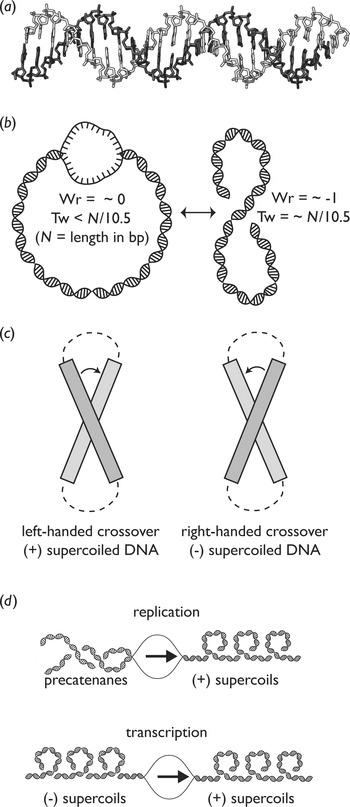
Twist and writhe are given negative or positive designations based on the handedness of the DNA crossings. By convention, a right-handed helix, such as that exhibited by B-form DNA, has positive twist (Fig. 1a), whereas a left-handed helix (e.g. Z-DNA) has negative twist. In contrast, right-handed writhe (in the form of plectonemic supercoils, Fig. 1b) has a negative value, whereas left-handed writhe is positive. The crossings formed by plectonemic supercoils have characteristic geometries, which may be exploited by some topoisomerases during substrate recognition (Fig. 1c) (Stone et al. Reference Stone, Bryant, Crisona, Smith, Vologodskii, Bustamante and Cozzarelli2003).
DNA topology. (a) Ideal B-form DNA is a right-handed double helix (figure generated using PyMOL and Coot). (b) Negative ΔLk in a closed circular DNA plasmid can manifest as a decrease in overall twist (a single-stranded bubble, left panel) or as negative writhe (a right-handed, negative plectonemic supercoil, right panel). These two forms can interconvert, although the stability of base-paired DNA generally maintains a relatively constant twist, which can be approximated as N/10·5, where N=number of base pairs in the DNA. (c) Definition of crossover handedness. Positively supercoiled DNA plectonemes form left-handed crossovers (left), whereas negatively supercoiled DNA plectonemes have right-handed crossovers (right). (d) During DNA replication (top), positive supercoils accumulate ahead of the replication fork, while the daughter chromosomes become interwound behind it. These interwindings, known as precatenanes, can become interlinked if left unresolved. During DNA transcription (bottom), positive supercoils accumulate ahead of the progressing fork, whereas negative supercoils accumulate behind it. Arrows indicate the direction of fork progression.

Since the structure of DNA is known, the ‘ground state’ linking number (Lk0) of any topologically constrained, nonsupercoiled DNA molecule is likewise known: it is approximately the number of base pairs (N) divided by the periodicity of the helix [average of 10·5 bp per turn] (Depew & Wang, Reference Depew and Wang1975; Rhodes & Klug, Reference Rhodes and Klug1980):

Deviations from Lk0 are expressed by the value ΔLk, where ΔLk is the sum of ΔWr and ΔTw. These values are applicable only in a closed system, like a circular bacterial chromosome or a segment of linear DNA that is bounded on both ends. In such a system, twist and writhe can interconvert to alter the structure of DNA while leaving linking number constant (Fig. 1b) (White, Reference White1969).
The relationship among twist, writhe, and linking number has important consequences for the topology of the genome. A progressing replication fork, for example, locally unwinds the duplex, generating compensatory overwinding (manifested as positive supercoils) in front of the fork and leaving interwound daughter duplexes in its wake (Peter et al. Reference Peter, Ullsperger, Hiasa, Marians and Cozzarelli1998). These interwound duplexes, or precatenanes, have the potential to become interlinked segments (catenanes) at the completion of replication and must be resolved before chromosome segregation to avoid DNA breakage. A transcription bubble similarly alters the local topology of DNA, creating waves of positive and negative supercoils ahead of and behind itself, respectively (Liu & Wang, Reference Liu and Wang1987; Wu et al. Reference Wu, Shyy, Wang and Liu1988). If left unchecked, these changes in supercoiling can interfere with proper gene expression. In addition, cellular DNA can become knotted and tangled; in fact, knotting is thermodynamically more favorable than supercoiling at high levels of ΔLk (Podtelezhnikov et al. Reference Podtelezhnikov, Cozzarelli and Vologodskii1999). Like excess supercoiling, knotting can interfere with gene expression and DNA replication (Deibler et al. Reference Deibler, Mann, Sumners and Zechiedrich2007). Knots and tangles also present a hazard to the separation of daughter chromosomes during replication.
The same topological properties that give rise to such problems also make possible certain topological states of DNA that are favorable to the cell. Because the twist characteristic of B-form DNA is so inherently stable (due to base pairing and stacking), deviations from Lk0 are mostly (75%) absorbed in writhe (Adrian et al. Reference Adrian, ten Heggeler-Bordier, Wahli, Stasiak, Stasiak and Dubochet1990; Boles et al. Reference Boles, White and Cozzarelli1990). Writhe is a highly compacting manifestation of linking number deviations since it represents the winding of the double helix upon itself. Thus, DNA can be compacted simply by changing its linking number, a useful property indeed for stuffing extremely long chromosomes into the tiny confines of a cell. In general, the circular genomes of mesophilic bacteria and archaea tend to be maintained with an excess of negative supercoils (Vinograd et al. Reference Vinograd, Lebowitz, Radloff, Watson and Laipis1965). This underwound state has the benefit of promoting DNA melting, which facilitates transcription and the initiation of DNA replication. In eukaryotes, histones are primarily responsible for DNA compaction, constraining supercoils in a solenoidal form (like a phone cord) rather than a plectonemic conformation (like a figure eight). Thus, in a remarkable example of convergent evolution, different organisms have evolved different strategies for maintaining superhelical homeostasis commensurate with their particular physiologies.
Within the context of this discussion, it is important to note that any deviation from Lk0 stores energy in DNA. For example, a 5·2-kbp plasmid containing the typical physiological level of negative supercoils found in Escherichia coli stores between 50 and 100 kcal mol−1 of free energy, depending on solution conditions and the mathematical model used to make the calculation (Vologodskii & Cozzarelli, Reference Vologodskii and Cozzarelli1994). A single nick in one strand of the double helix allows the rapid release of superhelical tension by rotation of one duplex around the other around the intact phosphodiester bond. It is this energy “in part” that topoisomerases have evolved to both harness and control. Through the specific and regulated creation of enzyme-linked DNA nicks and breaks, topoisomerases provide a means to store and release the free energy of supercoiling. The key to their control of these processes is the use of sophisticated protein–protein and protein–DNA interfacial interactions in mediating strand manipulations and preventing the inadvertent release of deleterious DNA breaks.
3. Introduction to topoisomerases: variety, classification, and specialization
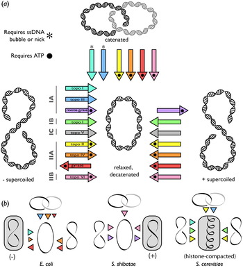
Topoisomerases create single- or double-stranded breaks in DNA by the nucleophilic attack of a tyrosine residue on the phosphodiester backbone, creating a covalent phosphotyrosyl linkage. This strand scission activity forms the basis of the major classification scheme for topoisomerases – types I and II – in which type I enzymes create a single-stranded break (Depew et al. Reference Depew, Liu and Wang1978; Liu & Wang, Reference Liu and Wang1979; Tse et al. Reference Tse, Kirkegaard and Wang1980; Champoux, Reference Champoux1981) and type II topoisomerases cleave both strands (Fig. 2a) (Brown et al. Reference Brown, Peebles and Cozzarelli1979; Morrison & Cozzarelli, Reference Morrison and Cozzarelli1979; Liu et al. Reference Liu, Rowe, Yang, Tewey and Chen1983; Sander & Hsieh, Reference Sander and Hsieh1983).
DNA topoisomerases have competing and overlapping roles in maintaining cellular DNA topology. (a) Activities of DNA topoisomerases. Members of the five DNA topoisomerase families are shown as arrows indicating their activities, labeled at left. DNA topoisomerases are capable of relaxing negative supercoils (from left to center), relaxing positive supercoils (from right to center), decatenating linked DNA (from top to center), and introducing positive or negative supercoils (from center to right or left). The requirement of an ATP cofactor or the presence of a single-stranded DNA gap or nick for a particular activity is indicated. (b) Example topoisomerase complements from three organisms. Colors of triangles correspond to the color key in (a); highlighted topological states are those found in vivo. Left: E. coli, a mesophilic bacterium, possesses the type IA topoisomerases topo I and topo III, and the type IIA topoisomerases topo IV and DNA gyrase. Its genome is maintained in a net negatively supercoiled state (Vinograd et al. Reference Vinograd, Lebowitz, Radloff, Watson and Laipis1965). Center: S. shibatae, a hyperthermophilic archaea, possesses the type IA topoisomerase reverse gyrase and the type IIB topoisomerase topo VI. Plasmids extracted from this organism are slightly positively supercoiled (Forterre et al. Reference Forterre, Bergerat and Lopez-Garcia1996). Right: The mesophilic eukaryote S. cerevisiae has type IB (topo I), IA (topo III), and IIA topoisomerases (topo II). Chromosome compaction in this organism is aided by histones, as indicated by the solenoidal supercoils.

There are multiple subclasses of each topoisomerase type, divided by both amino-acid sequence and reaction mechanism. The type I enzymes comprise three subfamilies: IA, IB, and IC. Type IB topoisomerases include eukaryotic topoisomerase I and poxvirus topoisomerase I, as well as homologous enzymes found in certain bacteria (Krogh & Shuman, Reference Krogh and Shuman2002a) and in Mimivirus (Benarroch et al. Reference Benarroch, Claverie, Raoult and Shuman2006). Type IB proteins form a 3′-phosphotyrosyl linkage to one DNA strand (Champoux, Reference Champoux1981), nicking the duplex in a metal-independent reaction (Dynan et al. Reference Dynan, Jendrisak, Hager and Burgess1981) to facilitate the relaxation of both negatively and positively supercoiled DNAs (Koster et al. Reference Koster, Croquette, Dekker, Shuman and Dekker2005). The recently established type IC family comprises only topoisomerase V and is found exclusively in a single genus of archaea (Slesarev et al. Reference Slesarev, Stetter, Lake, Gellert, Krah and Kozyavkin1993; Gadelle et al. Reference Gadelle, Filee, Buhler and Forterre2003). Much remains unknown about topo V, but it appears to be functionally similar to type IB topoisomerases in that the enzyme cleaves the 3′ end of DNA, does not require any cofactors for activity and relaxes both positive and negative supercoils (Slesarev et al. Reference Slesarev, Stetter, Lake, Gellert, Krah and Kozyavkin1993; Taneja et al. Reference Taneja, Schnurr, Slesarev, Marko and Mondragon2007). Both the IB and IC topoisomerase classes are believed to use a controlled rotation mechanism for relaxing DNA (see below) (Slesarev et al. Reference Slesarev, Stetter, Lake, Gellert, Krah and Kozyavkin1993; Taneja et al. Reference Taneja, Schnurr, Slesarev, Marko and Mondragon2007).
The type IA topoisomerase family includes bacterial and archaeal topoisomerase I (Wang, Reference Wang1971; Slesarev et al. Reference Slesarev, Zaitzev, Kopylov, Stetter and Kozyavkin1991) and topoisomerase III (Srivenugopal et al. Reference Srivenugopal, Lockshon and Morris1984), eukaryotic topoisomerase III (Wallis et al. Reference Wallis, Chrebet, Brodsky, Rolfe and Rothstein1989), and the yet-uncharacterized Mimivirus topoisomerase IA (Raoult et al. Reference Raoult, Audic, Robert, Abergel, Renesto, Ogata, La Scola, Suzan and Claverie2004). These enzymes create a 5′-phosphotyrosyl linkage to DNA (Depew et al. Reference Depew, Liu and Wang1978; Liu & Wang, Reference Liu and Wang1979; Tse et al. Reference Tse, Kirkegaard and Wang1980), forming an enzyme-bridged, single-stranded DNA gap through which a second DNA strand or duplex is passed (Brown & Cozzarelli, Reference Brown and Cozzarelli1981). This strand passage mechanism enables the efficient unknotting and decatenation of DNAs containing single-stranded regions or nicks, as well as relaxation of negatively supercoiled substrates. An interesting twist on the type IA family is reverse gyrase, an enzyme unique to hyperthermophiles that is capable of positively supercoiling DNA (Kikuchi & Asai, Reference Kikuchi and Asai1984; Bouthier de la Tour et al. Reference Bouthier, Portemer, Nadal, Stetter, Forterre and Duguet1990; Bouthier de la Tour et al. Reference Bouthier, Portemer, Huber, Forterre and Duguet1991; Brochier-Armanet & Forterre, Reference Brochier-Armanet and Forterre2007). All type IA topoisomerases are dependent on Mg2+ and Zn2+ for full activity (Tse-Dinh & Beran-Steed, Reference Tse-Dinh and Beran-Steed1988; Domanico & Tse-Dinh, Reference Domanico and Tse-Dinh1991); reverse gyrase further requires ATP (Kikuchi & Asai, Reference Kikuchi and Asai1984).
Type II topoisomerases are subclassified as IIA or IIB on the basis of sequence and structure. All type II topoisomerases form an enzyme-bridged break in double-stranded DNA and effect topological change by passing a second DNA duplex through the gap (Brown et al. Reference Brown, Peebles and Cozzarelli1979; Morrison & Cozzarelli, Reference Morrison and Cozzarelli1979; Brown & Cozzarelli, Reference Brown and Cozzarelli1981; Liu et al. Reference Liu, Rowe, Yang, Tewey and Chen1983; Sander & Hsieh, Reference Sander and Hsieh1983). The type IIA family includes: (1) eukaryotic topo II (Baldi et al. Reference Baldi, Benedetti, Mattoccia and Toccini-Valentini1980; Hsieh & Brutlag, Reference Hsieh and Brutlag1980; Miller et al. Reference Miller, Liu and Englund1981), (2) viral and bacteriophage topo II (Liu et al. Reference Liu, Liu and Alberts1979; Stetler et al. Reference Stetler, King and Huang1979; Lavrukhin et al. Reference Lavrukhin, Fortune, Wood, Burbank, van Etten, Osheroff and Lloyd2000; Raoult et al. Reference Raoult, Audic, Robert, Abergel, Renesto, Ogata, La Scola, Suzan and Claverie2004), (3) bacterial, archaeal, and chloroplast DNA gyrase (Gellert et al. Reference Gellert, Mizuuchi, O'Dea and Nash1976; Thompson & Mosig, Reference Thompson and Mosig1985; Sioud et al. Reference Sioud, Possot, Elie, Sibold and Forterre1988), and (4) bacterial topo IV (Kato et al. Reference Kato, Nishimura, Imamura, Niki, Hiraga and Suzuki1990). Topo II and topo IV can relax negative and positive supercoils, although topo IV is particularly effective at catalyzing the latter reaction (Peng & Marians, Reference Peng and Marians1993b; Crisona et al. Reference Crisona, Strick, Bensimon, Croquette and Cozzarelli2000). Gyrase is also capable of relaxing positive supercoils and is further distinguished by an ability to introduce negative supercoils into DNA (Gellert et al. Reference Gellert, Mizuuchi, O'Dea and Nash1976). All type IIA topos are also capable of decatenating and unknotting DNA, although topo IV again excels at this task (Adams et al. Reference Adams, Shekhtman, Zechiedrich, Schmid and Cozzarelli1992; Rybenkov et al. Reference Rybenkov, Ullsperger, Vologodskii and Cozzarelli1997).
In contrast to the widespread nature of the type IIA topoisomerases, type IIB enzymes are confined to archaea, plants, and certain algae. This topoisomerase family includes only topo VI (Gadelle et al. Reference Gadelle, Filee, Buhler and Forterre2003), which is similar in its activity profile to topo II, but seems to have a distinct evolutionary lineage (Bergerat et al. Reference Bergerat, De Massy, Gadelle, Varoutas, Nicolas and Forterre1997; Gadelle et al. Reference Gadelle, Filee, Buhler and Forterre2003). Both type IIA and IIB topoisomerases create phosphotyrosyl linkages to the 5′ end of DNA (Morrison & Cozzarelli, Reference Morrison and Cozzarelli1979; Sugino et al. Reference Sugino, Higgins and Cozzarelli1980; Tse et al. Reference Tse, Kirkegaard and Wang1980; Buhler et al. Reference Buhler, Lebbink, Bocs, Ladenstein and Forterre2001) and require ATP and Mg2+ for full activity (Gellert et al. Reference Gellert, Mizuuchi, O'Dea and Nash1976; Bergerat et al. Reference Bergerat, Gadelle and Forterre1994).
The phylogenetic distribution of topoisomerases is complex, but a unifying characteristic among cellular organisms is the presence of at least one type I and one type II enzyme. Many distinct combinations are possible (Fig. 2b). For example, in a mesophilic bacterium such as E. coli, DNA gyrase maintains a high level of negative supercoils in the circular chromosome to promote compaction and unwinding (Gellert et al. Reference Gellert, Mizuuchi, O'Dea and Nash1976; Zechiedrich et al. Reference Zechiedrich, Khodursky, Bachellier, Schneider, Chen, Lilley and Cozzarelli2000). Since this bacterium divides rapidly, the decatenation of daughter chromosomes must be equally facile, a need that has led E. coli (and most other bacteria) to also adopt topo IV, an enzyme specialized for this task (Adams et al. Reference Adams, Shekhtman, Zechiedrich, Schmid and Cozzarelli1992; Peng & Marians, Reference Peng and Marians1993a, Reference Peng and Mariansb). E. coli further contends with a number of single-stranded DNA recombination and replication intermediates, requiring topoisomerase III, which is particularly efficient at resolving such structures (DiGate & Marians, Reference Digate and Marians1988; Lopez et al. Reference Lopez, Yang, Deibler, Ray, Pennington, Digate, Hastings, Rosenberg and Zechiedrich2005). Positive supercoils generated by replication and transcription are handled by topo IV and gyrase, whereas supercoiling homeostasis is maintained by competition among gyrase, topoisomerase IV, and topoisomerase I, a type IA enzyme (Drlica, Reference Drlica1992; Zechiedrich et al. Reference Zechiedrich, Khodursky, Bachellier, Schneider, Chen, Lilley and Cozzarelli2000).
In contrast to E. coli, the hyperthermophilic organism Sulfolobus shibatae uses a very different topoisomerase complement. The presence of reverse gyrase and the absence of gyrase in this archaeon helps to maintain the chromosome in a slightly positively supercoiled state, which likely helps prevent temperature-induced unwinding (Forterre et al. Reference Forterre, Bergerat and Lopez-Garcia1996). Eukaryotes rely on yet another combination of topoisomerases. Here, because the genome is extensively coated with and compacted by histones, the resident topoisomerases (topo IB, topo III, and one or more topo II paralogs) must contend with DNA superstructures formed during replication and transcription events in the context of chromatin. It is important to note that although the particular set of topoisomerases endemic to a given organism plays a role in removing chromosome entanglements and maintaining appropriate levels of supercoiling, these enzymes function in a cellular context in which many other factors (such as protein expression levels and subcellular localization) also come in to play.
4. Type IB topoisomerases
4.1 Introduction
The heritage of the type IB topoisomerases is clearly distinct from that of all other topoisomerases. Although these enzymes, like other proteins, possess the ability to cleave DNA through a covalent phosphotyrosine intermediate, the catalytic tyrosine of the type IB family is provided by a helix–turn–helix (HTH) fold homologous to tyrosine recombinases and integrases (Cheng et al. Reference Cheng, Kussie, Pavletich and Shuman1998; Redinbo et al. Reference Redinbo, Stewart, Kuhn, Champoux and Hol1998). Until recently, type IB topoisomerases were thought to exist exclusively in eukaryotes and poxviruses, but orthologs have been discovered in certain bacterial species, suggesting that horizontal gene transfer has taken place between the kingdoms (Krogh & Shuman, Reference Krogh and Shuman2002a; Gadelle et al. Reference Gadelle, Filee, Buhler and Forterre2003; Patel et al. Reference Patel, Shuman and Mondragon2006).
4.2 Type IB topoisomerases form a protein clamp around DNA
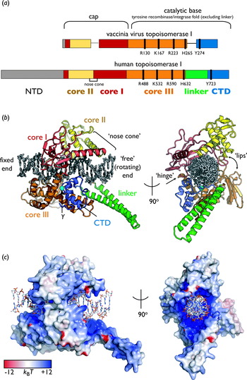
Eukaryotic type IB topoisomerases (topo IBs) consist of four major domains (Fig. 3a): the N-terminal domain (NTD), a ‘core’ region (which can be broken into three subdomains), an extended α-helical linker element and the C-terminal domain (CTD) (Stewart et al. Reference Stewart, Ireton and Champoux1996; Redinbo et al. Reference Redinbo, Stewart, Kuhn, Champoux and Hol1998). The NTD is a protease-sensitive, likely disordered region that contains nuclear localization signals. The first 190 amino acids of this domain are dispensable for activity in vitro (Stewart et al. Reference Stewart, Ireton and Champoux1997; Frohlich et al. Reference Frohlich, Andersen, Westergaard, Andersen and Knudsen2004, Reference Frohlich, Veigaard, Andersen, McClendon, Gentry, Andersen, Osheroff, Stevnsner and Knudsen2007). Subdomains I and II of the core form a ‘cap’ over bound DNA, contributing to a pair of α-helices, termed the ‘nose cone’, which jut out over the duplex (Fig. 3b). The cap is connected to core subdomain III by a short hinge helix, whereas core subdomain III and the CTD, which comprise the catalytic base of the enzyme, are split into two elements by the linker domain. The linker helices are dispensable for relaxation activity but play a role in the DNA cleavage-religation equilibrium (Stewart et al. Reference Stewart, Ireton and Champoux1997). Together, the cap and catalytic base form a clamp that encircles DNA in a positively charged central hole (Fig. 3c).
Type IB topoisomerase organization and structure. (a) The primary domain structures of poxvirus and eukaryotic topoisomerase I (type IB topoisomerases). Key catalytic residues are indicated by black bars; NTD: N-terminal domain, CTD: C-terminal domain. (b) Two views of the structure of human topoisomerase I bound to DNA. Domains are colored as in (a); the catalytic tyrosine (Y) is shown as a cyan sphere. DNA is colored gray. The covalently bound (fixed) and unbound (rotating) ends are indicated. The lips and hinge region are labeled in the orthogonal view at right (PDB ID 1A36, this and all molecular figures were generated in PyMOL (Stewart et al. Reference Stewart, Redinbo, Qiu, Hol and Champoux1998; DeLano, Reference Delano2002). (Structures are available on the protein data bank: http://www.rcsb.org; identification numbers are given.) (c) Electrostatic surface potential of human topoisomerase I, contoured at ±12k BT, where k B is Boltzmann constant and T is temperature. Orientations are the same as in (b). Note the highly positively charged nature of the DNA binding cavity, as well as the positive potential on the DNA-facing sides of the linker and nose cone helices. DNA is colored by atom (PDB ID 1A36); this and all other electrostatic calculations were carried out via APBS (Stewart et al. Reference Stewart, Redinbo, Qiu, Hol and Champoux1998; Baker et al. Reference Baker, Sept, Joseph, Holst and McCammon2001).

The poxvirus and bacterial topo IB orthologs are minimized versions of the eukaryotic enzyme (Fig. 3a). Poxvirus topo IB lacks the nose cone and linker helices and, because it does not require nuclear localization, is missing most of the NTD. This topoisomerase is further distinguished by an ability to recognize and cleave a specific sequence, 5′-(T/C)CCTT-3′, through a base-specific interaction between DNA and two structural elements: an α-helix (α5) in subdomain III of the core domain and a β strand (β5) in the cap (Shuman & Prescott, Reference Shuman and Prescott1990; Hwang et al. Reference Hwang, Wang and Bushman1998; Perry et al. Reference Perry, Hwang, Bushman and Van Duyne2006). Despite these differences, all type IB topoisomerases are thought to share a common strand cleavage and supercoil relaxation mechanism.
4.3 Type IB topoisomerase are swivelases
The basic mechanism of type IB topoisomerases follows a model for topoisomerase action proposed by Cairns (Reference Cairns1963) well before the discovery of any such enzyme, that of a ‘swivelase’. Clues to this behavior came first from bulk measurements, which showed that topo IB can change Lk in multiple integral steps, with an average ΔLk of 5 (Stivers et al. Reference Stivers, Harris and Mildvan1997). More recently, controlled rotation by topo IB has been directly confirmed by single-molecule measurements (Koster et al. Reference Koster, Croquette, Dekker, Shuman and Dekker2005). During the topo IB reaction, the free energy stored in DNA supercoils drives one duplex end to rotate about the intact phosphodiester linkage opposite the enzyme-induced nick, relaxing both positive and negative supercoils (Champoux, Reference Champoux1981). Higher supercoiling levels lead to an increase in the mean number of turns accomplished by the enzyme per cleavage/religation cycle, indicating that topo IB is sensitive to the torque stored in under- or overwound DNA (Koster et al. Reference Koster, Croquette, Dekker, Shuman and Dekker2005).
4.4 The topo IB active site and DNA cleavage mechanism
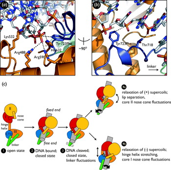
Multiple crystal structures of human, bacterial, and viral topo IBs have revealed a conserved active site constellation used for strand scission (Fig. 4a). Tyr723 (human topo I numbering; Tyr274 in vaccinia virus topo I) serves as the catalytic tyrosine and is responsible for forming a covalent link with the phosphodiester backbone of the cleaved DNA segment (Redinbo et al. Reference Redinbo, Stewart, Kuhn, Champoux and Hol1998). The covalently attached end of the DNA occupies the majority of a large cavity formed by the base and the cap, whereas the duplex end that is free to rotate extends out from this cavity between the nose cone and linker helices (Redinbo et al. Reference Redinbo, Stewart, Kuhn, Champoux and Hol1998). Extensive work on the poxvirus enzymes, synthesized with structural information, has revealed the cleavage mechanism for topo IB in detail (Redinbo et al. Reference Redinbo, Stewart, Kuhn, Champoux and Hol1998, Reference Redinbo, Stewart, Champoux and Hol1999, Reference Redinbo, Champoux and Hol2000; Krogh & Shuman, Reference Krogh and Shuman2000, Reference Krogh and Shuman2002b; Krogh et al. Reference Krogh, Claeboe, Hecht and Shuman2001; Staker et al. Reference Staker, Hjerrild, Feese, Behnke, Burgin and Stewart2002; Interthal et al. Reference Interthal, Quigley, Hol and Champoux2004; Tian et al. Reference Tian, Claeboe, Hecht and Shuman2004, Reference Tian, Claeboe, Hecht and Shuman2005; Davies et al. Reference Davies, Mushtaq, Interthal, Champoux and Hol2006; Patel et al. Reference Patel, Shuman and Mondragon2006; Perry et al. Reference Perry, Hwang, Bushman and Van Duyne2006). Arg488, Arg590, and His632 (Arg130, Arg223, and His265 in vaccinia virus topo I, respectively) all contact the scissile phosphate oxygens in the uncleaved, cleaved, and transition states (Fig. 4a), suggesting that they play a role in transition state stabilization (Stewart et al. Reference Stewart, Redinbo, Qiu, Hol and Champoux1998; Tian et al. Reference Tian, Claeboe, Hecht and Shuman2004, Reference Tian, Claeboe, Hecht and Shuman2005; Mondragon, Reference Mondragon2005; Davies et al. Reference Davies, Mushtaq, Interthal, Champoux and Hol2006). The role of general acid is thought the be played by Lys532 (Lys167 in vaccinia virus topo I), since the mutation of this residue to alanine can be rescued by the use of a phosphorothiolate-modified nucleotide at the cleavage site (Krogh & Shuman, Reference Krogh and Shuman2000; Interthal et al. Reference Interthal, Quigley, Hol and Champoux2004; Tian et al. Reference Tian, Claeboe, Hecht and Shuman2004). In vaccinia virus, Arg130 (Arg488 in human topo I) has also been implicated as a general acid (Krogh & Shuman, Reference Krogh and Shuman2002b). Structural information (Davies et al. Reference Davies, Mushtaq, Interthal, Champoux and Hol2006) reveals that both residues interact with the 5′ bridging oxygen in the transition state, suggesting that they act in concert (Yakovleva et al. Reference Yakovleva, Chen, Hecht and Shuman2008) rather than through, a proton-relay mechanism (Krogh & Shuman, Reference Krogh and Shuman2002b). The role of the catalytic base may be played by Arg587 (Arg220 in poxvirus), which interacts with the phosphotyrosyl bridging oxygen in the structure of the covalent DNA–protein complex of vaccinia virus topo I (Perry et al. Reference Perry, Hwang, Bushman and Van Duyne2006; Yakovleva et al. Reference Yakovleva, Chen, Hecht and Shuman2008).
Type IB topoisomerase mechanism. (a) A close-up view of the active site of human topo I. DNA is colored by atom. Protein is colored by domain (CTD, blue; core III, orange; linker; green). In this structure, the active site tyrosine (Tyr723) has been mutated to phenylalanine. His632 contacts one of the nonbridging oxygens on the scissile phosphate; the other is coordinated by Arg488 and Arg590. Lysine 532 contacts the 5′ bridging oxygen of the scissile phosphate (PDB ID 1A36) (Stewart et al. Reference Stewart, Redinbo, Qiu, Hol and Champoux1998). (b) Contacts between the ‘free’ (rotatable) DNA end (colored by atom) and human topoisomerase I [coloring as in (a)]. In this structure, the catalytic tyrosine (Tyr723) has been mutated to phenylalanine. Thr 718 forms a hydrogen bond with the +1 phosphate of the cleaved strand (PDB ID 1A36) (Stewart et al. Reference Stewart, Redinbo, Qiu, Hol and Champoux1998). (c) Mechanism for relaxation of positive and negative supercoils by human topoisomerase I. Domains are colored as in Fig. 3. An ‘open’ state of the enzyme (1) is competent to bind and clamp around DNA (2). DNA cleavage occurs, resulting in a 3′ phosphotyrosyl linkage to the enzyme (cyan star) (Champoux, Reference Champoux1981) and an increase in linker flexibility (indicated by the wavy line) (3) (Stewart et al. Reference Stewart, Ireton and Champoux1996; Chillemi et al. Reference Chillemi, Redinbo, Bruselles and Desideri2004, Reference Chillemi, Bruselles, Fiorani, Bueno and Desideri2007). If positive supercoils are being relaxed, the free DNA helix rotates in a counterclockwise direction, contacting the core I nose cone helix (yellow cylinder) and causing slight separation of the lips (4a) (Sari & Andricioaei, Reference Sari and Andricioaei2005). If negative supercoils are being relaxed, the free DNA helix rotates in a clockwise direction, contacting the core II nose cone helix (red cylinder) and stretching the hinge helix (4b) (Sari & Andricioaei, Reference Sari and Andricioaei2005).

4.5 Topoisomerase IB maintains a closed conformation during DNA relaxation
A conformation commonly observed in crystal structures of type IB topos bound to DNA is that of a ‘closed-clamp’, in which the enzyme envelops ~10 bp of the duplex (Redinbo et al. Reference Redinbo, Stewart, Kuhn, Champoux and Hol1998). The clamp is sealed by a pair of loops, termed the ‘lips’, which extend from core domains I and III (Fig. 3b) (Redinbo et al. Reference Redinbo, Stewart, Kuhn, Champoux and Hol1998). Rotation of the free DNA has been proposed to occur in the closed-clamp state based on the observation that cross-linking the lips together through engineered cysteines at positions Ala499 and His367 (human topo I numbering) has little to no effect on relaxation activity (Carey et al. Reference Carey, Schultz, Sisson, Fazzio and Champoux2003). A cross-link formed between positions Gly365 and Ser534, which lie closer to the active site, does significantly impair relaxation (Woo et al. Reference Woo, Losasso, Guo, Pattarello, Benedetti and Bjornsti2003), although this effect has been attributed to the close proximity of the cross-link to the active site tyrosine (Sari & Andricioaei, Reference Sari and Andricioaei2005). In further support of a model in which relaxation occurs in the closed state, molecular dynamics simulations suggest that the closed conformation can be maintained during DNA rotation (Sari & Andricioaei, Reference Sari and Andricioaei2005). The simulations indicate that rotation is accommodated through a slight (~10–14 Å) separation of the lips during positive supercoil relaxation and stretching of the hinge during negative supercoil relaxation.
In contrast to cleavage and rotation, the binding and release of duplex DNA must be accompanied by opening of the clamp. Molecular dynamics simulations, again, have provided some clues as to how this process might occur. In silico modeling suggests that electrostatic repulsion at the lips can lead to their opening, followed by a 180° rotation about the hinge helix residue Glu445 to open a 100-Å gap between the cap and the catalytic core (Chillemi et al. Reference Chillemi, Bruselles, Fiorani, Bueno and Desideri2007). Despite this large interdomain movement, the active site does not appear to change conformation, in agreement with crystallographic observations of bacterial topo IB showing that the active site constellation adopts a cleavage-competent configuration even when DNA is absent (Patel et al. Reference Patel, Shuman and Mondragon2006).
4.6 The nose cone helices may engage rotating DNA
The nose cone helices, located at the core I–core II interface in the cap element of topo IB, meet at an almost 90° angle over the DNA duplex. Although it has not been observed to directly contact DNA in any crystal structure available to date, the nose cone displays a positively charged face with which to potentially engage nucleic acid. Consistent with this idea, modeling studies indicate that even a slight (~10°) shift in the position of the DNA can bring the phosphate backbone into contact with this face (Stewart et al. Reference Stewart, Redinbo, Qiu, Hol and Champoux1998). Protein footprinting analyses have further shown that the lysines in the nose cone are protected by DNA binding to vaccinia topo IB (Hanai & Wang, Reference Hanai and Wang1994). Moreover, molecular dynamics simulations indicate that during positive or negative supercoil relaxation, the rotating DNA contacts either the core I or core II nose cone helices (Sari & Andricioaei, Reference Sari and Andricioaei2005). This contact pushes or pulls on the cap domain and has been postulated to facilitate or trigger the stretching of the hinge helix or separation of the lips that is needed to accommodate duplex rotation in a closed-clamp conformation (Sari & Andricioaei, Reference Sari and Andricioaei2005).
4.7 Linker domain dynamics govern the DNA religation rate
Although dispensable for relaxation activity, the linker domain appears to play an important role in the topo IB catalytic cycle by tuning the DNA cleavage-religation equilibrium. A functional, linker-less topoisomerase can be formed by reconstitution of the core and the CTD from separately expressed polypeptides (Stewart et al. Reference Stewart, Ireton and Champoux1997); however, this reconstituted enzyme has reduced relaxation efficiency, increased fluctuations in the α-carbon backbone, and faster DNA religation kinetics (Stewart et al. Reference Stewart, Ireton and Champoux1997; Chillemi et al. Reference Chillemi, Redinbo, Bruselles and Desideri2004). An increase in religation rate appears to be a consistent effect of perturbing the linker, particularly when those perturbations (such as mutating a proline to alanine, or creating a discontinuity) are likely to cause a concomitant increase in helix flexibility (Stewart et al. Reference Stewart, Ireton and Champoux1999; Fiorani et al. Reference Fiorani, Bruselles, Falconi, Chillemi, Desideri and Benedetti2003). Mutations that decrease linker flexibility seem to have the opposite effect (Chillemi et al. Reference Chillemi, Fiorani, Castelli, Bruselles, Benedetti and Desideri2005). For example, substitution of alanine for Thr718, which contacts the +1 phosphate of the cleaved strand (Fig. 4b) (Stewart et al. Reference Stewart, Redinbo, Qiu, Hol and Champoux1998), decreases linker flexibility in molecular dynamics simulations and favors DNA cleavage in biochemical assays (Chillemi et al. Reference Chillemi, Fiorani, Castelli, Bruselles, Benedetti and Desideri2005). Other computational and biochemical observations have suggested that linker flexibility can increase after DNA is cleaved (Stewart et al. Reference Stewart, Ireton and Champoux1996; Fiorani et al. Reference Fiorani, Bruselles, Falconi, Chillemi, Desideri and Benedetti2003; Chillemi et al. Reference Chillemi, Redinbo, Bruselles and Desideri2004, Reference Chillemi, Bruselles, Fiorani, Bueno and Desideri2007). Together, these studies indicate that contacts between the linker helices and the DNA may tune the relative mobility of the linker, which may in turn help specify the religation rate.
4.8 The protein–DNA interface exerts frictional control on relaxation velocity
During supercoil relaxation, topo IB allows the free end of the duplex to rotate around the covalently bound segment. To religate the break, however, the enzyme must correctly align the free 5′ end of DNA with the phosphotyrosine intermediate, holding the two duplexes rotationally motionless long enough to accomplish religation chemistry. One mechanism through which topo IB may be able to stabilize such a state is by slowing the rotation of DNA through friction. This idea has been supported by single-molecule measurements of the relaxation velocities of human and vaccinia virus topo IB (Koster et al. Reference Koster, Croquette, Dekker, Shuman and Dekker2005). Human topo IB, which makes more extensive protein–DNA contacts, is slower than the more streamlined vaccinia enzyme, which is in turn slower than a simple nicking enzyme (Koster et al. Reference Koster, Croquette, Dekker, Shuman and Dekker2005). The degree of rotational impedance is proportional to the surface area of the protein–DNA interface and can be modeled as a frictional coefficient (Koster et al. Reference Koster, Croquette, Dekker, Shuman and Dekker2005). Consistent with this model, mutation of a conserved hydrophobic cavity residue (Tyr70) to a smaller amino acid (alanine) in the vaccinia virus topo IB results in an increased reaction velocity, implying that the reduced protein–DNA contact area leads to less friction and thus faster duplex rotation (Koster et al. Reference Koster, Croquette, Dekker, Shuman and Dekker2005).
4.9 Type IB topoisomerases mediate topological change through controlled rotation
Together, this wealth of structural, biochemical, and computational analyses has provided a fairly detailed picture of supercoil relaxation by type IB topoisomerases (Fig. 4c). To begin, the enzyme adopts an open conformation, with the cap and catalytic base spread apart. Upon binding DNA, these domains close to form a clamp around the duplex. DNA cleavage covalently secures the 3′ phosphate (and one duplex DNA end) to the enzyme, permitting the free and tethered duplex segments to rotate with respect to one another as a means to relax positive or negative supercoils. Rotation is slowed by contacts with the inner cavity of the enzyme, while the torsional tension generated by these contacts is accommodated by slight movements in the hinge and lips. The linker and nose cone helices found in eukaryotic topo IB provide additional interactions between the protein and DNA, further regulating the cleavage/religation equilibrium and adding an additional layer of control over the rate of rotation. Religation occurs upon a brief cessation of rotation due to stochastic pausing events and/or the exhaustion of torque (i.e. when the DNA is fully relaxed). Thus, type IB topos harness the energy stored in supercoiled DNA to drive supercoil relaxation, but they constrain the release of this energy through friction at defined protein–DNA interfaces.
5. Type IC topos
5.1 Type IC topoisomerases constitute a distinct family of enzymes
Topoisomerase V is the exclusive member of the newly established group of type IC topoisomerases. Thus far, topo V has been identified solely in the Methanopyrus genus of archaea (Gadelle et al. Reference Gadelle, Filee, Buhler and Forterre2003). In addition to possessing topoisomerase activity, this protein processes apurinic/apyrimidinic (AP) sites to remove abasic lesions in DNA (Belova et al. Reference Belova, Prasad, Kozyavkin, Lake, Wilson and Slesarev2001). The two activities are structurally segregated from one another, and it is not clear whether they collaborate in any way. Befitting its distribution in a hyperthermophilic genus, topo V is incredibly tolerant of high temperatures, acting maximally at 108°C and maintaining activity up to 122°C (Kozyavkin et al. Reference Kozyavkin, Pushkin, Eiserling, Stetter, Lake and Slesarev1995). Aside from its temperature tolerance, topo V is functionally similar to topo IB: the monomeric enzyme relaxes both positive and negative supercoils, forms a phosphotyrosine link to the 3′ end of a single DNA strand, and requires neither Mg2+ nor ATP as a cofactor (Slesarev et al. Reference Slesarev, Stetter, Lake, Gellert, Krah and Kozyavkin1993). However, these similarities are superficial because the structural, phylogenetic, and mechanistic properties of topo V strongly differentiate it from every other type of topoisomerase.
5.2 Topoisomerase V has a complex and repetitive structure
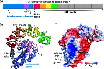
Topo V is a relatively large enzyme (110 kDa) comprising an N-terminal topoisomerase domain and a C-terminal region with 12 tandem repeats of a helix–hairpin–helix (HhH) motif (Fig. 5) (Taneja et al. Reference Taneja, Patel, Slesarev and Mondragon2006). The topoisomerase domain bears no sequence or structural homology to any other topoisomerase, although the active site tyrosine is located on a HTH motif, similar to the active site elements of other topoisomerases (Berger et al. Reference Berger, Gamblin, Harrison and Wang1996; Grishin, Reference Grishin2000b; Taneja et al. Reference Taneja, Patel, Slesarev and Mondragon2006). The HhH motifs appear to be more flexible than the topoisomerase domain, exhibiting variable positions in the four different crystal structures of the enzyme that have been solved to date (Taneja et al. Reference Taneja, Patel, Slesarev and Mondragon2006). The AP site processing activity is housed in the HhH domains, but the precise location and nature of this function is largely unknown (Belova et al. Reference Belova, Prasad, Kozyavkin, Lake, Wilson and Slesarev2001).
Type IC topoisomerase organization and structure. (a) The primary domain structure of topoisomerase V from Methanopyrus kandleri (a type IC topoisomerase). The catalytic tyrosine (Tyr226) resides on a HTH domain (purple). (b) Structure of M. kandleri topo V. The topoisomerase domain (blue) and the HTH domain (purple) are packed closely together. The catalytic tyrosine is shown as a cyan sphere. Four tandem HhH motifs observed in the structure are shown in red, orange, yellow, and green (PDB ID 2CSB) (Taneja et al. Reference Taneja, Patel, Slesarev and Mondragon2006). (c) Electrostatic surface potential of topoisomerase V contoured at ±5k BT, showing the putative DNA binding region (PDB ID 2CSB) (Taneja et al. Reference Taneja, Patel, Slesarev and Mondragon2006).

5.3 Topoisomerase V employs a controlled rotation mechanism
Bulk studies and recent single-molecule experiments have suggested that topo V behaves as a controlled swivelase (Slesarev et al. Reference Slesarev, Stetter, Lake, Gellert, Krah and Kozyavkin1993; Taneja et al. Reference Taneja, Schnurr, Slesarev, Marko and Mondragon2007). Like topo IB, topo V relaxes DNA not in discrete steps of one or two windings, but with a Gaussian distribution of step sizes displaying a mean |ΔLk| value greater than 1 (Taneja et al. Reference Taneja, Schnurr, Slesarev, Marko and Mondragon2007). Another similarity to topo IB is that topo V processivity and velocity increases at greater superhelical densities (Taneja et al. Reference Taneja, Schnurr, Slesarev, Marko and Mondragon2007).
Despite these commonalities, there are functional differences suggesting that the topo V mechanism is different from that of topo IB in certain important ways. Most notably, the velocity of topo V is significantly slower than that of topo IB (Taneja et al. Reference Taneja, Schnurr, Slesarev, Marko and Mondragon2007). Currently, it is not clear whether this distinction is due to intrinsic properties of the two classes of enzymes or whether the reaction conditions used in the study, particularly the temperature (40°C), account for some of the difference (Taneja et al. Reference Taneja, Schnurr, Slesarev, Marko and Mondragon2007). It is also possible that the slower relaxation velocity exhibited by topo V is due to greater friction between the enzyme and DNA, but if so, the source of the impedance is unknown (Taneja et al. Reference Taneja, Schnurr, Slesarev, Marko and Mondragon2007). The putative DNA binding groove of topo V, as proposed by inspection of the electrostatic surface potential of the enzyme (Fig. 5c), shows no obvious cavity in which DNA could be encircled (Taneja et al. Reference Taneja, Patel, Slesarev and Mondragon2006). It is clear, however, that a conformational change must take place upon DNA binding or cleavage, since the topo V active site tyrosine is buried in the crystal structure (Taneja et al. Reference Taneja, Patel, Slesarev and Mondragon2006). This sequestration suggests that topo V likely undergoes a large structural rearrangement to expose the active site tyrosine and create a more extensive DNA binding surface; however, it remains to be seen exactly how this unique enzyme employs protein–DNA contacts to control DNA rotation (Taneja et al. Reference Taneja, Patel, Slesarev and Mondragon2006).
6. Type IA topoisomerases
6.1 Type IA topoisomerases are found in all three domains of life
The sequence, structure, mechanism, and distribution of type IA topoisomerases all distinguish this group of enzymes from the type IB and IC families. Bacteria typically possess two type IA paralogs: topo I and topo III (Wang, Reference Wang1971; Srivenugopal et al. Reference Srivenugopal, Lockshon and Morris1984). In contrast, eukaryotes tend only to retain topo III, although higher eukaryotes can possess multiple isoforms of this enzyme (Wallis et al. Reference Wallis, Chrebet, Brodsky, Rolfe and Rothstein1989; Kim & Wang, Reference Kim and Wang1992). Some archaea also possess a type IA topoisomerase annotated as topo I or topo III, although these isoforms have not been extensively characterized (Forterre et al. Reference Forterre, Gribaldo, Gadelle and Serre2007). Topo I is specialized for relaxing negatively supercoiled DNA, whereas topo III primarily resolves complex single-stranded DNA juxtapositions, such as those arising from replication and recombination events (DiGate & Marians, Reference Digate and Marians1988; Wallis et al. Reference Wallis, Chrebet, Brodsky, Rolfe and Rothstein1989; Hiasa et al. Reference Hiasa, Digate and Marians1994; Harmon et al. Reference Harmon, Digate and Kowalczykowsji1999; Lopez et al. Reference Lopez, Yang, Deibler, Ray, Pennington, Digate, Hastings, Rosenberg and Zechiedrich2005). Hyperthermophiles exclusively possess a fourth type IA protein, called reverse gyrase (Kikuchi & Asai, Reference Kikuchi and Asai1984; Gadelle et al. Reference Gadelle, Filee, Buhler and Forterre2003).
6.2 Type IA topoisomerase structure
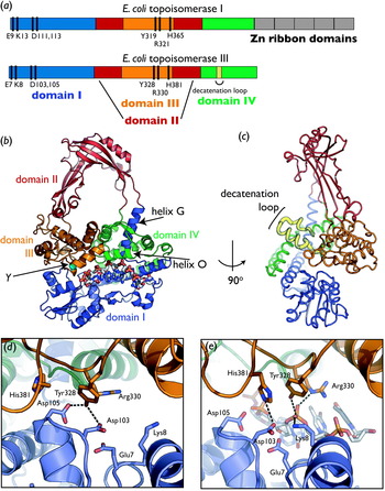
The structure of all type IA topoisomerases is globally reminiscent of a padlock, with a large (27 Å) cavity in the center bounded by four major conserved domains (Fig. 6a, b) (Lima et al. Reference Lima, Wang and Mondragon1994; Mondragon & DiGate, Reference Mondragon and Digate1999). Domain I adopts a topoisomerase–primase (TOPRIM) fold, a divalent metal ion binding element found in type IA, IIA, and IIB topoisomerases, as well as bacterial primases (Aravind et al. Reference Aravind, Leipe and Koonin1998). Domain II consists of an all-β fold that arches over the rest of the enzyme, defining the central hole (Lima et al. Reference Lima, Wang and Mondragon1994; Mondragon & DiGate, Reference Mondragon and Digate1999). Domain III, which houses the active site tyrosine, comprises a winged-helix domain (WHD) fold, as does a portion of domain IV. The interface of domains I, III, and IV forms a positively charged groove responsible for binding single-stranded DNA and also constitutes a protein ‘gate’ for controlling access to the interior of the enzyme (Changela et al. Reference Changela, Digate and Mondragon2001).
Type IA topoisomerase organization and structure. (a) The primary domain structures of E. coli topoisomerase I and III (type IA topoisomerases). Domains are indicated by color and labeled catalytic residues are shown as black bars. (b) Structure of E. coli topoisomerase III complexed with DNA. Domains are colored as in (a). Bound DNA is colored by atom and shown as sticks. The catalytic tyrosine (Y, cyan sphere) is located at the domain III–domain I interface. (PDB ID 1I7D) (Changela et al. Reference Changela, Digate and Mondragon2001). (c) Rotation of the structure in (b) by 90° reveals the decatenation loop projecting from the ‘back’ of the enzyme. (PDB ID 1I7D) (Changela et al. Reference Changela, Digate and Mondragon2001). (d) Close-up view of the E. coli topo III active site in the absence of DNA. Protein is colored by domain as in (b). The active site tyrosine forms hydrogen bonds with two conserved aspartates (PDB ID 1D6M) (Mondragon & DiGate, Reference Mondragon and Digate1999). (e) Close-up of the active site of E. coli topoisomerase III bound to DNA in a ‘pre-cleavage’ conformation. Protein is colored as in (b). DNA is colored by atom (carbons are in white). The active site tyrosine is poised to interact with the scissile phosphate, while a conserved arginine and lysine coordinate the nonbridging oxygens. One of the aspartates that formerly interacted with the tyrosine now forms a hydrogen bond with a conserved histidine. (PDB ID 2O59) (Changela et al. Reference Changela, Digate and Mondragon2007).

In addition to the conserved catalytic core, sometimes referred to as a ‘topofold’ (Duguet et al. Reference Duguet, Serre and Bouthier De La Tour2006), type IA toposiomerases are often appended with auxiliary elements important for their function. For example, most type IA topoisomerases contain one or more zinc-binding domains that belong to the zinc ribbon fold family (Yu et al. Reference Yu, Allen, Andreeva, Fersht and Bycroft2006). These modules may assist in binding DNA and, at least in E. coli topo I, are necessary for full activity (Tse-Dinh & Beran-Steed, Reference Tse-Dinh and Beran-Steed1988; Yu et al. Reference Yu, Zhu, Tse-Dinh and Fesik1995). In addition, bacterial topoisomerase III contains an insertion into its core topofold called the decatenation loop (Fig. 6c). This small, positively charged helical hairpin is necessary for robust unlinking of single-stranded DNA substrates (see below) (Li et al. Reference Li, Mondragon, Hiasa, Marians and Digate2000).
6.3 The catalytic center transitions to an active state upon binding DNA
Structures of E. coli topo I and topo III captured in conformational states with and without bound DNA have revealed that the type IA topoisomerase catalytic center adopts an active state through a series of incremental changes that accompany DNA recognition (Fig. 6d, e). The active site is located at one end of the single-stranded DNA binding groove and is formed at the interface of domains III and I, both of which contribute amino acids required for catalysis. When DNA is absent or only partially bound by the groove, type IA topoisomerases adopt a closed state in which the active site tyrosine (Tyr328, E. coli topo III numbering) in domain III hydrogen bonds to conserved aspartates (Asp103 and Asp105) in domain I (Lima et al. Reference Lima, Wang and Mondragon1994; Changela et al. Reference Changela, Digate and Mondragon2007). The complete binding of single-stranded DNA to the groove induces a 20° rotation of domain III, shifting these residues and breaking their hydrogen bonding interactions (Changela et al. Reference Changela, Digate and Mondragon2007). This movement allows Asp103 to form a new interaction with a conserved histidine in domain III (His381) that is important for relaxation (Chen & Wang, Reference Chen and Wang1998; Feinberg et al. Reference Feinberg, Changela and Mondragon1999a; Perry & Mondragon, Reference Perry and Mondragon2002; Changela et al. Reference Changela, Digate and Mondragon2007). The domain III shift further allows a conserved TOPRIM residue in domain I, Glu7, to contact the 3′ bridging oxygen of the scissile phosphate. This amino acid has been proposed to act as the general base during cleavage (Changela et al. Reference Changela, Digate and Mondragon2001, Reference Changela, Digate and Mondragon2007), although its substitution to glutamine has no effect on the rate of strand scission (Chen & Wang, Reference Chen and Wang1998; Zhu et al. Reference Zhu, Roche, Papanicolaou, Dipietrantonio and Tse-Dinh1998).
Another player in the conversion of the catalytic center from an inactive to active state is the conserved residue Arg330 (Chen & Wang, Reference Chen and Wang1998). When only six nucleotides are bound in the single-stranded DNA binding groove, Arg330 contacts the base of the nucleotide adjacent to the scissile phosphate (Changela et al. Reference Changela, Digate and Mondragon2007). In contrast, once the single-stranded DNA binding groove is occupied with eight nucleotides, Arg330 shifts to ligand the scissile phosphate itself (Changela et al. Reference Changela, Digate and Mondragon2007). The role of the residue at this latter stage likely is to stabilize the negative charge of the transition state, a task that may be aided further by Lys8 (Changela et al. Reference Changela, Digate and Mondragon2007). The position of Arg330, along with the sequestration of the catalytic tyrosine, seems to ensure that a cleavage competent state cannot be achieved until the single-stranded DNA binding groove is occupied by at least eight bases of substrate (Perry & Mondragon, Reference Perry and Mondragon2003).
The role of divalent metals in the type IA topoisomerase reaction is somewhat unclear. Although magnesium ions have been shown to bind to domain I and are required for full activity, they are not required for cleavage and have not been observed to be associated with this region in crystal structures obtained to date (Domanico & Tse-Dinh, Reference Domanico and Tse-Dinh1991; Lima et al. Reference Lima, Wang and Mondragon1994; Mondragon & DiGate, Reference Mondragon and Digate1999; Zhu & Tse-Dinh, Reference Zhu and Tse-Dinh2000). Interestingly, however, mutation of conserved TOPRIM domain residues in E. coli topoisomerase I (Asp 113, Asp113, and Glu115 in topo I; Asp103, Asp105, and Glu107 in topo III) leads to reduction in DNA cleavage as well as a loss of Mg2+ binding (Zhu & Tse-Dinh, Reference Zhu and Tse-Dinh2000). This suggests that the biochemical requirement for Mg2+ is linked to liganding of those ions by the TOPRIM domain. Whether metal ions play a direct part in the phosphoryl transfer reaction catalyzed by type IA topoisomerases, as they often do in other TOPRIM-fold enzymes, remains to be determined.
6.4 DNA binding induces small conformational changes outside of the active site
In addition to rearrangement of the active site constellation upon binding, the DNA binding groove also alters its conformation upon engaging substrate. Most notably, helix O of domain IV (residues 497–509 in E. coli topo I), which forms a positively charged ceiling over the single-stranded DNA binding groove, interacts with the phosphodiester backbone of the bound nucleic acid segment (Perry & Mondragon, Reference Perry and Mondragon2003). As a greater length of single-stranded DNA progressively binds in the groove (from six to eight nucleotides), helix O shifts slightly, widening the groove and increasing its contact area with DNA (Perry & Mondragon, Reference Perry and Mondragon2003). This movement also alters the position of the decatenation loop in topo III (Changela et al. Reference Changela, Digate and Mondragon2007). An additional spatial adjustment is made by helix G, an element that connects domain I to domain II and interacts with bound substrate, resulting in a small rotation of domain II (Changela et al. Reference Changela, Digate and Mondragon2001).
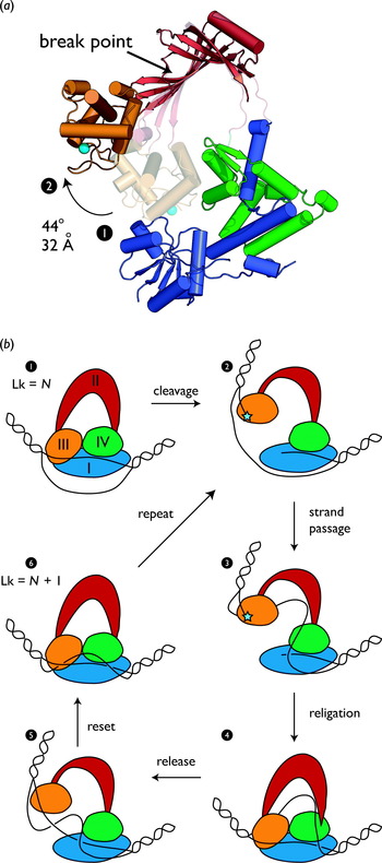
The point at which helix G meets domain II initially was proposed to serve as the primary hinge point for opening a ‘gateway’ into the interior chamber of type IA topisomerases. However, structures of isolated domain II/III fragments of E. coli topo I have suggested that an alternative hinge region residing in the center of three β strands in domain II may allow domain III to disengage from the domain I/IV interface (Feinberg et al. Reference Feinberg, Lima and Mondragon1999b) (Fig. 7a). A model in which this so-called ‘break point’ is enforced on the full-length enzyme suggests that this hinge movement is sufficient to lift domain III away from the domain I/IV body of the enzyme by ~30 Å without disturbing the structures of other elements (Fig. 7a) (Feinberg et al. Reference Feinberg, Lima and Mondragon1999b). Such a movement would allow the topoisomerase to create a wide, enzyme-bridged gap in a cleaved strand, leaving the active site tyrosine on domain III covalently attached to one segment and the DNA binding groove associated with the free 3′ end.
Type IA topoisomerase mechanism. (a) Gate opening in type IA topoisomerases. The fully closed structure of E. coli topoisomerase I (domain I, blue; domains II, red; domain III, orange; domain IV, green) is superposed with an alternate conformation captured for domains II and III (transparent red and orange); the superposition is through half of domain II. Active site tyrosines are shown as cyan spheres. The closed-to-open conformational change results in a 44°, 32-Å shift in domain III about the ‘break point’ indicated by the arrow. These two states likely correspond to states 1 and 2 in (b) (PDB IDs 1ECL and 1CYY) (Lima et al. Reference Lima, Wang and Mondragon1994; Feinberg et al. Reference Feinberg, Lima and Mondragon1999b). (b) Strand passage mechanism for type IA topoisomerases (Lima et al. Reference Lima, Wang and Mondragon1994). Domains are colored as in (a); the covalent attachment of DNA to the active site tyrosine is indicated by a cyan star. The enzyme binds a single-stranded region of DNA (1). Once the DNA binding groove is fully occupied, cleavage occurs and domain III lifts away from the domain I–IV interface (2). The uncleaved strand is passed into the central cavity (3), following which the gate closes and the cleaved strand is religated (4). The gate reopens without cleavage, releasing the passed strand from the central cavity (5). The linking number of the substrate DNA has now increased by 1, and the enzyme is able to perform the next round of strand passage (6).

6.5 The type IA strand passage mechanism
Together, these disparate pieces of data give rise to a model for topology modification by type IA topoisomerases that is quite different from the rotary mechanism used by type IB/IC enzymes. By cleaving DNA and opening the domain III gate, type IA topoisomerases can separate a DNA strand sufficiently wide enough to allow the passage of a second DNA segment through the gap (Fig. 7b). In this reaction, single-stranded DNA first binds at the groove formed by the interface of domains I, III, and IV. DNA binding accompanies remodeling of the enzyme active site and, once complete, sets the stage for DNA cleavage and the formation of a 5′ phosphotyrosine intermediate with the catalytic tyrosine in domain III (Tse et al. Reference Tse, Kirkegaard and Wang1980; Perry & Mondragon, Reference Perry and Mondragon2003). A conformational change at the break point in domain II permits domain III to separate from domains I and IV (Feinberg et al. Reference Feinberg, Lima and Mondragon1999b), pulling the covalently attached DNA strand with it. This event opens the gate to the central cavity, allowing entry of the unpaired partner strand. Subsequent closure of the gate traps the passed strand in the cavity and permits religation of the cleaved strand; a second gate-opening event in the absence of DNA scission serves to release the trapped strand, now on the opposite side of the cleaved segment from whence it started. The result of this reaction is a net change in Lk of 1, precisely the value observed from both bulk biochemical and single-molecule studies (Brown & Cozzarelli, Reference Brown and Cozzarelli1981; Dekker et al. Reference Dekker, Rybenkov, Duguet, Crisona, Cozzarelli, Bensimon and Croquette2002). Because cleavage does not occur until the DNA binding groove is fully occupied, single-stranded DNA is required for activity (Kirkegaard & Wang, Reference Kirkegaard and Wang1985). This property accounts for type IA topoisomerases' preference for negatively supercoiled substrates, as underwound duplexes are more readily denatured. This behavior also explains why type IA enzymes only exhibit strand passage activity on positively supercoiled substrates containing an artificially unpaired region (Kirkegaard & Wang, Reference Kirkegaard and Wang1985), since overwound DNA is more resistant to melting and thus less likely to provide the enzyme with a suitable substrate.
The creation of a physical gap in DNA is highly useful for unlinking DNA strands, but has the potential to generate full-fledged DNA breaks. To minimize this risk, type IA topoisomerases have evolved multiple protein–DNA and protein–protein interfaces that ensure that both ends of the protein bridge are firmly secured to their nucleic acid partner. The extensive primary DNA binding groove on one domain and the covalent attachment point on the other are linked by a protein scaffold to prevent accidental dissociation of the anchor sites. Attainment of a competent active site configuration is dependent on DNA fully occupying the binding groove, ensuring that the phosphodiester backbone cannot be cleaved unless the noncovalently bound strand is firmly in place. Thus, although their means are different, type IA topoisomerases, like type IB enzymes, rely on dynamic interactions between macromolecular surfaces to mitigate aberrant DNA cleavage events and control topology modification.
6.6 Zinc ribbon domains assist in DNA binding
One way type IA topoisomerases may further stabilize their association with single-stranded DNA regions is through the use of zinc ribbon domains (Fig. 6a) (Tse-Dinh & Beran-Steed, Reference Tse-Dinh and Beran-Steed1988; Yu et al. Reference Yu, Zhu, Tse-Dinh and Fesik1995). The number of zinc ribbon folds found in different type IA family members varies, with some orthologs having as many as five repeats and others lacking the domain entirely (Grishin, Reference Grishin2000a). For example, E. coli topo I has three typical zinc ribbons as well as two that have lost the tetracysteine motif required to coordinate a zinc ion (Grishin, Reference Grishin2000a). Despite this sequence heterogeneity, all five E. coli topo I zinc-binding domains are capable of binding single-stranded DNA (Yu et al. Reference Yu, Zhu, Tse-Dinh and Fesik1995). Zinc ribbon elements are usually found at the C terminus of the catalytic core, but in some orthologs (such as Thermotoga maritima topo I) they can reside at the N terminus (Hansen et al. Reference Hansen, Harrenga, Wieland, Schomburg and Reinemer2006). Regardless of their position in the amino-acid sequence, the tertiary structure of the type IA topoisomerase core is such that the zinc ribbon domains typically lie near the base of the DNA binding groove, opposite the active site (Hansen et al. Reference Hansen, Harrenga, Wieland, Schomburg and Reinemer2006). This location may enable the zinc ribbons to interact with single-stranded DNA regions, both to facilitate strand passage and improve enzyme processivity.
6.7 A unique insertion mediates decatenation activity in topoisomerase III
Although topo I and topo III are relatively similar in sequence, they are functionally quite different both in vitro and in vivo. Topo I is a very effective relaxase and a somewhat poor decatenase, whereas the reverse is true for topo III (DiGate & Marians, Reference Digate and Marians1988; Hiasa et al. Reference Hiasa, Digate and Marians1994). The decatenation loop of topo III, which helps mediate its DNA unlinking activity, consists of a 17-amino-acid insertion (Fig. 6c), which projects from the back face of the enzyme at the base of the central cavity (Li et al. Reference Li, Mondragon, Hiasa, Marians and Digate2000). Decatenation of double-stranded DNA is thought to occur when a duplex segment occupies the central cavity before the binding of substrate to the single-stranded DNA binding groove (Li et al. Reference Li, Mondragon, Hiasa, Marians and Digate2000; Champoux, Reference Champoux2001). If a single-stranded DNA segment with a nicked complementary strand is subsequently engaged by the binding site, cleavage results in a double-stranded gap that can allow passage of the bound duplex segment out of the central cavity. Alternatively, the decatenation of single-stranded substrates can occur if the cleaved and transported segments derive from different DNA molecules, rather than from two strands of one duplex. Precisely how the decatenation loop assists in these reactions is currently unknown.
7. Reverse gyrase
7.1 Reverse gyrase is a hybrid hyperthermophilic type IA enzyme
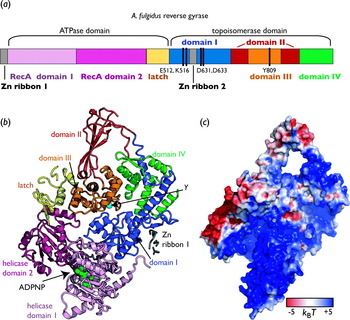
Although composed of two fairly common catalytic folds, reverse gyrase is somewhat of an enigma (Fig. 8). The enzyme is classified as a type IA topoisomerase because one of its constituent parts consists of the core catalytic fold found in topo I and topo III. The other portion of reverse gyrase consists of a helicase-like domain that binds and hydrolyzes ATP. Reverse gyrase is found exclusively in hyperthermopilic archaea and bacteria, where its use of ATP enables it to introduce positive supercoils into DNA (Gadelle et al. Reference Gadelle, Filee, Buhler and Forterre2003; Brochier-Armanet & Forterre, Reference Brochier-Armanet and Forterre2007).
Reverse gyrase organization and structure. (a) Primary domain structure of A. fulgidus reverse gyrase. The topoisomerase region is similar to E. coli topoisomerase I, except for the insertion of a zinc ribbon fold in domain I. The active site tyrosine (Tyr809) is indicated. (b) Tertiary structure of A. fulgidus reverse gyrase. Domains are colored as in (a). The active site tyrosine (Y) is indicated as a cyan sphere. The binding site for ATP (green spheres) lies between the two helicase domains, as indicated (PDB ID 1GL9) (Rodriguez & Stock, Reference Rodriguez and Stock2002). (c) Surface electrostatic potential of A. fulgidus reverse gyrase, contoured at ±5k BT. Orientation is the same as in (b) (PDB ID 1GKU) (Rodriguez & Stock, Reference Rodriguez and Stock2002).

7.2 The structure of reverse gyrase is similar to that of other type IA topos
The C-terminal half of reverse gyrase constitutes the topoisomerase region. The fold is very similar overall to E. coli topoisomerase I (1·4 Å rmsd), albeit with a somewhat smaller central cavity (Fig. 8b) (Rodriguez & Stock, Reference Rodriguez and Stock2002). The enzyme has one zinc ribbon domain inserted into domain I and another at the extreme N terminus of the protein. These domains are not well ordered in the crystal structure, possibly due to the absence of substrate DNA (Rodriguez & Stock, Reference Rodriguez and Stock2002). Consistent with its homology to type IA topoisomerases, the isolated C-terminal region can relax DNA, although this activity is somewhat inefficient compared to E. coli topo I (Declais et al. Reference Declais, Marsault, Confalonieri, De La Tour and Duguet2000).
The N-terminal helicase domain of reverse gyrase comprises two RecA-type ATP binding folds (Fig. 8) (Rodriguez & Stock, Reference Rodriguez and Stock2002). Sequence- and structure-based comparisons indicate that this region is most closely related to superfamily 2 (SF2) DNA helicases/translocases and RNA remodeling enzymes (Rodriguez & Stock, Reference Rodriguez and Stock2002). The RecA elements have no helicase activity in isolation or in the context of the full-length topoisomerase, although they bind and hydrolyze ATP and are necessary for positive supercoiling activity (Declais et al. Reference Declais, Marsault, Confalonieri, De La Tour and Duguet2000). In the ADPNP (5′-adenylyl-β,γ-imidodiphosphate)-bound structure of Archaeoglobus fulgidus reverse gyrase, only one of the RecA domains appears to make contact with bound nucleotide, and the active site is largely solvent-exposed (Rodriguez & Stock, Reference Rodriguez and Stock2002).
Inserted between the second RecA fold and the topoisomerase core is a small, α-helical bundle termed the ‘latch’. This element is structurally homologous to a portion of the E. coli transcription termination factor Rho, although the significance of this similarity is unclear (Rodriguez & Stock, Reference Rodriguez and Stock2002). The latch makes contact with the topoisomerase module, specifically domain III, and appears to play an important role in regulating reverse gyrase activity (see below) (Rodriguez, Reference Rodriguez2002, Reference Rodriguez2003).
7.3 Reverse gyrase displays diverse activities in vitro
Reverse gyrase is capable of relaxing negative supercoils, introducing positive supercoils and annealing single-stranded plasmids. All of these activities are nucleotide-dependent, with the supercoiling and annealing functions further requiring hydrolysis of a nucleoside triphosphate (preferably ATP) (Rodriguez, Reference Rodriguez2002, Reference Rodriguez2003; Hsieh & Capp, Reference Hsieh and Capp2005; Hsieh & Plank, Reference Hsieh and Plank2006; Jungblut & Klostermeier, Reference Jungblut and Klostermeier2007). In addition to these more traditional topoisomerase functions, reverse gyrase may also act as a ‘DNA chaperone’ by binding preferentially to nicked DNA and preventing temperature-related DNA degradation (Kampmann & Stock, Reference Kampmann and Stock2004). The role these activities play in vivo is currently unclear. Reverse gyrase is not strictly necessary for high-temperature growth, although its absence results in slower growth (Atomi et al. Reference Atomi, Matsumi and Imanaka2004). The chaperone activity of the enzyme may be responsible for the high-temperature genome stability observed in Pyrococcus furiosos (Peak et al. Reference Peak, Robb and Peak1995), whereas its positive supercoiling activity may help renature DNA regions melted by high temperature and maintain a net positive superhelical density generally seen in the plasmids of hyperthermophilic organisms (Forterre et al. Reference Forterre, Bergerat and Lopez-Garcia1996).
7.4 Substrate and temperature preferences suggest a single-stranded bubble requirement
The physical mechanisms by which reverse gyrase accomplishes its myriad actions on DNA remain unclear, although mutagenesis studies and analysis of the enzyme's temperature and nucleotide dependence have provided some clues. First, in topological footprinting assays, the presence of reverse gyrase leads to products with a reduced linking number, suggesting that the enzyme creates and/or stabilizes single-stranded regions of DNA (Declais et al. Reference Declais, Marsault, Confalonieri, De La Tour and Duguet2000). In agreement with this observation is the finding that A. fulgidus reverse gyrase is only capable of positively supercoiling DNA efficiently at 80°C if it is given a substrate plasmid containing a noncomplementary, 20-bp single-stranded bubble (Hsieh & Plank, Reference Hsieh and Plank2006). If the bubble is replaced with a metastable region of AT repeats, or the temperature increased to 85°C, the activity of reverse gyrase likewise increases (Hsieh & Plank, Reference Hsieh and Plank2006). These results, combined with the observation that the enzyme binds single-stranded DNA more efficiently when it is joined to a short (10 bp) double-stranded region, suggest that reverse gyrase prefers to associate with locally denatured DNA near single-strand/double-strand DNA junctions (Rodriguez, Reference Rodriguez2002).
7.5 The latch may couple ATP turnover with strand passage
The latch, comprising residues 352–427 in A. fulgidus reverse gyrase, interacts with a short helix in domain III (residues 857–866) (Fig. 8b) (Rodriguez & Stock, Reference Rodriguez and Stock2002). This interaction appears to anchor domain III to domain I, a conformation that may block unregulated strand passage by preventing reverse gyrase from opening a gap in the cleaved strand. When the association is abrogated by removal of either the latch or its domain III interaction element, the result is a 13-fold increase in the ATPase rate, but no change in the supercoiling efficiency (Rodriguez, Reference Rodriguez2002, Reference Rodriguez2003). In addition, DNA relaxation, which typically requires the presence of a nucleotide, can now occur at wild-type levels even in the absence of ATP (Rodriguez, Reference Rodriguez2002; Jungblut & Klostermeier, Reference Jungblut and Klostermeier2007). The latch thus appears to couple nucleotide binding and hydrolysis to topology modification events. The normal role of this element may be to link these two activities by transmitting conformational changes from the helicase domain to the domain III/domain I gate in the topoisomerase region. In support of this idea, the ADPNP-bound structure of A. fulgidus reverse gyrase indicates that nucleotide binding by the RecA domains does induce a small conformational change in the latch (Rodriguez & Stock, Reference Rodriguez and Stock2002).
7.6 An incomplete model for reverse gyrase action
Taken together, these data suggest a general, albeit incomplete, model for reverse gyrase function. The helicase and topoisomerase domains likely work in concert to generate and/or stabilize single-stranded DNA bubbles. The formation of such substrates is an inherently favorable event with negatively supercoiled substrates, but requires the energy of ATP in positively supercoiled substrates. Consistent with this model is the observation that reverse gyrase relaxes negatively supercoiled DNA in a fast and processive burst and then introduces positive supercoils more slowly (Hsieh & Capp, Reference Hsieh and Capp2005). ATP binding or hydrolysis may trigger the controlled separation of domain III from the topoisomerase core by virtue of the latch element, thereby linking strand passage to nucleotide hydrolysis. Whether the ATPase domains actively provide substrates to the type IA topoisomerase domain of reverse gyrase remains unclear. In this regard, it is interesting that the RecA folds of reverse gyrase are most closely related to SF2 helicases, which tend to be either processive double-stranded DNA translocases or duplex remodeling enzymes that distributively melt short duplex or multiplex structures (Singleton et al. Reference Singleton, Dillingham and Wigley2007; Pyle, Reference Pyle2008).
8. Type IIA topoisomerases
8.1 Type II topoisomerases employ a strand passage mechanism
Similar to type IA topoisomerases, type IIA enzymes modify topology by creating an enzyme-bridged gap in DNA and passing a second DNA segment through the break (Brown & Cozzarelli, Reference Brown and Cozzarelli1979; Mizuuchi et al. Reference Mizuuchi, Fisher, O'Dea and Gellert1980). Type IIA topoisomerases differ, however, by cleaving both strands of DNA, creating a double-stranded break through which they pass a second duplex (Morrison & Cozzarelli, Reference Morrison and Cozzarelli1979; Liu et al. Reference Liu, Rowe, Yang, Tewey and Chen1983; Sander & Hsieh, Reference Sander and Hsieh1983). This reaction inverts double-stranded DNA crossovers, changing Lk in steps of two (Brown & Cozzarelli, Reference Brown and Cozzarelli1979; Mizuuchi et al. Reference Mizuuchi, Fisher, O'Dea and Gellert1980). In addition, type II topoisomerases use ATP to drive and control strand passage (Gellert et al. Reference Gellert, Mizuuchi, O'Dea and Nash1976), even when acting on a supercoiled substrate whose energetic state favors relaxation. To manage the progression of strand passage, type IIA topoisomerases rely on the regulated opening and closing of dimeric protein gates, the actuation of which is controlled by nucleotide and/or DNA.
8.2 Type IIA topoisomerases have a twofold symmetric, multi-gate structure
Type IIA topoisomerases are the most widely distributed topoisomerase family, with members in the bacterial, eukaryal, archaeal, bacteriophage, and viral lineages (Gadelle et al. Reference Gadelle, Filee, Buhler and Forterre2003). The active type IIA holoenzyme can take the form of a homodimer (eukaryotic and viral topo II), an A2B2 heterotetramer (bacterial and archaeal gyrase and bacterial topo IV) or even an A2B2C2 heterohexamer (bacteriophage T4 topo II) (Fig. 9a). Despite these differences, type IIA topoisomerases are always twofold symmetric. The quaternary assembly of type IIA enzymes results in the formation of at least three major subunit interfaces, or gates, which cordon off two separate cavities within the enzyme (Fig. 9b). The N-terminal gate (termed the N or ATP gate) is formed by two elements: an ATP binding and hydrolysis fold and an adjacent domain called the transducer (Wigley et al. Reference Wigley, Davies, Dodson, Maxwell and Dodson1991; Classen et al. Reference Classen, Olland and Berger2003; Corbett & Berger, Reference Corbett and Berger2003b; Bellon et al. Reference Bellon, Parsons, Wei, Hayakawa, Swenson, Charifson, Lippke, Aldape and Gross2004). The ATPase region is a member of the GHKL superfamily, so named because it is shared by all type II topoisomerases (including DNA gyrase), the molecular chaperone Hsp90, CheA-type histidine kinases, and the DNA mismatch repair enzyme MutL (Dutta & Inouye, Reference Dutta and Inouye2000). The transducer domain contains a rare left-handed α/β crossover connection (Wigley et al. Reference Wigley, Davies, Dodson, Maxwell and Dodson1991) and is thought to transmit signals from the N gate to the central gate where DNA is bound and cleaved (the DNA gate) (Bjergbaek et al. Reference Bjergbaek, Kingma, Nielsen, Wang, Westergaard, Osheroff and Andersen2000; Corbett & Berger, Reference Corbett and Berger2003b). The DNA gate is formed by a divalent metal-binding TOPRIM domain and a WHD similar to that typified by the catabolite activation protein (McKay & Steitz, Reference McKay and Steitz1981; Harrison & Aggarwal, Reference Harrison and Aggarwal1990; Berger et al. Reference Berger, Gamblin, Harrison and Wang1996; Aravind et al. Reference Aravind, Leipe and Koonin1998; Gajiwala & Burley, Reference Gajiwala and Burley2000). In bacterial and archaeal type IIA enzymes, these two folds reside on separate subunits, whereas in eukaryotic topo II, they lie within a single polypeptide chain. The WHD houses the catalytic tyrosine and cooperates with the TOPRIM fold to cleave DNA, generating a pair of 5′ cuts staggered 4 bp apart from each other on opposite strands (Morrison & Cozzarelli, Reference Morrison and Cozzarelli1979; Liu et al. Reference Liu, Rowe, Yang, Tewey and Chen1983; Sander & Hsieh, Reference Sander and Hsieh1983). Adjacent to the WHD is an α/β fold termed the ‘shoulder’ or ‘tower’ (Morais Cabral et al. Reference Morais Cabral, Jackson, Smith, Shikotra, Maxwell and Liddington1997), which also participates in binding DNA (Dong & Berger, Reference Dong and Berger2007). From the shoulder extends a coiled-coil element capped at its distal end with a small globular domain, which forms a third dimerization interface of the protein, the C gate. This latter interface is well conserved and serves as the primary dimer interface of the protein (Berger et al. Reference Berger, Gamblin, Harrison and Wang1996; Morais Cabral et al. Reference Morais Cabral, Jackson, Smith, Shikotra, Maxwell and Liddington1997; Fass et al. Reference Fass, Bogden and Berger1999; Corbett et al. Reference Corbett, Schoeffler, Thomsen and Berger2005; Dao-Thi et al. Reference Dao-Thi, Van Melderen, De Genst, Afif, Buts, Wyns and Loris2005; Laponogov et al. Reference Laponogov, Veselkov, Sohi, Pan, Achari, Yang, Ferrara, Fisher and Sanderson2007). In bacterial type IIA topos, an all-β DNA binding domain is further appended to the C terminus of the DNA cleavage core of the enzyme (Reece & Maxwell, Reference Reece and Maxwell1989, Reference Reece and Maxwell1991; Qi et al. Reference Qi, Pei and Grishin2000; Corbett et al. Reference Corbett, Shultzaberger and Berger2004).
Type IIA topoisomerases structure and mechanism. (a) Primary domain structures of representative eukaryotic and bacterial type IIA topoisomerases. Domains are indicated by color, and important residues are indicated by black bars. Gene names are shown above each primary structure along with the subunit compositions of the holoenzymes. (b) Structures of the ATPase and DNA binding and cleavage regions of S. cerevisiae topoisomerase II and E. coli DNA gyrase (inset). One protomer of each dimer is shown in gray for clarity. The GHKL and transducer domains are shown in yellow and orange [topo II: PDB ID 1PVG; gyrase (inset): PDB ID 1EI1]. Bound ADPNP molecules are shown as green spheres. TOPRIM, winged-helix, tower, and coiled-coil domains are shown in red, purple, teal, and blue, respectively [topo II: PDBID 1BGW; gyrase (inset): PDB ID 1AB4]. The catalytic tyrosines (Y) are shown as cyan spheres. Gate regions are indicated to the left (Berger et al. Reference Berger, Gamblin, Harrison and Wang1996; Classen et al. Reference Classen, Olland and Berger2003). (c) The type IIA topoisomerase two-gate mechanism. Domains are colored as in (a). The enzyme first binds the G segment at the DNA gate (1). The binding of ATP (black stars) by each GHKL domain closes the N gate, capturing a T segment (2). Hydrolysis of one ATP and the release of inorganic phosphate occur concomitantly with passage of the T segment through the DNA gate (3). The remaining ATP remains intact, preventing N-gate opening and backwards escape of the T segment. Upon G segment closure and religation, the C gate opens, allowing the T segment to pass out of the enzyme (4). Hydrolysis of the remaining ATP and release of the ADP and inorganic phosphate triggers N-gate opening, resetting the topoisomerase for another round of strand passage.

8.3 Type IIA topos use a two-gate mechanism to accomplish DNA strand passage
The protein gates described above regulate DNA strand passage by a more general ‘two-gate’ mechanism, first proposed in the early 1980s (Mizuuchi et al. Reference Mizuuchi, Fisher, O'Dea and Gellert1980; Wang & Kirkegaard, Reference Wang and Kirkegaard1981) and since validated in many of its essentials. The model posits that a minimum of two protein interfaces control access to a central cavity. One of these gates binds and cleaves a DNA segment. A second segment of DNA then enters the central cavity through one gate and exits through the other, passing through the cleaved segment (and through the topoisomerase) to effect strand transport and alter linking number (Wang & Kirkegaard, Reference Wang and Kirkegaard1981; Roca & Wang, Reference Roca and Wang1994; Roca et al. Reference Roca, Berger, Harrison and Wang1996).
Although structural studies of type IIA topoisomerases have revealed the existence of more than two physical gates, the enzymes are nonetheless believed to operate by the general principles of the two-gate model (Fig. 9c). First, the topoisomerase binds a segment of double-stranded DNA, termed the gate or G segment, that will be cleaved (Roca & Wang, Reference Roca and Wang1992). Binding takes place at the DNA gate, within the so-called ‘breakage–reunion’ or A′ region of the enzyme (Dong & Berger, Reference Dong and Berger2007). Next, the topoisomerase associates with a second segment of DNA (the transport or T segment), an event that may be favored by its preferential binding to DNA crossovers or by DNA wrapping (Liu & Wang, Reference Liu and Wang1978; Brown & Cozzarelli, Reference Brown and Cozzarelli1979; Zechiedrich & Osheroff, Reference Zechiedrich and Osheroff1990b; Howard et al. Reference Howard, Lee, Hsieh and Griffith1991; Roca et al. Reference Roca, Berger and Wang1993b; Kampranis et al. Reference Kampranis, Bates and Maxwell1999). Association of the enzyme with ATP induces dimerization of the GHKL motifs (Wigley et al. Reference Wigley, Davies, Dodson, Maxwell and Dodson1991), closing the N gate around the T segment (Roca & Wang, Reference Roca and Wang1992; Ali et al. Reference Ali, Orphanides and Maxwell1995; Bjergbaek et al. Reference Bjergbaek, Jensen, Westergaard and Andersen1999; Olland & Wang, Reference Olland and Wang1999). Hydrolysis of one molecule of ATP to adenosine diphosphate (ADP) and release of the resultant phosphate triggers a conformational change coupled to DNA-gate opening (Harkins & Lindsley, Reference Harkins and Lindsley1998; Harkins et al. Reference Harkins, Lewis and Lindsley1998; Baird et al. Reference Baird, Harkins, Morris and Lindsley1999), which occurs via separation of the WHDs through flexion of the coiled coil arms (Berger et al. Reference Berger, Gamblin, Harrison and Wang1996; Morais Cabral et al. Reference Morais Cabral, Jackson, Smith, Shikotra, Maxwell and Liddington1997; Fass et al. Reference Fass, Bogden and Berger1999; Dong & Berger, Reference Dong and Berger2007). The T segment is propelled through this gap and the G segment is closed and resealed, following which the T segment escapes through a transient opening of the C gate (Roca & Wang, Reference Roca and Wang1994; Roca et al. Reference Roca, Berger, Harrison and Wang1996; Williams & Maxwell, Reference Williams and Maxwell1999; Dong & Berger, Reference Dong and Berger2007). Hydrolysis of the second ATP and release of all hydrolysis products results in the opening of the N gate (Wigley et al. Reference Wigley, Davies, Dodson, Maxwell and Dodson1991), resetting the enzyme to catalyze another round of strand passage.
Through this action, type IIA topoisomerases unidirectionally pass one DNA duplex through another to modify DNA topology. This remarkable function is powered by ATP turnover and the free energy stored in DNA superstructures, and it is controlled by the timed sequential opening and closing of protein gates. Recent biochemical and structural studies have now begun to provide us with a multifaceted view of precisely how type II topos harness this energy to regulate protein gate opening and DNA movement.
8.4 GHKL domains bind ATP in a cavity sequestered from solvent
One of the key functions of the N gate is the binding of ATP by the GHKL domains. Many of the contacts between nucleotide and protein are made by the ATP lid (Wigley et al. Reference Wigley, Davies, Dodson, Maxwell and Dodson1991; Lamour et al. Reference Lamour, Hoermann, Jeltsch, Oudet and Moras2002; Classen et al. Reference Classen, Olland and Berger2003; Corbett & Berger, Reference Corbett and Berger2003b; Bellon et al. Reference Bellon, Parsons, Wei, Hayakawa, Swenson, Charifson, Lippke, Aldape and Gross2004; Wei et al. Reference Wei, Ruthenburg, Bechis and Verdine2005), a flexible loop hinged with conserved glycine motifs termed the G1 and G2 boxes (Dutta & Inouye, Reference Dutta and Inouye2000). Glycines 113, 114, 117, and 119 (E. coli DNA GyrB numbering) at one end of the ATP lid are clustered at the base of a short helix (Fig. 10a) (Wigley et al. Reference Wigley, Davies, Dodson, Maxwell and Dodson1991). Their small size grants space to the phosphates of bound nucleotide and further permits a positive helix dipole to stabilize negative charge on the phosphates. Glycine-rich loops that engage the phosphates of a bound nucleotide are common motifs among highly diverse types of mononucleotide binding proteins. For example, the phosphate-binding loops (P-loops) of Ras-type protein GTPases (Pai et al. Reference Pai, Kabsch, Krengel, Holmes, John and Wittinghofer1989) and RecA ATPases (Story & Steitz, Reference Story and Steitz1992) have a similar interaction with bound nucleotide even though the tertiary structures of these proteins are entirely distinct from the GHKL fold (Dutta & Inouye, Reference Dutta and Inouye2000; Iyer et al. Reference Iyer, Leipe, Koonin and Aravind2004). The ATP lid also provides a conserved lysine (Lys103) for liganding the β-phosphate, as well as several backbone nitrogens, which further coordinate the γ-phosphate (Wigley et al. Reference Wigley, Davies, Dodson, Maxwell and Dodson1991). A conserved aspartate (Asp73) appears to enforce specificity for ATP over guanosine triphosphate (GTP) by contacting the exocyclic amino group of the adenine base (Wigley et al. Reference Wigley, Davies, Dodson, Maxwell and Dodson1991).
Interactions between nucleotide and the type II topoisomerase N gate. (a) Interactions between nucleotide and protein in the dimerized S. cerevisiae topo II ATPase domains. ADPNP is shown as a stick model, and the magnesium ion is shown as a green sphere. In the GHKL domain (yellow), four conserved glycines (small cyan spheres) surround the γ-phosphate, furthering charge stabilization by the positive dipole of the adjacent helix (red arrow). The strap from the partner GHKL domain (gray) interacts with the binding site, allowing Tyr5 to make an interaction with the adenine ring. The ‘switch lysine’ (Lys367) from the transducer domain (orange) also interacts with the γ-phosphate (PDB ID 1PVG) (Classen et al. Reference Classen, Olland and Berger2003). (b) The ATPase domain of S. shibatae topoisomerase VI in two conformations, aligned by the GHKL domains. The ‘closed’ conformation (gray) is formed by the presence of ATP analogs and transition state mimics, as well as elevated concentrations of ADP and ADP-Pi (product state). This structure contains bound ADP and inorganic phosphate (blue stick models). The switch lysine (light blue, labeled ‘in’) makes contact with the inorganic phosphate by protruding into the nucleotide binding pocket. The open, or unrestrained, conformation (yellow and orange) can be formed in the absence of nucleotide or with ADP alone. This structure has only ADP bound (pink stick model). In this conformation, the switch lysine (light pink, labeled ‘out’) has moved out of the ATP binding pocket and is now solvent-exposed. The two structures were aligned through their GHKL domains, highlighting the 11° movement of the transducer domain (shown at bottom) (PDB IDs 1Z5C and 1Z59) (Corbett & Berger, Reference Corbett and Berger2005).

8.5 An N-terminal ‘strap’ mediates nucleotide-dependent dimerization of the ATP gate
Although each GHKL domain can bind a single-molecule of ATP independently, it is the ATP-dependent dimerization of partner GHKL domains that grants the N gate the ability to regulate the topoisomerase reaction. The molecular basis for dimerization derives in part from the ATP lid, which forms a portion of the subunit–subunit interface upon ATP binding, and in part from an element termed ‘the strap’, a segment of approximately 15 amino acids that extends off the N terminus of the GHKL domain. Upon GHKL dimerization, the strap of each protomer reaches over to interact with the nucleotide binding pocket of its partner (Wigley et al. Reference Wigley, Davies, Dodson, Maxwell and Dodson1991; Ali et al. Reference Ali, Jackson, Howells and Maxwell1993, Reference Ali, Orphanides and Maxwell1995; Lamour et al. Reference Lamour, Hoermann, Jeltsch, Oudet and Moras2002; Classen et al. Reference Classen, Olland and Berger2003; Corbett & Berger, Reference Corbett and Berger2003b; Bellon et al. Reference Bellon, Parsons, Wei, Hayakawa, Swenson, Charifson, Lippke, Aldape and Gross2004; Wei et al. Reference Wei, Ruthenburg, Bechis and Verdine2005). Removal of the strap or mutation of its conserved hydrophobic residues (Tyr5 and Ile10, E. coli gyrase numbering) reduces or destroys dimerization propensity and also eliminates the dimerization dependence of ATPase activity (Brino et al. Reference Brino, Urzhumtsev, Mousli, Bronner, Mitschler, Oudet and Moras2000). Tyr5 is notable in that it directly contacts the adenine ring of bound nucleotide (Fig. 10a).
8.6 The switch loop couples release of inorganic phosphate to strand passage
Although the N gate is closed by a homodimeric ATPase domain interaction, pre-steady-state kinetic studies on Saccharomyces cerevisiae topoisomerase II have revealed that ATP hydrolysis is asynchronous (Harkins & Lindsley, Reference Harkins and Lindsley1998; Harkins et al. Reference Harkins, Lewis and Lindsley1998). Kinetic competition analyses using vanadate and ADP as competitive inhibitors have further indicated that phosphate release, as opposed to ADP escape, accompanies strand passage (Harkins & Lindsley, Reference Harkins and Lindsley1998; Harkins et al. Reference Harkins, Lewis and Lindsley1998). The ability of a single hydrolysis event to accelerate strand passage is supported by studies of a mutant topoisomerase II (S. cerevisiae E66Q), which is capable of binding ATP but exhibits slow hydrolysis behavior (Baird et al. Reference Baird, Harkins, Morris and Lindsley1999). Compared to wild-type topo II, the decatenation rate of this mutant is reduced by 20-fold, a value similar to that obtained in the presence of nonhydrolyzable ATP analogs. This finding indicates that type II topoisomerases use hydrolysis to accelerate strand passage (Baird et al. Reference Baird, Harkins, Morris and Lindsley1999). Consistent with this model, a wild-type/E66Q topo II heterodimer decatenates DNA at half the wild-type rate, indicating that one ATP hydrolysis event is sufficient for this stimulatory effect (Baird et al. Reference Baird, Harkins, Morris and Lindsley1999).
Although the molecular mechanisms associated with Pi release are not fully understood, clues have come from crystallographic studies. The structure of the GyrB ATPase domain bound to novobiocin shows an asymmetry in the relative positions of the GHKL and transducer folds due to the nonequivalent occupancy of nucleotide between partner GHKL folds (Lamour et al. Reference Lamour, Hoermann, Jeltsch, Oudet and Moras2002). The human topo II ATPase region likewise adopts different conformations in ADP- and ATP-bound states, with both of the transducer domains shifting in position relative to each other and the GHKL domains (Wei et al. Reference Wei, Ruthenburg, Bechis and Verdine2005). A complete structural map of the ATPase cycle of S. shibatae topo VI, a type IIB topoisomerase whose ATPase elements are related to those of type IIA enzymes (see below), suggests that the physical link between strand passage and phosphate release may reside in the region known as the switch loop, a flexible element containing a conserved lysine that assists with ATP hydrolysis (Lys427 of S. shibatae topo VI) (Corbett & Berger, Reference Corbett and Berger2003b, Reference Corbett and Berger2005). When an ATP analog or a transition state mimic is bound to the domain, the switch lysine protrudes into the ATP binding pocket of the GHKL fold, where it contacts the γ-phosphate (Fig. 10a). In ADP-bound or apo forms of the protein, however, the switch loop changes its conformation dramatically, swinging the lysine out of the active site and leaving a solvent-accessible channel to the catalytic center. Movement of the switch loop further coincides with an 11° outward swivel of the transducer region (Fig. 10b), an event proposed to communicate the status of the ATPase site to the DNA gate for controlling G segment opening. Mutagenesis studies targeting the linkage between the transducer domain and the downstream TOPRIM fold in yeast topo II have provided support for this model, although the exact nature of the coupling is still unknown (Bjergbaek et al. Reference Bjergbaek, Kingma, Nielsen, Wang, Westergaard, Osheroff and Andersen2000).
8.7 Strain due to ATP gate closure may facilitate strand passage
The impetus for transducer domain movement, and hence DNA-gate opening, is unclear; however, some lines of evidence suggest that closure of the N gate around a T segment may play a role in gate communication. Structural studies have shown that the interior of the closed N gate of topo II is only 7–14 Å wide, a diameter too small to comfortably accommodate DNA (Classen et al. Reference Classen, Olland and Berger2003; Wei et al. Reference Wei, Ruthenburg, Bechis and Verdine2005). In E. coli DNA gyrase, the central N gate cavity is about 20 Å in diameter on average (Wigley et al. Reference Wigley, Davies, Dodson, Maxwell and Dodson1991), but is constricted to 13 Å at one point by a conserved arginine, Arg286, which resides on the transducer domain (Tingey & Maxwell, Reference Tingey and Maxwell1996). Interestingly, mutating this arginine to glutamine abrogates DNA-dependent ATPase activity and abolishes the ability of gyrase to supercoil DNA, suggesting that the transducer can engage a T segment and that this event influences downstream topoisomerase activities (Tingey & Maxwell, Reference Tingey and Maxwell1996). Consistent with this idea, biochemical studies have suggested that T segment binding directly stimulates G segment cleavage (Corbett et al. Reference Corbett, Zechiedrich and Osheroff1992). Further support for a DNA-dependent link between the N and DNA gates comes from detailed studies monitoring the ATPase activity of yeast topo II as a function of DNA occupancy at the G and T segment binding sites (Mueller-Plantiz & Herschlag, Reference Mueller-Plantiz and Herschlag2006). This analysis revealed that both G and T segment binding have a stimulatory effect in ATPase activity, but that the effect of T segment binding is much greater (Mueller-Plantiz & Herschlag, Reference Mueller-Plantiz and Herschlag2006).
One model that could potentially account for these findings posits that after the ATPase domains bind a T segment and dimerize, the T segment may sterically push on the transducer domains. ATP hydrolysis would release this strain by encouraging the switch lysine to move out of the active site, an event that in turn would facilitate Pi release and trigger strand passage. Alternatively, T segment binding may prevent the switch loops of both transducers from engaging the active site until a secondary event, perhaps due to stochastic fluctuations, permits one of the two domains to swing inward and initiate ATP hydrolysis, following which Pi release and strand passage could occur. Additional experiments are needed to better understand these events.
8.8 Release of hydrolysis products resets the enzyme
Whereas the first ATP hydrolysis event is linked to strand passage (Baird et al. Reference Baird, Harkins, Morris and Lindsley1999), the second is involved in resetting the enzyme for a subsequent round of catalysis (Harkins & Lindsley, Reference Harkins and Lindsley1998; Harkins et al. Reference Harkins, Lewis and Lindsley1998). At high protein concentrations, such as those found in crystallographic conditions, binding of any adenine nucleotide, including ADP, can be sufficient to induce ATPase domain dimerization (Lamour et al. Reference Lamour, Hoermann, Jeltsch, Oudet and Moras2002; Corbett & Berger, Reference Corbett and Berger2005; Wei et al. Reference Wei, Ruthenburg, Bechis and Verdine2005). Thus, even after hydrolysis of both bound ATPs, both resulting ADP molecules need to be released either before or concomitant with N gate reopening. Kinetic analyses have suggested that the hydrolysis and release of the second nucleotide does not occur until after strand passage, with product release being rate-limiting (Harkins et al. Reference Harkins, Lewis and Lindsley1998). This finding suggests that the second ATP hydrolysis event may serve as a timing mechanism, ensuring that the N gate stays shut until the T segment has passed through the DNA gate and possibly out of the enzyme entirely (Fig. 9c). The ability of nonhydrolyzable analogs, which trap the N gate in a closed state, to support a single round of strand passage is consistent with this idea (Roca & Wang, Reference Roca and Wang1994), as is the similar behavior of topo II mutants that bind but do not hydrolyze ATP (Lindsley & Wang, Reference Lindsley and Wang1993; Harkins & Lindsley, Reference Harkins and Lindsley1998; Harkins et al. Reference Harkins, Lewis and Lindsley1998; Baird et al. Reference Baird, Harkins, Morris and Lindsley1999). Likewise there is evidence to suggest that ATP hydrolysis is the rate-limiting step of the Bacillus subtilis gyrase reaction cycle, although this study did not specifically measure phosphate release (Gottler & Klostermeier, Reference Gottler and Klostermeier2007).
8.9 Structural changes accompany DNA-gate opening
The DNA gate is a complex structure, comprising two different domains – the TOPRIM and winged-helix folds – in addition to the G segment DNA. The TOPRIM and WHDs either reside on separate subunits (gyrase, topo IV) or are connected by an extended linker (topo II), allowing their relative positions to change with respect to one another. This conformational plasticity is apparent in two different structures of the DNA binding and cleavage core of S. cerevisiae topoisomerase II determined in the absence of DNA (see Fig. 11a, left and center panels) (Berger et al. Reference Berger, Gamblin, Harrison and Wang1996; Fass et al. Reference Fass, Bogden and Berger1999). In one structure, the WHDs lie about 13 Å apart, and each TOPRIM domain makes extensive contact with the WHD, tower and TOPRIM of its dimer mate (Fass et al. Reference Fass, Bogden and Berger1999). In a second, more open conformation, the WHDs are separated by ~33 Å, and the contacts between the two TOPRIM domains are less extensive (Berger et al. Reference Berger, Gamblin, Harrison and Wang1996). These movements are mediated in large part by rotation of the TOPRIM domains and by a 30° swiveling of the WHD head regions with respect to the dimerized C gate about a hinge point in the coiled-coil arms (Morais Cabral et al. Reference Morais Cabral, Jackson, Smith, Shikotra, Maxwell and Liddington1997; Fass et al. Reference Fass, Bogden and Berger1999). Despite this dramatic domain reorganization, the partner catalytic tyrosines remain separated by a relatively constant distance (25–28 Å). The flexibility of the WHD and catalytic tyrosine positions, coupled with the dimerized C gate, has been observed in several other structures of type IIA topoisomerases determined in the absence of DNA as well (Morais Cabral et al. Reference Morais Cabral, Jackson, Smith, Shikotra, Maxwell and Liddington1997; Corbett et al. Reference Corbett, Schoeffler, Thomsen and Berger2005; Laponogov et al. Reference Laponogov, Veselkov, Sohi, Pan, Achari, Yang, Ferrara, Fisher and Sanderson2007).
Conformational changes in the type IIA topoisomerase DNA and C gates. (a) Three conformations of the S. cerevisiae DNA binding and cleavage core. WHD, tower, and coiled coil regions are shown in blue. The main body of the TOPRIM domain is shown in red, and the smaller ‘hook’ domain is shown in orange to aid visualization of the domain's relative position. One protomer of the homodimer is shown in gray for clarity, but both catalytic tyrosines are highlighted as green spheres. At left is an ‘open’ conformation of the DNA gate (PDB ID 1BGW) (Berger et al. Reference Berger, Gamblin, Harrison and Wang1996). The diagram below the structure represents the conformation of the gate when viewed from above (looking down the dyad axis); the WHD helices are separated by a 33-Å gap (blue crescents/dotted line) and the phenolic oxygens of the catalytic tyrosines by 28 Å (green dumbbell/solid line). In an intermediate structure (PDB ID 1BJT) (Fass et al. Reference Fass, Bogden and Berger1999), the WHDs have moved closer together (13 Å apart), but the phenolic oxygens of the catalytic tyrosines maintain a similar distance from one another (25 Å). The rightmost structure is of a DNA-bound state of the topo II DNA binding and cleavage region (PDB ID 2RGR) (Dong & Berger, Reference Dong and Berger2007). In this conformation, the WHDs are nearly touching (6 Å apart), whereas the catalytic tyrosines remain separated by a similar distance as seen in the other structures (26 Å). Interestingly, the C gate is open in this state, creating a 17-Å gap between the primary dimerization domains. (b) Two views of the structure of the S. cerevisiae DNA binding and cleavage core bound to DNA. Protein domains are colored as in Fig. 9; DNA is shown as a green surface. The total bend angle imparted to DNA by the protein is ~150°. The rotated view shows the DNA channel formed by the tower and TOPRIM domains (PDB ID 2RGR) (Dong & Berger, Reference Dong and Berger2007). (c) Ile833 (see arrow) of the tower domain intercalates between two base pairs of the DNA, imparting a 75° bend to this region. Protein domains are colored as in (b); DNA is colored by atom (PDB ID 2RGR) (Dong & Berger, Reference Dong and Berger2007).

8.10 DNA binding triggers dramatic conformational changes in the DNA gate and C gate
A recently determined structure of the DNA binding and cleavage core of S. cerevisiae topoisomerase II bound to DNA offers the first picture of a type IIA topoisomerase–DNA complex and demonstrates how DNA binding radically alters the DNA gate interface (Fig. 11a, right panel) (Dong & Berger, Reference Dong and Berger2007). In this structure, each TOPRIM domain rotates by up to ~80° from its position in the apo state to make substantial contacts with the tower and WHD of its dimer mate (Dong & Berger, Reference Dong and Berger2007). The motion creates a deep, positively charged groove, which channels DNA from the tower domain into the cleavage center (Fig. 11b) (Dong & Berger, Reference Dong and Berger2007). This reorganization also places metal-binding residues of the TOPRIM domains (Glu449, Asp526, and Asp528, yeast topo II) near the catalytic tyrosines of the WHDs (Dong & Berger, Reference Dong and Berger2007), a condition necessary for cleavage to occur (Bahng et al. Reference Bahng, Mossessova, Nurse and Marians2000; West et al. Reference West, Meczes, Thorn, Turnbull, Marshall and Austin2000; Noble & Maxwell, Reference Noble and Maxwell2002). As seen in the DNA-free structures, the distance between the catalytic tyrosines is maintained at about 26 Å, although the distance between dimer-related WHDs narrows to ~6 Å (Fig. 11a) (Dong & Berger, Reference Dong and Berger2007). This conformation is similar to, but subtly distinct from, that determined for the DNA breakage–reunion domain of gyrase (Morais Cabral et al. Reference Morais Cabral, Jackson, Smith, Shikotra, Maxwell and Liddington1997) and, effectively closes the DNA gate (Dong & Berger, Reference Dong and Berger2007).
Another striking aspect of the DNA-bound structure of topo II is seen at the C-gate interface. Unexpectedly, the two halves of this region are separated, producing an approximately 17-Å gap between the globular dimerization domains that form the base of the coiled coil region (Fig. 11a) (Dong & Berger, Reference Dong and Berger2007). Since the DNA gate is sealed, it is possible that this conformation represents a state of the enzyme immediately before G segment cleavage and opening, or an intermediate following strand passage, when the T segment exits the enzyme. Regardless, the structure provides direct visual evidence of a key conformational state predicted to exist in the two-gate model of strand passage.
Recent single-molecule fluorescence resonance energy transfer (FRET) experiments have also provided evidence for gate opening. Using doubly labeled fluorescent DNA substrates, Simley et al. (Reference Smiley, Collins, Hammes and Hsieh2007) were able to visualize an ATP-dependent change between the FRET probes, consistent with DNA gate opening and closing. Surprisingly, their analyses suggest that the DNA gate spends an equal amount of time open as closed, although this result may be influenced by aspects of the experimental setup, such as absence of the T segment DNA during the measurements (Smiley et al. Reference Smiley, Collins, Hammes and Hsieh2007; Wang, Reference Wang2007). Future studies will be needed to further probe the dynamics of this and other gating events.
8.11 Topoisomerase II dramatically bends the G segment
Just as DNA binding triggers conformational changes in the DNA and C gates, the enzyme also reshapes DNA. About 26 bp of DNA are bound by the DNA gate, in agreement with footprinting data for eukaryotic topo II (Thomsen et al. Reference Thomsen, Bendixen, Lund, Anderson, Sorensen and Westergaard1990) and topo IV (Peng & Marians, Reference Peng and Marians1995). The DNA adopts an A-form conformation in the center of the enzyme, but reverts to B form as it exits the primary binding site (Dong & Berger, Reference Dong and Berger2007). As DNA transitions between these two structural states, each protomer imparts a 75° bend in its associated segment, resulting in a 150° bend overall (Fig. 11b) (Dong & Berger, Reference Dong and Berger2007). The scope of this bend is strikingly close to that determined for topo IV (130°) on the basis of J-factor analysis (Vologodskii et al. Reference Vologodskii, Zhang, Rybenkov, Podtelezhnikov, Subramanian, Griffith and Cozzarelli2001). DNA deformation appears to be induced by a conserved isoleucine (Ile833, S. cerevisiae topo II numbering), which intercalates between two base pairs approximately eight nucleotides away from the scissile phosphate (Fig. 11c). Remarkably, this mechanism for kinking DNA is structurally analogous to that used by integration host factor (Rice et al. Reference Rice, Yang, Mizuuchi and Nash1996), a DNA remodeling protein (Craig & Nash, Reference Craig and Nash1984).
The bending of DNA by a type II topoisomerase may explain the preferential association of these enzymes with highly curved DNA regions, such as crossovers and the apices of plectonemic supercoils (Zechiedrich & Osheroff, Reference Zechiedrich and Osheroff1990a; Howard et al. Reference Howard, Lee, Hsieh and Griffith1991; Roca et al. Reference Roca, Berger and Wang1993a; Vologodskii et al. Reference Vologodskii, Zhang, Rybenkov, Podtelezhnikov, Subramanian, Griffith and Cozzarelli2001). DNA bending may also account for the preferential cleavage of certain DNA sequences by topo II. Consistent with this idea, kinetic and thermodynamic analyses of the ability of topo II to associate with short DNA duplexes of varying sequence reveal that cleavage specificity occurs after DNA binding, but before cleavage. This result suggests that the enzyme–DNA complex engages in sequence-specific conformational changes that favor cleavage (Mueller-Plantiz & Herschlag, Reference Mueller-Plantiz and Herschlag2007). The topo II-DNA structure points to DNA bending as a candidate state for this cleavage-promoting conformation (Dong & Berger, Reference Dong and Berger2007).
8.12 Bacterial type IIA topoisomerases have specialized domains and activities
Most bacteria possess two distinct type IIA topoisomerase paralogs: topoisomerase IV and DNA gyrase. These enzymes share with other type IIA topoisomerases the basic structural core and the two-gate mechanism, but they also serve more specialized functions in the cell. DNA gyrase is notable for its ability to introduce negative supercoils into DNA (Gellert et al. Reference Gellert, Mizuuchi, O'Dea and Nash1976), an activity that may help maintain the relatively high negative superhelicity of some bacterial genomes (Drlica, Reference Drlica1992; Zechiedrich et al. Reference Zechiedrich, Khodursky, Bachellier, Schneider, Chen, Lilley and Cozzarelli2000). Topoisomerase IV resembles eukaryotic topoisomerase II in that it relaxes supercoiled DNA (Kato et al. Reference Kato, Nishimura, Imamura, Niki, Hiraga and Suzuki1990); however, the enzyme is particularly adept at removing positive supercoils and decatenating plasmids (Peng & Marians, Reference Peng and Marians1993a; Zechiedrich et al. Reference Zechiedrich, Khodursky and Cozzarelli1997; Crisona et al. Reference Crisona, Strick, Bensimon, Croquette and Cozzarelli2000).
The specialized activities of gyrase and topo IV appear to be dependent on unique C-terminal DNA binding domains (CTDs) appended to the GyrA and ParC subunits that are not found in eukaryotic or phage type IIA enzymes. For example, removal of the GyrA CTD abolishes gyrase's ability to negatively supercoil DNA, but not its ability to decatenate DNA in an ATP-dependent manner (Kampranis & Maxwell, Reference Kampranis and Maxwell1996). Topo IV's preferential relaxation activity on positively supercoiled substrates and catenanes likewise is lost upon ablation of the ParC CTD (Corbett et al. Reference Corbett, Schoeffler, Thomsen and Berger2005). In isolation, the GyrA CTD is able to bind DNA and bend it by 180° (Corbett et al. Reference Corbett, Shultzaberger and Berger2004). This domain has also been shown to stabilize positive supercoils in closed circular plasmids, indicating that the bend is chiral (Reece & Maxwell, Reference Reece and Maxwell1991; Ruthenburg et al. Reference Ruthenburg, Graybosch, Huetsch and Verdine2005).
The ParC CTD is considerably less efficient than the GyrA CTD at bending DNA (Corbett et al. Reference Corbett, Shultzaberger and Berger2004), and it does not appear to be able to stabilize significant numbers of supercoils in the context of full-length ParC (Peng & Marians, Reference Peng and Marians1995). The role of this element in the topo IV holoenzyme may instead derive in part from its spatial position with respect to the DNA breakage–reunion domain of ParC (Fig. 12a). The CTD is connected to the core of the enzyme by a well-ordered, ~15-amino-acid segment that runs along the tower domain and docks at the upper, outer edge of the protein (Fig. 12a, inset) (Corbett et al. Reference Corbett, Schoeffler, Thomsen and Berger2005). A four-residue linker, typically containing one or two prolines, extends from a hydrophobic anchor point on the tower to the globular CTD, localizing the CTD to this region (Fig. 12a, inset) (Corbett et al. Reference Corbett, Schoeffler, Thomsen and Berger2005). The linker is ordered, but unrestrained, suggesting that the CTD may be free to rotate about its tether (Corbett et al. Reference Corbett, Schoeffler, Thomsen and Berger2005). Interestingly, modeling a 180° rotation of the CTD about the tetrapeptide linker allows the domain to be oriented so that its positively charged outer rim could potentially engage an incoming T segment, particularly one juxtaposed via a left-handed crossing angle relative to the G segment (Figs 1c and 12b) (Corbett et al. Reference Corbett, Schoeffler, Thomsen and Berger2005). This arrangement may help topo IV more effectively recognize and act on positively supercoiled DNA (Fig. 12b), since left-handed crossovers are predominant in such substrates (Stone et al. Reference Stone, Bryant, Crisona, Smith, Vologodskii, Bustamante and Cozzarelli2003). Single-molecule experiments have supported the idea that the preference of topo IV for positive supercoils stems from its ability to recognize local crossover geometry rather than the global topology of a substrate DNA (Stone et al. Reference Stone, Bryant, Crisona, Smith, Vologodskii, Bustamante and Cozzarelli2003).
The CTDs of bacterial type IIA topoisomerases govern holoenzyme activity. (a) Structure of the E. coli ParC dimer. The WHD is shown in purple, the tower in teal, the coiled coil domain in blue, and the CTD in pink. The catalytic tyrosines are shown as cyan spheres. One subunit is shown in gray for clarity. This structure represents the DNA gate in its most open state yet observed, with the catalytic tyrosines separated by 38 Å. Left inset: close-up view of the linker connecting the CTD and the tower. The consensus sequence (PSEP) is shown as a sequence LOGO (Crooks et al. Reference Crooks, Hon, Chandonia and Brenner2004), generated from a MAFFT alignment (Katoh et al. Reference Katoh, Misawa, Kuma and Miyata2002) of all available ParC sequences. At least one of the prolines is present in all topoisomerase IV orthologs. Right inset: electrostatic surface representation of the tower domain. Two methionines (at least one of which is present in all topo IV orthologs) dock into the tower domain, anchoring the CTD (PDB ID 1ZVU) (Corbett et al. Reference Corbett, Schoeffler, Thomsen and Berger2005). (b) Model for the mechanism of topoisomerase IV. Domains are colored as in Fig. 9. The CTDs are docked high on the tower domains, ideally positioning them to interact with an incoming T segment, particularly one with a left-handed crossover geometry (inset). These crossovers are prevalent in positively supercoiled DNA; the CTD juxtaposition may explain the heightened ability of topo IV to relax these substrates over negatively supercoiled DNAs (Peng & Marians, Reference Peng and Marians1995; Crisona et al. Reference Crisona, Strick, Bensimon, Croquette and Cozzarelli2000; Stone et al. Reference Stone, Bryant, Crisona, Smith, Vologodskii, Bustamante and Cozzarelli2003). (c) SAXS model of E. coli GyrA (Costenaro et al. Reference Costenaro, Grossmann, Ebel and Maxwell2005), superposed with crystal structures of the N- and C-terminal regions (PDB IDs 1AB4 and 1ZI0) (Morais Cabral et al. Reference Morais Cabral, Jackson, Smith, Shikotra, Maxwell and Liddington1997; Ruthenburg et al. Reference Ruthenburg, Graybosch, Huetsch and Verdine2005). (d) Model for the mechanism of DNA gyrase. The CTDs are attached to the tower domains via flexible linkers. At least one CTD binds the exiting G segment, introducing a positive (right-handed), solenoidal wrap into the duplex. This event reintroduces the DNA to the N gate as a T segment in cis, enforcing a left-handed crossover geometry (see inset). Inversion of this crossover through strand passage results in a negative change in linking number (negative supercoiling) (Gellert et al. Reference Gellert, Mizuuchi, O'Dea and Nash1976; Brown & Cozzarelli, Reference Brown and Cozzarelli1979).

Although the ParC CTD appears to be stably associated with the rest of the enzyme core, the position of the homologous domain in GyrA may be less static. In small-angle X-ray scattering studies (SAXS) of E. coli GyrA, ab initio reconstructions suggest that the CTDs can reside near the C gate (Fig. 12c) (Costenaro et al. Reference Costenaro, Grossmann, Ebel and Maxwell2005). Given the ability of gyrase to wrap DNA and stabilize a left-hand crossover in cis, the hallmark of its interaction with DNA (Liu & Wang, Reference Liu and Wang1978; Brown & Cozzarelli, Reference Brown and Cozzarelli1979; Kampranis & Maxwell, Reference Kampranis and Maxwell1996), this finding suggests that the GyrA CTDs could transition between multiple positional states throughout the strand passage cycle (Fig. 12d). In support of this idea, DNA footprinting experiments using the gyrase holoenzyme show that nucleotide-dependent changes in protection patterns occur in the CTD binding region (Orphanides & Maxwell, Reference Orphanides and Maxwell1994; Heddle et al. Reference Heddle, Mitelheiser, Maxwell and Thomson2004).
The importance of DNA wrapping in gyrase's supercoiling mechanism has been further established by single-molecule studies, which show that at high DNA tensions, gyrase loses its ability to wrap DNA and its ability to introduce supercoils, but not its ability to relax positive supercoils (Nollmann et al. Reference Nollmann, Stone, Bryant, Gore, Crisona, Hong, Mitelheiser, Maxwell, Bustamante and Cozzarelli2007b). DNA tension has also been shown to affect the processivity of the enzyme in single-molecule assays, with greater tensions increasing the probability that the enzyme will pause and decreasing the probability that it will initiate another round of activity (Gore et al. Reference Gore, Bryant, Stone, Nollmann, Cozzarelli and Bustamante2006). These studies corroborate the idea that DNA wrapping is essential for gyrase's supercoiling function.
All of these observations fit with a model for gyrase action in which at least one of the GyrA CTDs binds to the G segment as it exits the DNA gate and introduces a chiral bend in the duplex that wraps the DNA back into the enzyme interior. This deformation thus provides a T segment to the N gate in cis (Fig. 12d), while the directionality of the bend ensures that only left-handed (positive node) crossovers are presented to the enzyme. Upon strand passage, such crossovers would become inverted, resulting in a negative change in linking number and the introduction of negative supercoils (Brown & Cozzarelli, Reference Brown and Cozzarelli1979). Thus, the interaction between the GyrA CTD and DNA is specialized to promote supercoiling by enforcing crossover geometry and selecting against the binding of distal T segments.
8.13 Structural diversity of the CTDs
Most of the studies concerning gyrase and topo IV have been conducted on E. coli proteins; however, emerging structural and biochemical evidence suggests that homologous bacterial and archaeal enzymes may, in some instances, have different CTD structures and perhaps different activity profiles. Thus far, the structures of four CTDs have been solved (Fig. 13a) (Corbett et al. Reference Corbett, Shultzaberger and Berger2004, Reference Corbett, Schoeffler, Thomsen and Berger2005; Hsieh et al. Reference Hsieh, Farh, Huang and Chan2004; Ruthenburg et al. Reference Ruthenburg, Graybosch, Huetsch and Verdine2005), revealing a remarkable architectural diversity compared to the close homology of the type IIA topoisomerase ATPase domains and DNA binding/cleavage core. CTDs have been imaged as either open or closed rings and in planar or subtly spiral (lock-washer) configurations. Currently, it is unclear how this structural variety contributes to the overall activity of a given gyrase or topo IV, since there has yet to emerge a consistent sequence or architectural pattern that relates CTD structure to holoenzyme function. Nonetheless, there are some suggestive clues.
Structural diversity of bacterial type IIA topoisomerase CTDs. (a) Structures of bacterial type IIA topo CTDs (GyrA and ParC). Face-on (upper) and side (lower) views are shown for the indicated proteins. Side views are shown aligned by blade one (gold), highlighting the different spiral and flat shapes of the CTDs (PDB IDs 1SUU, 1ZVT, 1ZI0, 1WP5) (Corbett et al. Reference Corbett, Shultzaberger and Berger2004, Reference Corbett, Schoeffler, Thomsen and Berger2005; Hsieh et al. Reference Hsieh, Farh, Huang and Chan2004; Ruthenburg et al. Reference Ruthenburg, Graybosch, Huetsch and Verdine2005). (b) Loop region homology of each CTD blade. Sequence LOGOs (Crooks et al. Reference Crooks, Hon, Chandonia and Brenner2004) were generated from a MAFFT alignment (Katoh et al. Reference Katoh, Misawa, Kuma and Miyata2002) for all gyrase and topo IV sequences available at publication. Blade one contains the GyrA box (consensus QRRGGKG), which is present in gyrase but not in topo IV. The remaining blades all contain degenerate GyrA box repeats that are present in both gyrase and topo IV.

The most obvious and consistent difference among the CTDs is the number of blades they possess. Phylogenetic comparisons suggest that all GyrA orthologs possess six-bladed CTDs, whereas ParC CTDs are considerably more diverse (Huang, Reference Huang1996; Corbett et al. Reference Corbett, Schoeffler, Thomsen and Berger2005). ParC CTDs with five and six blades have been observed structurally, but secondary structure analyses suggest that the range of blade number may extend from three (genus Borrelia) to seven (genus Clostridium). There also exist ParC genes that appear to lack a CTD altogether (genus Chlamydiae). Spiral-shaped and flat CTDs have been observed in both gyrase and topo IV paralogs. Only one closed CTD structure has been observed, that of the Borrelia burgdorferi gyrase (Corbett et al. Reference Corbett, Shultzaberger and Berger2004). It is not yet clear what significance, if any, blade number has on the interaction of the CTD with DNA, or on holoenzyme activity.
8.14 The GyrA box is critical for gyrase function
Unlike CTD shape, the GyrA box has well-established importance for gyrase's supercoiling reaction. The GyrA box is a conserved motif (consensus QRRGGKG) located in the N-terminal-most GyrA CTD blade (Ward & Newton, Reference Ward and Newton1997) and is absent in ParC CTDs. Deleting the GyrA box or replacing it with alanines has little to no effect on relaxation and decatenation by E. coli gyrase, but supercoiling activity is reduced more than 300-fold (Kramlinger & Hiasa, Reference Kramlinger and Hiasa2006). A way in which the GyrA box may function is that the conserved arginines in the motif could insert into the minor groove of DNA, creating kinks much like those used by the nucleosome to wrap DNA. Consistent with this model, footprinting experiments have revealed three strong, distinct kinking sites in DNA regions where the CTD likely binds (Oram et al. Reference Oram, Travers, Howells, Maxwell and Pato2006).
Surprisingly, an alignment of GyrA and ParC CTDs reveals sequence elements similar to the GyrA box in every blade (Fig. 13b), suggesting that multiple blades arose through duplication events of a single ancestral fold. These GyrA box repeats vary in their degree of conservation, but all of them tend to possess at least one arginine, as well as a distal glycine residue (RxxxG). The importance of these GyrA box repeats remains largely untested, although a single arginine-to-alanine mutation of the blade 5 motif of Mycobacterium tuberculosis GyrA results in a loss of DNA binding, supercoiling, and relaxation activity (Huang et al. Reference Huang, Deng, Gu, Zhang, Maxwell, Bi, Chen, Zhou, Yu and Zhang2006).
8.15 C-terminal regions of human topoisomerase II isoforms confer substrate specificity
Vertebrates, like bacteria, possess two type IIA topoisomerases with specialized functions: topoisomerases IIα and IIβ (Drake et al. Reference Drake, Zimmerman, McCabe, Bartus, Per, Sullivan, Ross, Mattern, Johnson and Crooke1987, Reference Drake, Hofmann, Bartus, Mattern, Crooke and Mirabelli1989). Topoisomerase IIα is essential for cell survival and proliferation (Grue et al. Reference Grue, Grasser, Sehested, Jensen, Uhse, STraub, Ness and Boege1998; Carpenter & Porter, Reference Carpenter and Porter2004) and is thought to function in DNA replication and chromosome segregation (Heck & Earnshaw, Reference Heck and Earnshaw1986; Hsiang et al. Reference Hsiang, Wu and Liu1988; Woessner et al. Reference Woessner, Mattern, Mirabelli, Johnson and Drake1991; Bauman et al. Reference Bauman, Holden, Brown, Harker and Perkins1997). By contrast, topoisomerase IIβ is nonessential (Dereuddre et al. Reference Dereuddre, Delaporte and Jacquemin-Sablon1997), but has been linked both to proper neural development (Yang et al. Reference Yang, Li, Prescott, Burden and Wang2000) and the regulation of transcription (Ju et al. Reference Ju, Lunyak, Perissi, Garcia-Bassets, Rose, Glass and Rosenfeld2006). Neither enzyme appears to possess a globular C-terminal DNA binding domain, but instead retains a poorly conserved, unstructured C-terminal region containing nuclear localization signals (Mirski et al. Reference Mirski, Gerlach, Cummings, Zirngibl, Greer and Cole1997) and phosphorylation sites (Isaacs et al. Reference Isaacs, Davies, Sandri, Redwood, Wells and Hickson1998); The physical makeup of this C-terminal is typical of eukaryotic topo IIs (Caron et al. Reference Caron, Watt and Wang1994). Commensurate with its proposed role in DNA replication, human topoisomerase IIα relaxes positive supercoils more rapidly than negative supercoils (McClendon et al. Reference McClendon, Rodriguez and Osheroff2005). Topoisomerase IIβ does not display such specificity (McClendon et al. Reference McClendon, Rodriguez and Osheroff2005) and cannot compensate for the loss of topo IIα in vivo (Grue et al. Reference Grue, Grasser, Sehested, Jensen, Uhse, STraub, Ness and Boege1998). Interestingly, the C-terminal region of topoisomerase IIα may by responsible for its substrate specificity, since a chimeric enzyme comprising the topo IIβ core and the topo IIα C-terminal region can support cell growth in the absence of native topo IIα (Linka et al. Reference Linka, Porter, Volkov, Mielke, Boege and Christensen2007). Moreover, there is evidence that the topo IIα C-terminal region can regulate the cleavage activity of the enzyme and its response to drugs (Dickey & Osheroff, Reference Dickey and Osheroff2005; McClendon & Osheroff, Reference McClendon and Osheroff2006). Neither yeast nor Drosophila topoisomerase II shows any preference for the handedness of supercoiled DNA, indicating that, as with topoisomerase IIβ, the C-terminal tails of eukaryotic topos are not universally involved in topology discrimination (Osheroff et al. Reference Osheroff, Shelton and Brutlag1983; Roca & Wang, Reference Roca and Wang1996). Enzymes that naturally lack a C-terminal tail, such as Chlorella virus topoisomerase II, similarly display no difference in relaxation rates of different substrates, although they can display a natural proclivity to cleave negatively supercoiled DNAs (McClendon et al. Reference McClendon, Dickey and Osheroff2006). Additional studies are needed to more fully understand the influence of DNA topology and the role of the eukaryotic C-terminal region on the type II topoisomerase reaction.
9. Topology simplification
9.1 Type IIA topoisomerases produce nonequilibrium distributions of topoisomers
After DNA synthesis, it is critical that daughter chromosomes be completely disentangled to avoid DNA breaks and chromosome partitioning defects. The achievement of this state appears to be coupled to an interesting property of type IIA topoisomerases, namely, their ability to not just alter, but also ‘simplify’, the topological complexity of DNA (Rybenkov et al. Reference Rybenkov, Ullsperger, Vologodskii and Cozzarelli1997). More specifically, these enzymes are capable of compressing the final distribution of supercoils, catenanes, and knots in closed, circular DNAs to levels below that observed at thermodynamic equilibrium.
To illustrate this activity, consider first a circular DNA plasmid population that has been relaxed by a type IB topoisomerase. The result of this reaction is a Boltzmann distribution of plasmid topoisomers that are identical except for their linking numbers, which can be distinguished by their differing levels of residual supercoiling (Pulleyblank et al. Reference Pulleyblank, Shure, Tang, Vinograd and Vosberg1975). The most highly populated species in the group will be plasmids with no supercoils, followed in frequency by plasmids with one or two supercoils (ΔLk=0, ±1, ±2, …). Such a distribution is easily visualized by gel electrophoresis (Fig. 14). Since topo IB does not simplify topology, the distribution mirrors the population that would be obtained if a nicked plasmid had been ligated (Fig. 14a). In contrast, type IIA topoisomerases can compress this distribution so that the population of topoisomers is more sharply clustered around the mean (Rybenkov et al. Reference Rybenkov, Ullsperger, Vologodskii and Cozzarelli1997) (Fig. 14b). In thermodynamic terms, this process is easily explained: the energy to alter the product equilibrium comes from the turnover of ATP. The molecular mechanism by which type IIA topos alter equilibrium, however, has stirred considerable debate.
Topoisomer product distributions. (a) Distributions of plasmid topoisomers relaxed by wheat germ topo IB. A 3-kbp plasmid was relaxed to equilibrium and the reaction products run out on a TBE/chloroquine/agarose gel. Topoisomers were visualized by staining with ethidium bromide under ultraviolet transillumination. An intensity graph of the bands is shown above the gel image with different topoisomer species labeled. The highest peak is labeled ‘0’. (b) The same plasmid as in (a), relaxed with E. coli topoisomerase IV in the presence of ATP. Note the higher intensity and compressed distribution of topoisomers (figure generated in ImageQuant).

9.2 Molecular models for topology simplification
Computational analysis of theoretical strand passage events suggests that simply inverting DNA crossovers – even with the energy of ATP – is insufficient to simplify the product distribution (Rybeknov, Reference Rybenkov, Ullsperger, Vologodskii and Cozzarelli1997). Topoisomer distributions may be altered, however, if the enzyme can sense the global topology of the substrate DNA and selectively act on crossovers that favor compression of the topoisomer distribution. Thus far, five models have been put forth to explain how type IIA topoisomerases may accomplish this task (for reviews, see Maxwell et al. Reference Maxwell, Costenaro, Mitelheiser and Bates2005; Bates & Maxwell, Reference Bates and Maxwell2007). One proposes that these enzymes interact with intramolecular DNA crossovers, stabilizing a DNA loop. This loop helps to capture a distal T segment within itself, which aids the subsequent expulsion of the T segment by the enzyme (Rybenkov et al. Reference Rybenkov, Ullsperger, Vologodskii and Cozzarelli1997). A second model posits that type IIA topoisomerases induce a sharp bend in the bound G segment, which favors strand passage out of the bend. Such an event favors the production of relaxed topoisomers and decatenated rings (Vologodskii et al. Reference Vologodskii, Zhang, Rybenkov, Podtelezhnikov, Subramanian, Griffith and Cozzarelli2001). A third alternative is that the topoisomerase interacts with three DNA segments in a manner that favors strand passage on DNA crossovers formed by negative supercoils (Trigueros et al. Reference Trigueros, Salceda, Bermudez, Fernandez and Roca2004). A fourth proposal invokes a kinetic proofreading mechanism in which two T segment collision events occur. The first collision primes or ‘proofreads’ the configuration of the DNA crossover for an orientation that will yield a simplification of topology when passed, and the second actually accomplishes in strand passage (Yan et al. Reference Yan, Magnasco and Marko2001). Finally, modeling studies suggest that topologically interlinked DNA segments are more likely to form DNA crossovers with a particular spatial relationship in which the two duplexes curve toward and around each other and that type IIA topoisomerases can recognize and act preferentially on these ‘hooked juxtapositions’ (Buck & Zechiedrich, Reference Buck and Zechiedrich2004). Recently, a combination of the hooked juxtaposition and kinetic proofreading models has been invoked to explain the experimentally observed reduction in knotting levels achieved by type II topoisomerases (Burnier et al. Reference Burnier, Weber, Flammini and Stasiak2007).
Although various lines of data and reasoning support each of these models, structural and biochemical evidence has recently emerged that agrees well with the G segment bending and hooked juxtaposition schemes. For example, J-factor analyses and single-molecule studies of topo IV have indicated that the enzyme can significantly bend DNA (Vologodskii et al. Reference Vologodskii, Zhang, Rybenkov, Podtelezhnikov, Subramanian, Griffith and Cozzarelli2001; Charvin et al. Reference Charvin, Strick, Bensimon and Croquette2005a). Electron microscopy images of topo IV and topo II have shown that both proteins preferentially associate with sharply bent regions of DNA (Zechiedrich & Osheroff, Reference Zechiedrich and Osheroff1990b; Howard et al. Reference Howard, Lee, Hsieh and Griffith1991; Vologodskii et al. Reference Vologodskii, Zhang, Rybenkov, Podtelezhnikov, Subramanian, Griffith and Cozzarelli2001), while the recently determined structure of the S. cerevisiae topo II DNA binding and cleavage core in complex with DNA demonstrates that a bound G segment is indeed markedly bent (Dong & Berger, Reference Dong and Berger2007). Interestingly, the structure of E. coli ParC shows that its CTDs are positioned in a manner that may enable the recognition of curved DNA crossovers, an arrangement predicted by the hooked-juxtaposition model (Corbett et al. Reference Corbett, Schoeffler, Thomsen and Berger2005). Moreover, recent work on topo VI, a type IIB topoisomerase that lacks the isoleucine element and tower domain responsible for bending DNA in type IIA topos, has shown that this enzyme does not simplify topology (Corbett et al. Reference Corbett, Benedetti and Berger2007). Clearly, additional studies will be needed to more fully understand this fascinating behavior and its consequences for the cell.
10. Topoisomerase VI
10.1 Introduction
Type IIB topoisomerases constitute a relatively recent addition to the topoisomerase collection. Discovered initially in archaea (Bergerat et al. Reference Bergerat, Gadelle and Forterre1994, Reference Bergerat, De Massy, Gadelle, Varoutas, Nicolas and Forterre1997), then later in plants and certain bacteria and algae (Hartung & Puchta, Reference Hartung and Puchta2001; Corbett & Berger, Reference Corbett and Berger2003a; Malik et al. Reference Malik, Ramesh, Hulstrand and Logsdon2007), the only member of the type IIB topoisomerase family is topoisomerase VI (Gadelle et al. Reference Gadelle, Filee, Buhler and Forterre2003). Like type IIA topoisomerases, topo VI generates 5′-phosphotyrosine links to DNA and requires ATP and Mg2+ for activity (Bergerat et al. Reference Bergerat, Gadelle and Forterre1994). Topo VI also makes a double-stranded DNA break, albeit with 2-bp rather than 4-bp overhangs (Buhler et al. Reference Buhler, Lebbink, Bocs, Ladenstein and Forterre2001), and alters DNA topology by passing an intact DNA duplex through a transient, enzyme-bridged DNA gap (Bergerat et al. Reference Bergerat, Gadelle and Forterre1994). As with its type IIA counterparts, topo VI is capable of relaxing both negative and positive supercoils and decatenating DNA (Bergerat et al. Reference Bergerat, Gadelle and Forterre1994).
10.2 Topo VI consists of two protein gates
The overall architecture of topo VI is globally akin to that of a type IIA topoisomerase, but without the coiled-coil domain and its affiliated C gate (Fig. 15a, b). The enzyme forms an A2B2 heterotetramer with a GHKL-type ATP binding and hydrolyzing N gate similar to that of type IIA topoisomerases housed in the B subunit (Bergerat et al. Reference Bergerat, Gadelle and Forterre1994, Reference Bergerat, De Massy, Gadelle, Varoutas, Nicolas and Forterre1997; Corbett & Berger, Reference Corbett and Berger2003b). A DNA gate is formed by the A subunit, which is homologous to the meiotic recombination factor Spo11 (Bergerat et al. Reference Bergerat, De Massy, Gadelle, Varoutas, Nicolas and Forterre1997; Keeney et al. Reference Keeney, Giroux and Kleckner1997). Interestingly, the A subunit contains a TOPRIM fold and a WHD that bears the catalytic tyrosine (Aravind et al. Reference Aravind, Leipe and Koonin1998; Nichols et al. Reference Nichols, Deangelis, Keck and Berger1999), but their order is reversed from the arrangement retained by type IIA topoisomerases. The structure of the topo VI holoenzyme shows that each N gate subunit connects to one subunit of the DNA gate through a four-helix bundle (Corbett et al. Reference Corbett, Benedetti and Berger2007). A 50-Å α-helix extends from this interface to the body of the transducer domain, creating an internal cavity in the assembled heterotetramer large enough (20×40 Å) to accommodate two DNA duplexes (Corbett et al. Reference Corbett, Benedetti and Berger2007).
Type IIB topoisomerase structure and mechanism. (a) Primary structure of Methanosarcina mazei topoisomerase VI. The active site tyrosine is indicated. (b) Crystal structure of the M. mazei topo VI holoenzyme. One monomer of each dimer in the heterotetramer is shown in gray for clarity. Domains are colored as in (a); the active site tyrosine is shown as a cyan sphere, and the gate regions are indicated at right (PDB ID 2Q2E) (Corbett et al. Reference Corbett, Benedetti and Berger2007). (c) Open conformation of topoisomerase VI. Ab initio SAXS model of S. shibatae topoisomerase VI in the absence of nucleotide, superposed with the crystal structure of M. mazei domains. The ATPase domains have been rotated to fit into the SAXS envelope (Corbett et al. Reference Corbett, Benedetti and Berger2007). (d) Closed conformation of topoisomerase VI. Ab initio SAXS model of S. shibatae topoisomerase VI in the presence of a nonhydrolyzable ATP analog, superposed with the crystal structure of M. mazei domains (Corbett et al. Reference Corbett, Benedetti and Berger2007). The AMPPNP-bound dimer state of the isolated B subunit was used to model the closed ATP gate (Corbett & Berger, Reference Corbett and Berger2003b). (e) Type IIB topoisomerase reaction. Like type IIA topos, topo VI begins by binding a double-stranded DNA segment that will be cleaved (the G segment) at a DNA gate formed by the TOPRIM and WHDs (1). ATP binding (black stars) dimerizes the ATP gate, allowing the capture of the T segment (2). Cleavage of the G segment and opening of the DNA gate allows escape of the T segment, resulting in a change in linking number of two (3).

10.3 Unique domains decorate the topoisomerase VI core
In addition to the familiar catalytic folds, some type IIB enzymes are decorated with additional domains unique among topoisomerases. For example, the B subunit of topoisomerase VI has a small helix–2 turn–helix (H2tH) domain inserted between its GHKL and transducer domains. This element protrudes from the back of the GHKL region and does not appear to be linked to any specific GHKL–transducer interactions (Corbett & Berger, Reference Corbett and Berger2003b). The function of the H2tH domain is largely unknown, although when ADPNP is bound by the GHKL domains, they make up a significant fraction of the resultant dimer interface (Corbett & Berger, Reference Corbett and Berger2003b). Since topoisomerase VI lacks a C gate, this added interaction may serve to stabilize the holoenzyme against inadvertent subunit dissociation when the DNA gate opens during strand passage.
Some archaeal families (including Methanomicrobia, Halobacteria, and Archaeoglobi) also possess a CTD appended to their topo VI B subunit (Corbett et al. Reference Corbett, Benedetti and Berger2007). This region adopts an immunoglobulin (Ig)-like fold that sits at the lower extremity of the transducer domain, next to the DNA gate. The function of the Ig domain is not fully understood, as its removal reduces DNA relaxation activity by only approximately fivefold (Corbett et al. Reference Corbett, Benedetti and Berger2007). However, Ig domains are common protein–protein interaction modules. Such a function could be useful for allowing topo VI to associate with specific protein partners, a property frequently seen in type IIA topoisomerases. For example, eukaryotic topoisomerase II contains nuclear localization signals (Crenshaw & Hsieh, Reference Crenshaw and Hsieh1993; Caron et al. Reference Caron, Watt and Wang1994; Mirski et al. Reference Mirski, Gerlach, Cummings, Zirngibl, Greer and Cole1997), along with phosphorylation sites that recruit a variety of auxiliary factors (Isaacs et al. Reference Isaacs, Davies, Sandri, Redwood, Wells and Hickson1998). Similarly, bacterial topoisomerase IV associates with both replication fork proteins (Wang & Shapiro, Reference Wang and Shapiro2004) and the FtsK translocase (Espeli et al. Reference Espeli, Lee and Marians2003).
10.4 Topoisomerase VI likely follows a two-gate strand passage mechanism
Despite its architectural and biochemical differences with type IIA topoisomerases, topo VI is nonetheless thought to use a two-gate mechanism for duplex DNA transport. Structural studies of topo VI have supported this idea, providing a moderate-resolution view of the conformational changes that the enzyme undergoes in response to ATP. Crystallographic, SAXS and electron microscopy studies of S. shibatae topo VI in the absence of nucleotide show that the enzyme can adopt a splayed-open, V-shaped conformation in which the ATP-gate subunits are separated and the DNA gate is closed (Fig. 15c) (Corbett et al. Reference Corbett, Benedetti and Berger2007; Graille et al. Reference Graille, Cladiere, Durand, Lecointe, Gadelle, Quevillon-Cheruel, Vachette, Forterre and van Tilbeurgh2008). This structural state provides access to the interior chamber of the enzyme and appears sufficiently spacious to bind both G and T segments. By contrast, in the presence of a nonhydrolyzable ATP analog, topo VI dramatically transitions into a new conformation in which the ATPase domains dimerize, closing the N gate (Fig. 15d). Interestingly, inspection of SAXS and crystallographic models suggests that the WHDs may rotate inward toward the dimer axis by 20–30° during this conformational change (Corbett et al. Reference Corbett, Benedetti and Berger2007). Such a movement may help reposition the catalytic tyrosines close to the Mg2+ binding centers of the TOPRIM domains as part of the allosteric signal that allows ATP to stimulate DNA cleavage (Bergerat et al. Reference Bergerat, Gadelle and Forterre1994; Corbett et al. Reference Corbett, Benedetti and Berger2007). Although the precise mechanism for this stimulation is not clear, it is worth noting that EM studies of a type IIA topoisomerase (yeast topo II) show similar en bloc movements of its ATPase domains in response to ATP binding (Schultz et al. Reference Schultz, Olland, Oudet and Hancock1996). An open DNA gate has not yet been observed for topo VI; however, modeling of such an intermediate on the basis of other structural conformations available for the isolated ATPase domains (Corbett & Berger, Reference Corbett and Berger2005) suggests a mechanism for the enzyme to transport one DNA duplex through another (Fig. 15e). Future studies will be needed to test this model and understand how topo VI engages and cleaves DNA.
11. The interfacial inhibitor paradigm
The structural transitions observed in disparate topoisomerase families demonstrate that the precise control of dynamic macromolecular interfaces is essential for their function. A particularly critical step in all topoisomerase reactions is the covalent attachment of the enzyme to DNA, an event that creates a potentially deleterious DNA strand break. This capability, coupled with the use of complex protein–protein and protein–DNA interfaces that must be properly aligned and configured, offers excellent opportunities for cytotoxic agents to interfere with topoisomerase function. Indeed, rather than simply competitively occupying a catalytic pocket, many topoisomerase drugs in current clinical use act as interfacial inhibitors, directly disrupting key macromolecular contacts (Pommier & Cherfils, Reference Pommier and Cherfils2005). The current literature on topoisomerase inhibitors lies outside the scope of this review, and the reader is recommended to a number of treatises on the topic (Drlica & Zhao, Reference Drlica and Zhao1997; Burden & Osheroff, Reference Burden and Osheroff1998; Levine et al. Reference Levine, Hiasa and Marians1998; Hooper & Rubinstein, Reference Hooper and Rubinstein2003; Maxwell & Lawson, Reference Maxwell and Lawson2003; Pommier, Reference Pommier2006). Instead, we will briefly consider a few inhibitors that derail topoisomerase chemomechanics by perturbing vital interfacial interactions and conformational transitions.
11.1 Topo IB poisons alter the interface between protein and DNA
Camptothecin and its chemical relatives (e.g. topotecan, irinotecan) target human topo IB (Hsiang et al. Reference Hsiang, Hertzberg, Hecht and Liu1985). These agents are widely used in chemotherapy and are founding examples of the interfacial inhibitor paradigm (Pommier & Cherfils, Reference Pommier and Cherfils2005). Camptothecins perturb the topoisomerase–DNA interface by intercalating into DNA at the site of cleavage, pushing apart reactive groups by ~8 Å (Fig. 16a, b) (Staker et al. Reference Staker, Hjerrild, Feese, Behnke, Burgin and Stewart2002). This action inhibits religation by preventing the free 5′ DNA end from initiating nucleophilic attack on the covalent 3′-phosphotyrosyl enzyme–DNA linkage, thereby increasing the lifetime of cytotoxic DNA strand breaks. In addition, camptothecin binding slows the rate of positive supercoil relaxation by topo I (Koster et al. Reference Koster, Palle, Bot, Bjornsti and Dekker2007), an effect that may lead to the accumulation of positive supercoils ahead of progressing replication forks to help induce fork arrest. By altering the protein–DNA interface, camptothecin thus perturbs both the relaxation activity and the religation functions of topo IB, with catastrophic consequences for the cell. For their part, several classes of topoisomerase IIA inhibitors are thought to also induce cell death by perturbing religation of cleaved DNA (e.g. fluoroquinolones, epipodophylloids, anthracyclines); however, the precise mechanisms of action of these compounds remain to be fully established (Drlica & Zhao, Reference Drlica and Zhao1997; Burden & Osheroff, Reference Burden and Osheroff1998; Levine et al. Reference Levine, Hiasa and Marians1998).
Topoisomerase inhibitors function at macromolecular interfaces. (a and b) Interaction of topotecan with the human topo IB–DNA interface. (a) The covalent complex between human topo I and DNA is shown (PDB ID 1A31) (Redinbo et al. Reference Redinbo, Stewart, Kuhn, Champoux and Hol1998). The protein is colored by domain as in Fig. 3, and the DNA is shown in blue. The distance between the 3′ phosphate (linked to tyrosine) and the 5′ hydroxyl is shown by the dotted line. (b) Same view, but with topotecan (magenta spheres) bound (PDB ID 1RRJ) (Chrencik et al. Reference Chrencik, Staker, Burgin, Pourquier, Pommier, Stewart and Redinbo2004). The drug intercalates into DNA at the cleavage site, increasing the distance between the 3′ phosphotyrosyl linkage and the 5′ hydroxyl (dotted line) to disfavor religation. (c) Binding of the bisdioxopiperazine ICRF-187 at the S. cerevisiae topo II ATPase domain dimer interface. Protein is shown in yellow (GHKL domain) and orange (transducer domain), the bound ADPNP is shown in green, and drug is shown as magenta spheres (PDB ID 1QZR) (Classen et al. Reference Classen, Olland and Berger2003). (d) Structure of CcdB (magenta) bound to a fragment of the E. coli gyrase C gate (blue; boxed region of inset showing the dimerized GyrA DNA breakage–reunion domain). CcdB binding is thought to block closure of the DNA gate (PDB ID 1X75) (Dao-Thi et al. Reference Dao-Thi, Van Melderen, De Genst, Afif, Buts, Wyns and Loris2005).

11.2 Bisdioxopiperazines lock the N-gate interface of topo II
In addition to discrete protein–DNA interfaces, type IIA topoisomerases have other macromolecular contact surfaces that are susceptible to attack. One of these is the N gate, formed by the GHKL ATPase domains. Because ATP binding and hydrolysis play such important roles in governing the progression of strand passage, perturbation of this interface can be highly detrimental to topoisomerase function. Bisdioxopiperazines, which include the chemotherapeutic adjuvant ICRF-187 (dexrazoxane), fall into this category. These agents are specific inhibitors of the ATPase reaction of eukaryotic topoisomerase II (Roca et al. Reference Roca, Ishida, Berger, Andoh and Wang1994; Andoh & Ishida, Reference Andoh and Ishida1998; Chang et al. Reference Chang, Hu and Hsieh1998) and bind in a pocket formed between partner GHKL domains when ATP binding triggers their dimerization (Fig. 16c) (Classen et al. Reference Classen, Olland and Berger2003). Once bound, the drug locks the N gate closed, diminishing ATP turnover and trapping topo II in a closed-clamp conformation on DNA (Roca et al. Reference Roca, Ishida, Berger, Andoh and Wang1994; Morris et al. Reference Morris, Baird and Lindsley2000). Bisdioxopiperazines thus represent an unusual type of small molecule that inhibits enzyme function by stabilizing protein–protein interactions.
11.3 CcdB impairs DNA gate function
CcdB is a naturally occurring, proteinaceous inhibitor of gyrase expressed by the E. coli F-plasmid as part of a plasmid addiction system (Miki et al. Reference Miki, Chang and Horiuchi1984a, Reference Peng and Mariansb). Like camptothecin, CcdB kills cells by inducing its target topoisomerase to form DNA breaks; however, unlike small-molecule pharmaceuticals, CcdB requires several rounds of strand passage before it acts (Critchlow et al. Reference Critchlow, O'Dea, Howells, Couturier, Gellert and Maxwell1997; Smith & Maxwell, Reference Smith and Maxwell2006). A crystal structure of CcdB bound to a fragment of GyrA comprising the C gate and part of the coiled coil domains reveals that CcdB does not perturb the exit gate, but instead likely acts as a wedge to prevent proper closure of the DNA gate (Fig. 16d) (Dao-Thi et al. Reference Dao-Thi, Van Melderen, De Genst, Afif, Buts, Wyns and Loris2005). The need for strand passage to occur as a prerequisite for CcdB function may reflect the fact that the cavity bounded by the DNA gate and C gate is too small to accommodate CcdB when both interfaces are closed, necessitating that one or the other gate opens (Fig. 16d) (Dao-Thi et al. Reference Dao-Thi, Van Melderen, De Genst, Afif, Buts, Wyns and Loris2005). CcdB has thus evolved to take advantage of the controlled opening and closing of gates in gyrase, selectively inhibiting the enzyme when it is likely to be bound to a separated G segment.
12. Concluding remarks and future directions
12.1 Harnessing and controlling energy through interfacial interactions
Different types of topoisomerases are structurally and mechanistically diverse, but all are linked by their universal partnership with DNA. To manage the topology of the genome, these enzymes have evolved means of both harnessing and controlling the unique forces associated with extended and entwined DNA segments. Structure, biophysics, and biochemistry have collaborated to reveal that for each topoisomerase family, this control is exerted at subunit–subunit, interdomain, and protein–DNA interfaces. For type IB (and perhaps IC) topoisomerases, friction at protein–DNA contact points controls reaction speed, whereas the free energy of supercoiled DNA is used to propel supercoil relaxation. For type IA enzymes, an intramolecular interface controls access to an interior cavity of the enzyme. The opening in this interface is linked to the interaction between DNA and its binding site on the topoisomerase, while strand passage is driven by the energetics associated with the topological state of substrate DNA.
Functional connections between macromolecular interfaces are also apparent in type II topoisomerases. In these proteins, the rearrangement of intersubunit, ‘gating’ interfaces is intimately tied to the progression of strand passage, ensuring the unidirectional movement of DNA through the topoisomerase and connecting enzyme turnover with ATP hydrolysis. Topoisomerase-induced DNA deformations, the entropy of DNA populations and chemical energy all serve to regulate the type and extent of topological modification by type II topoisomerases and to prevent the formation of double-stranded DNA breaks. Even topoisomerase drugs fit into this paradigm – many antitopoisomerase agents work by perturbing interfacial interactions.
12.2 Outstanding questions
Despite major advances made thus far toward understanding topoisomerases, there remain a host of unsolved questions regarding the function and regulation of these enzymes. Particularly outstanding issues fall into three major categories: (1) the molecular basis and physiological purpose of topoisomerase specialization, (2) mechanisms for cleaving DNA and the perturbation of this reaction by certain classes of drugs and (3) the control of topoisomerase activity and localization through posttranslational modification and protein–protein interactions.
12.2.1 Specialization
Although we understand the fundamental structure and general reaction mechanism of most topoisomerase families, the means by which the central DNA manipulation activities of these proteins are modified to meet specific cellular demands is an active area of research. Understanding topoisomerase specialization is necessary to illuminate the evolutionary interplay between supercoiling homeostasis and the requirement for multiple topoisomerase activities to shepherd DNA through transcriptional, replication, and recombinational processes. For example:
• In bacteria, how are the GyrA and ParC CTDs differentially used to modulate substrate preference and govern the reaction profile of the enzyme? How has evolution fine-tuned the CTDs of these enzymes to meet the specific topological demands of particular organisms?
• In vertebrates, how does the C-terminal region of topoisomerase IIα contribute to its improved activity on positively supercoiled DNAs?
• Why are various type IIA topoisomerases differentially specialized for topology simplification? What is the physical basis for this ability?
• In type IA topoisomerases, how does the decatenation loop enable bacterial topoisomerase III to more efficiently decatenate DNA than topoisomerase I?
• How does ATP control the activity of reverse gyrase?
• What are the physiological roles of various distinct topoisomerase family members – for example, type IA and IIA topoisomerases specific to sperm (Har-Vardi et al. Reference Har-Vardi, Mali, Breietman, Sonin, Albotiano, Levitas, Potashnik and Priel2007), the ‘orphan’ type IIA topo of Mycobacterium smegmatis (Jain & Nagaraja, Reference Jain and Nagaraja2002, Reference Jain and Nagaraja2005), and the unique type IA topoisomerase found in Bacillus cereus (Li et al. Reference Li, Hiasa and Digate2006)? How are these enzymes unique? What are the structural and mechanistic bases for their activities?
• How are the AP lyase and DNA relaxation activities of topoisomerase V interconnected (or are they)? Why is the phylogenetic distribution of this protein so narrow?
• What are the roles of the auxiliary topoisomerase domains, such as helix–2-turn–helix and Ig folds in topoisomerase VI? Do they impart specific functionalities to the enzyme?
12.2.2 Cleavage and inhibition
The mechanism of DNA cleavage remains one of the most basic and poorly understood events in the topoisomerase reaction. Even for those topoisomerases in which DNA cleavage is believed to be fairly well understood, such as the type IB enzymes, there are still surprises. For example, recent single-molecule studies have found that the drug camptothecin functions not only by preventing DNA religation, but also by preferentially slowing positive supercoil relaxation (Koster et al. Reference Koster, Palle, Bot, Bjornsti and Dekker2007). For other systems, questions include:
• What are the chemical mechanisms of DNA cleavage in type IA, IC, and II topoisomerases? What purpose do divalent metal ions serve in the type IA and II families?
• How do poisons (e.g. drugs such as etoposide and ciprofloxacin) alter cleavage-religation processes in type II topoisomerases?
• What is the molecular basis for the specificity of antibacterial and chemotherapeutic topoisomerase inhibitors?
• What is the timing of gate opening in type IA and II topoisomerases, and which conformational events and physical elements specifically trigger and control this timing?
• How is DNA cleavage and religation controlled by specific structural events?
12.2.3 Regulation
The area of topoisomerase regulation offers many avenues for exploration, both in the posttranslational modification of eukaryotic topoisomerases and the interactions between topoisomerases and regulatory proteins. Most notably:
• How do posttranslational modifications such as phosphorylation (Isaacs et al. Reference Isaacs, Davies, Sandri, Redwood, Wells and Hickson1998) and SUMOylation (Mao et al. Reference Mao, Desai and Liu2000) control topoisomerase II localization function?
• What is the significance and nature of the interaction of topoisomerase II with proteins such as 14-3-3ε (Kurz et al. Reference Kurz, Leader, Kroll, Clark and Gieseler2000), Lim15/Dmc1 (Iwabata et al. Reference Iwabata, Koshiyama, Yamaguchi, Sugawara, Hamada, Namekawa, Ishii, Ishizaki, Chiku, Nara and Sakaguchi2005), TOPBP1 (Yamane et al. Reference Yamane, Kawabata and Tsuruo1997), and BRCA1 (Lou et al. Reference Lou, Minter-Dykhouse and Chen2005), to name but a few?
• How do proteins such as MurI/glutamate racemase (Sengupta et al. Reference Sengupta, Shah and Nagaraja2006), MfpA/Qnr (Tran & Jacoby, Reference Tran and Jacoby2002; Hegde et al. Reference Hegde, Vetting, Roderick, Mitchenall, Maxwell, Takiff and Blanchard2005), and GyrI (Nakanishi et al. Reference Nakanishi, Oshida, Matsushita, Imajoh-Ohmi and Ohnuki1998) interact with gyrase and inhibit or regulate its activity?
• Plant topoisomerase VI has been shown to interact with partners such as BIN4 (Breuer et al. Reference Breuer, Stacey, West, Zhao, Chory, Tsukaya, Azumi, Maxwell, Roberts and Sugimoto-Shirasu2007) and RHL1 (Sugimoto-Shirasu et al. Reference Sugimoto-Shirasu, Roberts, Stacey, McCann and Maxwell2005), both of which display sequence homology with the C-terminal regions of topoisomerase IIα. What is the physical basis for these proteins' interactions, and how do they affect topo IV activity?
• Topo III is assisted in the resolution of specific DNA crossovers by the helicase RecQ and the accessory factor RMI1/BLAP75. How do these enzymes collaborate (Harmon et al. Reference Harmon, Digate and Kowalczykowsji1999; Wu et al. Reference Wu, Davies, North, Goulaouic, Riou, Turley, Gatter and Hickson2000, Reference Wu, Bachrati, Ou, Xu, Yin, Chang, Wang, Li, Brown and Hickson2006)?
• How are topoisomerases properly localized to specific chromosomal regions in the cell, for example, topo IV with sites of chromosome partitioning or topo II with replication origins (Espeli et al. Reference Espeli, Lee and Marians2003; Aburashidova et al. Reference Aburashidova, Radulescu, Sandoval, Zahariev, Danailov, Demidovich, Santamaria, Biamonti, Riva and Falaschi2007)?
As the boundaries of our understanding of cellular biology consistently expand, we likewise discover additional diverse and specialized roles for topoisomerases. New questions will undoubtedly continue to surface as we learn more about how these fascinating molecular machines manage the persistent topological challenges that assault the genome, supporting the propagation of life in ever more diverse and unique environments.
Acknowledgments
We thank the members of the Berger Laboratory, especially Nancy Crisona and Ken Dong, and the referees for helpful comments and discussions. A.J.S. acknowledges prior support from the NSF, and J.M.B. from the NCI (CA077373).