Introduction
Recent immunohistochemical developments have allowed reassessment of a group of soft tissue tumours traditionally described as haemangiopericytomas. Sinonasal haemangiopericytomas, although rare, appear to be biologically distinct from their soft tissue counterparts. Newer techniques demonstrate clear immunohistological subtypes and allow a reclassification of these lesions. There is immunohistochemical evidence that sinonasal haemangiopericytomas are variants of glomus tumours and in view of their immunohistochemical features are best termed myopericytomas. Myopericytoma was first described as a rare distinct histological entityReference Granter, Badizadegan and Fletcher1 but currently the term myopericytoma describes a continuous morphological spectrum of lesions ranging from myofibromatosis to those which almost resemble a glomus tumour.Reference Calonje, Fletcher and Fletcher2 Management of these cases is compounded by their anatomical location, difficulty in obtaining adequate clearance margins and changing nomenclature.
Case report
An 18-year-old male presented to the head and neck lump clinic having been referred urgently by his general practitioner (on a national head and neck cancer pro-forma) with a four-week history of right nasal aperture swelling. The patient was asymptomatic apart from an obvious cosmetic deformity and had no family history of sarcomas or carcinogen exposure. On examination a firm soft tissue mass could be felt in the superior gingival sulcus, obscuring the right nasal aperture. This fleshy mass was visible inside his right nostril. The rest of his head and neck examination was normal. An urgent computed tomography (CT) scan of his facial nose and sinuses was arranged as were routine blood screen and fine needle aspiration cytology (FNAC). The FNAC was unhelpful and a biopsy under general anaesthetic was arranged. In theatre the mass was found to involve the anterior inferior turbinate and occupy the maxillary sinus invading the lateral nasal wall. It was not particularly vascular.
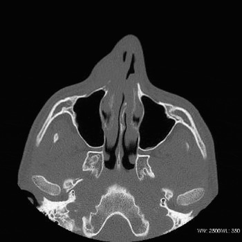
The CT scan was reviewed (Figure 1) as was the histology. The histology had initially been reported as a ‘spindle cell tumour of uncertain aetiology’. A decision was made to remove the tumour through a lateral rhinotomy/medial maxillectomy approach pending a second opinion on the histology. The second opinion revealed the lesion to be a myofibroma-like myopericytoma and the operation proceeded. The lesion was excised as planned, with bony excision margins extending laterally to the infra orbital foramen and medially to include the inferior turbinate. The operative specimen was also analysed and the features were in keeping with a diagnosis of myopericytoma. On this basis, and because it was felt the tumour was invasive, the patient was referred to the Myerstein Institute of Oncology at University College Hospital (London, UK) for consideration of adjuvant therapy. The advice from the Myerstein Institute was that this was unlikely to be an aggressive lesion and adjuvant therapy would be associated with unnecessary morbidity and a surveillance policy was adopted. Six months later, examination under anaesthetic of the nose and biopsy from the nasal vestibule suggested an area suspicious for recurrence. This required further excision with wider margins and composite tissue flap reconstruction. The operative specimen contained was clear of recurrent disease.
Axial CT showing right pyriform aperture involvement.

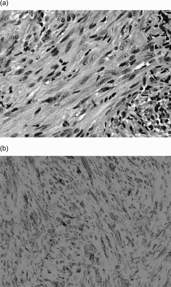
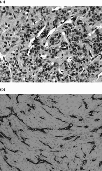
Histologically the tumour consisted of fascicles, or whorled nodules, of spindle cells with abundant eosinophilic cytoplasm and morphological features resembling smooth muscle cells. The nuclei were bland and cigar-shaped (Figure 2). There were also areas with more primitive spindle-shaped and oval cells associated with a haemangiopericytoma-like vascular pattern of thin-walled branching vessels. These cells differed from glomus cells in that they were larger and did not have a clearly demarcated cell membrane (Figure 3). In small areas the myoid spindle cells were in areas arranged concentrically around small vessels. The mitotic count was 2–3 per 10 high power field (HPF) and necrosis was not present. Small foci of hyalinisation were observed as well as a few multinucleated giant cells. The cells were immunoreactive for smooth muscle actin, muscle specific actin and vimentin but negative for desmin, myogenin, S-100, CD34, CD31, cytokeratins and epithelial membrane antigent (EMA). The features were those of a myofibroma-like myopericytoma.
(a) Fascicles of spindle-shaped tumour cells resembling smooth muscle cells. The cells have bland cigar-like nuclei and abundant eosinophilic cytoplasm (H&E; ×40), (b) strong immunopositivity for smooth muscle actin in most of the cells (SMA; ×40).

(a) Round and oval, smaller cells clustering around a haemangiopericytoma-like pattern of thin branching vessels (H&E; ×40), (b) positive CD34 immunostaining of numerous vessels but all tumour cells outside the vessels are negative (CD34; ×40).

Discussion
Haemangiopericytomas were first described by Stout and Murray in 1942 as richly vascularised tumours composed of Zimmerman's pericytes which were thought to be perivascular modified smooth muscle cells.Reference Stout and Murray3 Modern immunohistochemistry has failed to support this theory as the tumour cells surrounding the vessels do not stain for actin or other myoid markers and stain non-specifically for vimentin and CD34.Reference Calonje, Fletcher and Fletcher2 Haemangiopericytoma occurs in four different clinical settings: adult; infantile; sinonasal and myopericytoma. The term ‘myopericytoma’ embraces a spectrum of lesions that show distinct histological subtypes: adult and infantile myofibromatosis; glomangiopericytoma; myopericytoma and solitary myofibroma in adults.Reference Calonje, Fletcher and Fletcher2, Reference Requena, Kutzner, Hugel, Rutten and Furio4 On reflection many cases previously classified as adult haemangiopericytoma would now be reclassified as examples of solitary fibrous tumour. Despite this two groups of true haemangiopericytoma are still seen, namely haemangiopericytoma occurring principally in the female pelvis/retroperitoneum (associated with hypoglycaemia) and meningeal haemangiopericytoma (angioblastic meningioma).
Although sinonasal haemangiopericytoma accounts for only 1 per cent of sinonasal tumours they represent 10 per cent of all haemangiopericytoma presentations. The head and neck variant occur almost exclusively in the nose or paranasal sinuses of patients in their seventh decade and there is a slight female predominance.Reference Thompson, Miettinen and Wenig5, Reference Barnes and Barnes6 Sinonasal haemangiopericytomas are histologically similar to haemangiopericytomas of other soft tissues but are composed of more obviously myoid (actin-positive) cells, and appear to be biologically distinct by their tendency for local recurrence but not metastasis.Reference Eichhorn, Dickersin, Bhan and Goodman7 Soft tissue haemangiopericytomas have variable clinical behaviour ranging from indolent lesions to aggressive malignant behaviour. This behaviour does not readily correlate with any histopathological parameters. Also, soft tissue haemangiopericytomas have a common non distinctive architectural pattern that many other tumours mimic.Reference Backwinkel and Diddams8 It is unsurprising, therefore, that some pathologists have chosen to question the existence of haemangiopericytoma as an independent histological entity.Reference Thompson, Miettinen and Wenig5 Indeed there has been a shift within the histological community such that haemangiopericytoma is currently considered only as a diagnosis of exclusion when identifying soft tissue perivascular neoplasms.
The tumour described in this report did not have the typical features of a sinonasal haemangiopericytoma but falls into the fourth category, myopericytoma. Granter and his colleaguesReference Granter, Badizadegan and Fletcher1 reviewed 24 tumours showing perivascular myoid differentiation which would have been previously deemed haemangiopericytomas. They described three histologically distinct patterns and concluded that the tumours represent a spectrum of tumours. Myopericytoma (perivascular contractile cell) is the term currently used to embrace the three histologically similar lesions of: myofibromatosis; glomangiopericytoma and myopericytoma. This case predominantly had features of a myofibroma with perivascular myoid differentiation. Granter and associates reasoned that these tumours probably develop from a pluripotent periendothelial cell capable of differentiating along smooth muscle, pericytic and glomus lines. They, and authors elsewhere in the literature, have concluded that these lesions are best termed myopericytomas.Reference Mentzel, Hugel and Kutzner9, Reference McMenamin and Fletcher10 There is also a rare malignant variant of myopericytoma that has been recently described, with five cases displaying malignant properties resulting in disseminated disease and patient death in all but one case.Reference McMenamin and Fletcher10 No immunohistochemical marker for malignancy has yet been identified, only tumour behaviour such as bony invasion posing clues as to malignant potential. The sub-classification of myopericytomas is more in keeping with the variable biological outcome that had been previously noted.
• Sinonasal haemangiopericytoma is a rare nasal neoplasm
• Haemangiopericytomas are better termed as myopericytomas, representing a spectrum of neoplasms that demonstrate differentiation along pericytic and glomus cell lines
• There is a rare malignant subtype of myopericytoma
• Management requires surgical excision with further excision for local recurrence
• Local recurrence is likely (17 per cent) probably due to difficulty in obtaining adequate clearance margins
Whilst this distinction is now acceptable terminology for perivascular tumours with myoid differentiation outside the sinonasal region there is still some debate about the application of these distinctions to the sinonasal group of tumours. Some authors have noted that many of the immunohistochemical features of sinonasal haemangiopericytomas are also common to glomus tumours (although it is conventionally taught that true glomus tumours of the nose are extremely rare). Both glomus tumours and sinonasal haemangiopericytomas show pericytic differentiation with a perivascular arrangement of lesional cells (although glomus tumours are without the spindle cells or concentric growth pattern seen in sinonasal haemangiopericytoma). Sinonasal haemangiopericytomas and glomus tumours show immunoreactivity for smooth muscle actin and muscle specific actin but not for desmin and neither tumour expresses CD34. In contrast to sinonasal haemangiopericytomas, the soft tissue haemangiopericytomas do not show immunoreactivity for muscle specific actin, nor do they tend to show immunohistochemical evidence of myoid differentiation but up to 50 per cent will express CD34.Reference Tse and Chan11 The natural history of glomus tumours is similar to the sinonasal haemangiopericytomas. Both these tumours are considered benign with only very rare reports of malignancy. The rates of local recurrence are about 10 per cent with recurrence thought to reflect the difficulty of achieving complete excision due to anatomical constraints rather than malignant tendencies. Because of these observations it has been argued that sinonasal haemangiopericytomas are immunohistologically and biologically distinct from soft tissue haemangiopericytomas and more probably a biological variant of glomus tumour. Unfortunately, even when armed with this knowledge these tumours still pose a management dilemma. The largest series of cases reviewed 104 cases from 1970 to 1995 and suggests that surgery is the treatment of choice, either through endoscopic resection or through medial maxillectomy/lateral rhinotomy.Reference Thompson, Miettinen and Wenig5 Their recurrence rate was 17 per cent with time to recurrence ranging from five weeks to 12 years following resection. The only unique feature of irresectable disease was bony invasion. No patients in this series developed malignant disease.
Conclusion
Sinonasal haemangiopericytoma can be considered as an immunohistochemically and biologically different tumour to the soft tissue haemangiopericytomas. These benign tumours classically occur in women in their seventh decade and are immunohistochemically and biologically similar to glomus tumours. The overlapping histology of myopericytoma with myofibromatosis, solitary myofibroma and infantile haemangiopericytoma is discussed. All these ‘entities’ constitute a single morphological spectrum with differentiation towards perivascular myoid cells (pericytes). Myopericytoma is currently the most appropriate and accepted term for these entities. Myopericytomas are best treated by resection with further resection in cases of local recurrence.