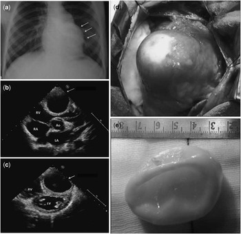
A 12-year-girl presented with persistent palpitations. On examination, a prominent systolic pulsation was seen at the left parasternal border. The chest X-ray also demonstrated a corresponding bulge on the left cardiac border (Fig 1a). Echocardiography demonstrated a cystic swelling of 3.6 cm × 4.5 cm arising from the right ventricular free wall and protruding into the right ventricular outflow tract, with homogeneous clear fluid (Fig 1b). The cyst wall had double echogenic lines separated by a hypoechogenic layer, suggestive of hydatid cyst (Fig 1c). Routine laboratory tests were normal. After a 4-week course of oral albendazole, the child underwent surgery with cardiopulmonary standby. A median sternotomy was performed. The cyst was almost entirely encased in muscle tissue (Fig 1d). The cyst wall was punctured with a hypodermic needle, and the contents were aspirated to avoid accidental spilling of the contents. Hypertonic 3% saline was then injected into the cyst to sterilise the contents, following which the cyst was enucleated and removed in its entirety by blunt dissection (Fig 1e). The post-operative course was uneventful. Sections of the surgical specimen confirmed the diagnosis of hydatid cyst. Hydatid disease, caused by the parasite echinococcus granulosus, has been reported worldwide, and is endemic in subtropical climates. Cardiac manifestations are rare. Typically, the cysts have a multivesicular appearance, although this is not invariable. Serological tests may be negative in the absence of a sufficient immune response, and surgical excision may be required to confirm the diagnosis.
( a ) Chest X-ray in the anteroposterior projection shows a prominent bulge on the left cardiac border (arrows); ( b ) transthoracic echocardiogram in the short-axis view, showing a cystic mass compressing the right ventricle (arrow); ( c ) the cyst wall has a double echo line, suggestive of a hydatid cyst (arrow); ( d ) appearance after median sternotomy. Except for the apex, the cyst is completely encased by muscle tissue; ( e ) operative specimen, confirming a typical hydatid cyst (scale in cm). RV = right ventricle; RA = right atrium; Ao = aorta; LA = left atrium.