I. INTRODUCTION
A. Surface topographies
The surface topography of a material is known to influence biological processes including protein adsorption and conformation, cell behavior, blood-contacting properties, and bacterial adhesion. This has implications for materials used for microfluidic devices, biosensors, and implant materials. Surface topography has been explored as a way to create both antifouling (e.g., protein, blood, or bacterial resistant) surfaces as well as surfaces with enhanced protein uptake properties without the need to change the bulk properties of the material or the surface chemistry. The power of surface topography can be seen by the ability to turn cell-repulsive polyethylene glycol (PEG) surfaces into cell-adhesive surfaces by the inclusion of surface topography.Reference Schulte, Diez, Moller and Lensen1 This effect was only possible in the presence of proteins. This demonstrates that protein interaction with surface topography is fundamentally important and can be studied independent of the material surface chemistry and hence is the focus of our review.
B. Topography on metals
Micro- and nanosized surfaces topographies have been engineered by a variety of methods to create both random and periodic structures in biomedical metal and polymeric materials. The study of metal surfaces often relates to titanium for dental,Reference Ota-Tsuzuki, Datte, Nomura, Cardoso and Shibli2 orthopedic,Reference Cei, Legitimo, Barachini, Consolini, Sammartino, Mattii, Gabriele and Graziani3, Reference Orsini, Salgarello, Martini, Bacchelli, Quaranta, Pisoni, Bellei, Joechler and Ottani4 and coronary stents.Reference Chai, Ochsenbein, Traisnel, Busch, Breme and Hildebrand5 Titanium is very stable and inert to the human body, and for this very reason, cell growth and healing around a smooth titanium implant is slow. As such, ways to enhance its bioactivity including engineering of surface topography have been explored.Reference Kammerer, Gabriel, Al-Nawas, Scholz, Kirchmaier and Klein6, Reference Stanford7 Nonperiodic surface texturing on titanium has been achieved by techniques such as plasma spraying, grit blasting, acid etching, or anodization as reviewed by Le Guehennec et al.Reference Le Guehennec, Soueidan, Layrolle and Amouriq8 Regular uniform patterns in the micrometer and nanometer rangeReference Khang, Lu, Yao, Haberstroh and Webster9 have also been studied. The addition of topographic features on to titanium improves tissue integration as well as influences the way in which cells proliferate and differentiate.
C. Topography on polymers
The use of polymers as medical implants is also highly extensive. Like metals, polymer surfaces have been modified to effect biological responses. Like metals, there are a number of techniques used to produce textured polymeric surfaces. These include electrostatic spinning,Reference Kim and Kim10 polymer demixing,Reference Song, Veiga, Custodio and Mano11, Reference Minelli, Kikuta, Tsud, Ball and Yamamoto12 microphase separation,Reference Leung, Hitchcock, Brash, Scholl and Doran13–Reference Leung, Hitchcock, Cornelius, Brash, Scholl and Doran15 plasma etching,Reference Shiu and Chen16, Reference Tsougeni, Vourdas, Tserepi, Gogolides and Cardinaud17 and imprinting.Reference Zhang and Low18
In our studies described herein, plasma etching and embossing engineering methods were chosen to understand random and ordered texture effects on biological responses to polymer surfaces. Plasma is used to etch the surface of a polymer to create random nano- or microtexturing. In some cases, an alteration in surface chemistry also occurs. For example, the oxygen plasma treatment of a polymer such as poly(methyl methacrylate) (PMMA) and polypropylene can induce high aspect ratio surface features in the nano- and microsize range but also increases the amount of polar groups on the surface.Reference Tsougeni, Vourdas, Tserepi, Gogolides and Cardinaud17 Alternatively, oxygen plasma has been used for the treatment of fluoropolymers to create surfaces with roughness from 10 to 65 nm with no change in surface chemistry.Reference Shiu and Chen16, Reference Shiu, Kuo, Whang and Chen19
Nanoimprinting on polymers is a direct patterning technique that uses a mold or template to emboss the topographic features by means of pressure on to a polymer. The polymer can be a thermoplastic material, which heated above its glass transition temperature becomes soft, or it can be a cross-linkable material, whereby either by heat or ultraviolet light, the polymer is solidified within the mold acquiring its relief.Reference Schift20 This process can create highly ordered regular patterns with sizes from hundreds of micrometers to tens of nanometers. Complex structures can also be fabricated using the imprinting process such as surfaces with hierarchical topographies whereby periodic nanoscale features can be imprinted onto micrometer-scale structures using a single polymer.Reference Zhang and Low18
II. CONTROL OF PROTEIN UPTAKE
Surface texturing has been used to influence the amount of protein uptake on surfaces. It provides a level of control over the total protein adsorption levels and also influences the ratio of different proteins, the spatial distribution, protein conformation, and surface binding affinity. Nanoscale topographies are thought to influence protein adsorption when the sizes of the surface features are in the same scale range as that of the dimensions of the proteins.Reference Roach, Farrar and Perry21 It has generally been considered that when proteins interact with topographies much larger that of their dimensions, such as micrometer-scale features, they view the surface as flat. However, there is evidence that even micrometer-sized topographies can be used to influence the way that proteins interact with a surface.
A. Reducing protein uptake using surface topography
The engineering of surface topography has been explored as a means of decreasing protein fouling on surfaces without changing the bulk properties of the material or altering the surface chemistry. Topography can be used to tune the wetting behavior of a surface as described by the Wenzel and Cassie–Baxter wetting models.Reference Dong, Nypelo, Osterberg, Laine and Alava22 Inherently, hydrophobic materials become more hydrophobic with the inclusion of surface topography due to the entrapment of air between the surface structures. Hydrophilic materials become more wettable with texturing due to the increased contact area of the liquid with the surface.
A reduction in protein adsorption can be achieved by engineering superhydrophobic materials through the addition of surface topography. The topography limits the contact of the protein solution to the tops of the structures giving a lower surface area for adsorption due to the Wenzel–Cassie wetting effect.Reference Koh, Rodriguez and Venkatraman23, Reference Koc, de Mello, McHale, Newton, Roach and Shirtcliffe24 When using a purely physical modification to change surface wettability, the surfactant nature of protein must be considered as well the pressure in the system, which causes the superhydrophobic wetting state to be unstable.Reference Koc, de Mello, McHale, Newton, Roach and Shirtcliffe24 This instability may be the reason for the somewhat limited literature reporting superhydrophobic topography as the sole strategy for nonprotein fouling applications.
Surface texturing can be used to switch a surface from protein resistant to protein adsorptive by changing the wettability of the surface structures. Shui and ChenReference Shiu and Chen16 engineered a Teflon surface roughened by oxygen plasma without an observed modification of surface chemistry. The rough superhydrophobic Teflon gave less Immunoglobulin G (IgG) uptake than the flat film due to decreased contact area of the liquid with the surface. The use of the surface topography gave a comparable reduction in protein adsorption as that of PEG-coated surfaces, a well-known antifouling layer. However, when the surface was made wettable by the process of electrowetting (modifying surface energy with an electric field), protein uptake was increased above flat Teflon due to the increase in available surface area for adsorption. When repeated cycles of protein deposition were performed on the roughened Teflon by cycling the electrode, the amount on the wettable areas continued to increase, where no increase was seen on the superhydrophobic surface during this time frame. However, the antifouling properties of the superhydrophobic surface do suffer from limitations. After a period of 30 min, an increase in the amount of protein on both the superhydrophobic and the PEG surfaces was observed as the protein solution begins to penetrate into the surface structures, likely due to instabilities in the wetting state. Thus, antifouling properties of such surfaces may be useful for short time frame applications only.
Using a combination of surface texturing and surface chemistry has been the most successful strategy for creating a protein-resistant surface. Zheng et al.Reference Zheng, Song, Huang and Chen25 engineered lotus-like surface structures on polyurethane. The lotus leaf template gave pillar-like structures with 7–10 μm base diameters. Uptake of fibrinogen and bovine serum albumin (BSA) increased on untreated lotus structures compared to the flat surface due to the increase in surface area provided. However, when the surfaces were modified with a pluronic known to be protein resistant, it was found that the amount of protein on pluronic-treated lotus structures was even lower than a flat pluronic surface despite the larger surface area. The increased surface area may provide a means to create a higher local surface concentration of the protein-resistant component, making it more protein resistant than the flat surface.
B. Increasing protein uptake using surface topography
Increasing the amount of protein uptake can be useful in on-chip devices to increase biosensor sensitivity or on biomaterials to promote favorable cell and tissue responses.Reference Wang, Hsieh and Lee26, Reference Brammer, Choi, Frandsen, Oh and Jin27 The surface engineering of random or periodic structures with different shapes and dimensions has been used as a strategy to increase the amount of protein adsorption.
Increasing the biomolecule loading capacity of microfluidic systems could be useful in applications such as DNA and protein biosensors such as that demonstrated by Shi et al.Reference Shi, Peroz, Peyrade, Salari, Belotti, Huang and Chen28 who fabricated dense high aspect ratio pillars used to immobilize higher quantities of enzymes. By selecting an appropriate engineering method, surface topographies can be included in more complex devises. LiuReference Liu29 integrated surface texturing into a three-dimensional microfluidic device to increase the surface area for DNA and protein analysis. They used a simple method to make hierarchical lotus leaf structures in polydimethylsiloxane (PDMS) using the natural leaf as a template. This technique provides a low cost and fast way to make structures without the need for microelectromechanical systems fabrication technology. After oxygen plasma treatment to change the superhydrophobic surface to hydrophilic, they found that fluidic devices with lotus surface gave 5 times higher sensitivity (lower detection limit) than the flat surfaces due to increase in loading capacity.
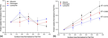
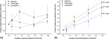
Systematic studies of surface topography allow a greater understanding of how to control protein uptake on a surface. Results from our laboratory have shown that micrometer-sized ordered textures provide a means by which to control the amount of protein uptake and that the uptake can be predicted by the amount of surface area. Using nanoimprint lithography methods, the dimensions of surface structures could be systematically varied to increase either the pillar height (Fig. 1) or the pillar spacing (Fig. 2) to study the effect on protein adsorption. It was found that the amount of protein uptake could be correlated in a linear manner to the increase in surface area provided by the surface structures. This could be achieved for a selection of proteins with different dimensions and charges tested on a biodurable PDMS polymer and a biodegradable poly(lactic-co-glycolic acid) (PLGA) polymer. This linear correlation between protein uptake and surface area provided by surface structures was only true when the surface structures were completely wetted by the protein solution (Figs. 3 and 4). Without surface modification, the surface structures increased the hydrophobicity of the surfaces (Tables I and II) due to the Wenzle–Cassie wetting effect, and no trend could be seen between protein uptake and surface area after an 18-h incubation. However, when the surfaces were treated with oxygen plasma to make them hydrophilic, the effect of surface area could be seen.
PDMS pillars fabricated by casting into silicon molds with 2 μm height, 2 μm spacing and heights of (a) 3, (b) 6, (c) 9, and (d) 12 μm giving an associated increase in total film surface area of 1.8, 2.4, 3.3, and 4.1 times respectively compared to a film with flat surfaces. Scale bar = 5 μm.

PLGA pillars fabricated by hot embossing of PLGA into silicon molds with 10 μm diameter, 10 μm height, and spacing of (a) 50, (b) 20, (c) 10, and (d) 6 μm, giving an associated increase in total film surface area of 1.05, 1.2, 1.4, and 1.6 times respectively compared to a film with flat surfaces. Scale bar = 10 μm.

Protein uptake on PDMS pillars normalized to the flat surface before and after treatment with oxygen plasma. (a) protein uptake on untreated films with pillars of increasing height show no correlation to surface area (b) after oxygen plasma treatment protein uptake correlates to surface area, increasing with pillar height.

Protein uptake on PLGA pillars normalized to the flat surface before and after treatment with oxygen plasma. (a) protein uptake on untreated films with increasing pillar density show no correlation to surface area (b) after oxygen plasma treatment protein uptake correlates to surface area, increasing as pillar spacing decreases.

Water contact angle of PDMS pillars with 2 μm height, 2 μm spacing, and varying height.

Water contact angles of PLGA pillars with 10 μm diameter, 10 μm height, and varying spacing.

The increase in surface area provided by surface topography is not always sufficient to explain observed increases in protein adsorption. The amount of protein uptake can be increased beyond the increase in surface area for both microReference Chen, Song, Zhou, Wu, Huang, Zhang, Lin and Yang30 and nanotopographies.Reference Sela, Badihi, Rosen, Steinberg and Kohavi31–Reference Gonzalez-Garcia, Sousa, Moratal, Rico and Salmeron-Sanchez36
Chen et al.Reference Chen, Song, Zhou, Wu, Huang, Zhang, Lin and Yang30 fabricated dot-like micrometer-sized protrusions in PDMS with a base diameter of 12 μm, an interval between protrusions of 7.5 μm, and a height of 1.27 μm, without significant nanometer-scale roughness. It was observed that an 8% increase in surface area led to a 46% increase in protein adsorption compared to flat surface. It was also found that protein distribution was affected by the micrometer-scale surface topography, with protein concentrated in the interspace regions between the domes and not at the peaks of the domes.
Scopelliti et al.Reference Scopelliti, Borgonovo, Indrieri, Giorgetti, Bongiorno, Carbone, Podesta and Milani32 provide a possible mechanism for how surface topography can contribute to higher adsorption than the increase in surface area alone. A highly sensitive fluorescence technique was used to quantify protein adsorption on titanium with different surface roughness ranging from 14 to 32 nm. Fibrinogen and BSA protein adsorption was 500% higher than the theoretical monolayer coverage on the roughest surface, indicating that surface roughness may promote protein–protein interaction to form multilayers. A study of the adsorbed protein layer by atomic force microscopy (AFM) showed that protein localized within pores of the nanorough surfaces with aspect ratios higher than 0.5. It is proposed that the proteins become trapped inside the pore area for a longer dwelling time, causing crowding to occur and nucleation of protein aggregation. It is this space-confined aggregation effect that facilitates higher protein surface coverage than surface area on surfaces with these types of dimensions. The preference of proteins to bind inside valley topographical regions has also been reported on different materials including siliconReference Elter, Lange and Beck35 and poly(L-lactide)/polystyrene nanoscale dimension grooved surfacesReference Gonzalez-Garcia, Sousa, Moratal, Rico and Salmeron-Sanchez36 when the bulk protein concentrations were sufficiently low. At low protein concentrations, the preference for binding in valleys has been described by the influence of short-range dispersion forces where the protein is contacted by two faces of the walls of the valley.Reference Elter, Lange and Beck37
These models for protein aggregation in the confined areas created by surface topography can also explain how topography influences protein distribution even on the micrometer-scaled surface described by Chen et al.Reference Chen, Song, Zhou, Wu, Huang, Zhang, Lin and Yang30 The preferential distribution of the proteins in the interspace between the surface protrusions could be due to aggregation in the confined space between the structures.
The increase in protein uptake above the surface area is also protein and material dependent. Sela et al.Reference Sela, Badihi, Rosen, Steinberg and Kohavi31 studied the adsorption of blood plasma proteins and individual proteins (albumin, fibronectin, fibrinogen, IgG) on roughened titanium with room mean square (RMS) roughness ranging from 77 to 230 nm with minimal change in surface chemistry. The roughest surface showed the highest levels of adsorption for all proteins. However, when protein uptake was normalized for surface area, a difference in the behavior of the proteins was observed. This may have to do with the way different proteins pack on a surface due to their individual size, shape, and unfolding upon adsorption.Reference Minelli, Kikuta, Tsud, Ball and Yamamoto12, Reference Rechendorff, Hovgaard, Foss, Zhdanov and Besenbacher34 Some proteins (fibronectin, fibrinogen, and IgG) showed only an increase in adsorption proportional to the increase in surface area. Albumin, on the other hand, showed an increase in adsorption greater than that of the increase in surface area. Conversely, Leung et al.Reference Leung, Hitchcock, Brash, Scholl and Doran13 showed polystyrene–polylactide block copolymer surfaces with rugosity ranging from 35 to 90 nm had no effect on albumin uptake. Rockwell et al.Reference Rockwell, Lohstreter and Dahn38 also found no differences between the uptake of fibrinogen or albumin across a roughness gradient despite the differences in protein structure, which was expected to influence the adsorption behavior on different scales topographies. It is apparent that the intrinsic nature of the protein in combination with the specific properties of the material may also govern the behavior.
C. Differential protein uptake on surface topography
Protein binding to surfaces is dynamic, and there is evidence that surface topography can be used as a way to preferentially bind specific proteins from complex biological fluids. This differential protein uptake may be important in blood contacting and other antifouling applications where it has been found that surfaces that preferentially bind albumin are more resistant to further protein adsorption and are less thrombogenic. Huang et al.Reference Huang, Lue, Ma and Huang39 found that a surface that bound a greater ratio of albumin than IgG gave less platelet adhesion, while a surface with equal albumin/IgG uptake showed grater platelet adhesion. On the surfaces compared (diamond-like carbon and titanium), the surface energy did not correlate to the protein uptake as reported in other studies, which is attributed to the effect of the greater surface roughness of the titanium surface compared to the carbon surface.
Changes in the ratio of proteins adsorbed from blood plasma have also been observed on surfaces with different roughness. Different topographical feature size may be preferential to proteins with different sizes and dimensions. Minelli et al.Reference Minelli, Kikuta, Tsud, Ball and Yamamoto12 studied the uptake of fibrinogen and the von Willebrand factor from platelet-poor plasma under flow conditions onto PMMA surfaces with nanoscale roughness between the range of 40 and 400 nm, which is in the same size range as these rod-like proteins (fibrinogen 47 × 4 nm, von Willebrand factor 120 nm length). The two proteins showed different trends in adsorption on surfaces with different roughness, thus eliminating surface area as the sole effect on protein adsorption behavior. It is thought that this is due to the Vroman effect of dynamic protein adsorption, with differential protein displacement occurring on different topographies.
D. Influence of surface topography on protein binding affinity
Surface roughness also influences the protein binding affinity to the surface as well as the absolute amount. Decreases in the strength of protein attachment to rough surfaces under shear conditions have been observed, which has important application in fluidic devices to prevent fouling. Under static conditions, Scopelliti et al.Reference Scopelliti, Borgonovo, Indrieri, Giorgetti, Bongiorno, Carbone, Podesta and Milani32 found an increase in Kd on rougher surfaces (titanium with 32 nm roughness). However, it is concluded that this is not due to changes in chemical affinity of the protein to the surface but rather from supersaturation of high protein concentrations inside protected pore areas of the topography. When fluid shear is brought into the system, it was found that protein is removed more effectively from textured surfaces than smooth surfaces. Koc et al.Reference Koc, de Mello, McHale, Newton, Roach and Shirtcliffe24 engineered a superhydrophobic surface through the fabrication of porous micro- and nanostructures where surface pores are not completely penetrated by the liquid due to the Wenzel–Cassie wetting effect. However, this superhydrophobic topography was not sufficient to make the surfaces more protein resistant than the flat surface. Surfaces with larger pore diameters gave the most protein binding due to the surfactant nature of protein solution and the pressure in the system allowing enough protein solution to penetrate into the pores and give access to the extra surface area. Although the textured surfaces bound more protein than flat surfaces, the difference was seen in the removal of protein from the surfaces under flow conditions. More protein was removed from structured surfaces compared to flat surfaces, with more protein being removed as structures became smaller (structure sizes 4 μm, 800 nm, and 10 nm). It is proposed that this is due to higher shear forces at the surface of structures under flow. The nanosized structure showed the greatest amount of protein desorption, with almost complete removal achieved. This is thought to be a combined effect of increased shear at the surface of smaller structures, as well as an effect of the curvature of the particles being in the same size range as that of the BSA protein studied.
E. Protein conformational changes
Surface topography affects protein unfolding upon adsorption,Reference Roach, Farrar and Perry21, Reference Jedlicka, McKenzie, Leavesley, Little, Webster, Robinson, Nivens and Rickus40–Reference Roach, Farrar and Perry42 and the size and shape of the protein determine how it responds to surface texture.Reference Penttinen, Silvennoinen, Hason and Silvennoinen43 Roach et al.Reference Roach, Farrar and Perry21 showed the effect of surface curvature on the unfolding of proteins with rod or globular shapes. Protein conformation is also important in regulating cell behavior such as attachment and differentiation. For example, fibronectin exposes its active cell binding sites [arginine-glycine-aspartate (RGD) sequence] when it unfolds upon adsorption to a surface, which in turn influences cell integrin binding.Reference Keselowsky, Collard and Garcia44, Reference Michael, Vernekar, Keselowsky, Meredith, Latour and Garcia45 Schricker et al.Reference Schricker, Palacio and Bhushan14 compared poly(methyl methacrylate)/poly(acrylic acid) block copolymer surfaces with the same chemical composition but with either porous structure or irregular surface features to study the level of RGD sequence exposure of adsorbed fibronectin. They found that the regular nanoporous structure had a higher amount of RGD exposed than the flat control or irregular surface features. Interestingly, they also observed a change in the mechanical properties of the adsorbed fibronectin layer, with the irregular surface showing lower stiffness compared to the regular porous structure. It is postulated that this is further evidence of a difference in protein conformation.
Protein morphology is also influenced by surface topography. For example, collagen forms elongated aggregates on smooth surfaces but not on textured surfaces even though the amount of protein bound was the same.Reference Denis, Hanarp, Sutherland, Gold, Mustin, Rouxhet and Dufrene46 This has important implications for tissue engineering applications where deposition of extracellular matrix proteins by cells is effected by surface topography.Reference Brydone, Dalby, Berry, Dominic Meek and McNamara47
III. CONTROL OF CELL/TISSUE INTERACTIONS
A. The effect of surface topography on blood clotting
Finding a correlation between surface texture and blood interaction is difficult due to different experimental conditions, e.g., whole blood versus plasma, and different shear conditions.Reference Ferraz, Carlsson, Hong and Ott48, Reference Milner, Snyder and Siedlecki49 Surface topography has been explored as a way to improve the blood compatibility of materials by controlling blood–cell interaction with the surface. Features that make the surface superhydrophobic or have features smaller than the size of a platelet can reduce platelet binding by reducing the available contact area,Reference Koh, Rodriguez and Venkatraman23, Reference Milner, Snyder and Siedlecki49–Reference Milner, Snyder and Siedlecki53 although these results can be conflictingReference Kikuchi, Park, Victor and Davies54 and are highly dependent on material, feature size, and experimental conditions. For example, Hou et al.Reference Hou, Wang, Zhu, Bao, Mao, Jiang and Shen55 fabricated a rough superhydrophobic (contact angle 150°) microporous polypropylene surface without a change in surface chemistry compared to the flat surface. It was found that clotting and blood cell binding were reduced from both platelet-rich plasma and whole blood due to the trapped air in the surface structures reducing the contact area for blood cells, platelets, and fibrin to adhere.
The ability of micrometer-sized surface topography to tune the surface energy of a material has also been used to reduce platelet adhesion. Zhou et al.Reference Zhou, Yang, Ye, Zheng, Li, Yang, Zhu and Cai50 engineered a PDMS surface with square pillars with width 10 μm, height 5 μm, and various spacing of 25, 35, or 45 μm. They found that the amount of platelets adsorbed from static incubation in platelet-rich plasma was decreased as the contact angle increased (the closer the spacing, the more hydrophobic the surface with the largest contact angle of 156°). This is attributed to different surface tension caused by the features, which leads to different platelet aggregation behavior. The group also studied periodic 200-nm grooves, that although not superhydrophobic (contact angle 120°), did significantly reduce platelet adhesion and also reduce platelet activation. It is thought that the small spacing of the features may inhibit the formation of focal contacts and prevent platelet aggregation, demonstrating that the structure dimensions as well as the surface energy may be important factors to consider. In addition to using surface topography to reduce the contact area for adhesion and to influence surface energy, Koh et al.Reference Koh, Rodriguez and Venkatraman23 suggest that using flexible high aspect ratio nanoscale pillars prevents platelets from making successful contact with the surface compared to stiffer more stable low aspect ratio structures.
Materials that will be used in blood-contacting applications may be faced with fluid flow conditions adding an additional level of complexity to the problem. The effect of shear forces on platelet binding to different surface topographies has been studied using rotational shear experimentsReference Milner, Snyder and Siedlecki49, Reference Milner, Snyder and Siedlecki53 and microfluidic devices.Reference Minelli, Kikuta, Tsud, Ball and Yamamoto12, Reference Milner, Snyder and Siedlecki53, Reference Minelli, Yamamoto and Kim56 Nanoscale topographies were able to reduce platelet adhesion at low shear stresses. Studies of blood clotting in microfluidic devices have shown that textured surfaces increase the formation of clots despite the fact that some types of blood protein adsorption can be reduced. These results highlight the difficulty of predicting the behavior of a surface in the complex blood environment and in different testing methods.
Protein adsorption happens immediately upon a materials exposure to blood, and this protein layer is thought to strongly influence the blood compatibility of a material. The effect of surface topography on blood protein adsorption has been suggested to play a large role in platelet and blood clot attachment. Platelet distribution on a patterned surface is indirectly influenced by the protein adsorption. Platelet attachment follows fibrinogen spatial distribution on a patterned surface.Reference Chen, Song, Zhou, Wu, Huang, Zhang, Lin and Yang30 Platelets adhere more on smaller feature sizes when not in the presence of protein. However, platelet adhesion followed the trend of protein adsorption in the presence of proteins.Reference Minelli, Kikuta, Tsud, Ball and Yamamoto12 Results are conflicting and platelet adhesion to a textured surface cannot be directly correlated to the amount of protein uptake alone.Reference Minelli, Kikuta, Tsud, Ball and Yamamoto12, Reference Koh, Rodriguez and Venkatraman23, Reference Park and Davies57, Reference Park, Gemmell and Davies58 The influence of topography on the differential uptake of certain blood proteins and the protein conformation need also to be taken into consideration. These factors may contribute to the varying results observed with platelet activation and morphology on textured surfaces.Reference Koh, Rodriguez and Venkatraman23, Reference Minelli, Yamamoto and Kim56 It has been suggested that different surface topography dimensions can activate platelet adhesions and clotting by different mechanisms, which could be due to changes in protein binding profiles. Controlling the pattern of different types of protein binding by changing surface texture may be a way to influence thrombogenicity of a surface.Reference Ferraz, Carlsson, Hong and Ott48
B. Bacterial attachment
Bacterial attachment to surface topography is governed by the type of bacteria and experimental conditions. Bacteria differ in their physiologies and modes of adhesion to surfaces, and it has been shown that nanoscale roughness can actually alter the morphological, genetic, and proteomic profiles of adhered bacteria.Reference Rizzello, Sorce, Sabella, Vecchio, Galeone, Brunetti, Cingolani and Pompa59 Komaromy et al.Reference Komaromy, Li, Zhang, Nicolau, Boysen and Hearn60 studied Staphylococcus aureus and Escherichia coli attachment to systematically varied surface topographies with ordered textures. Dots and lines with increasing nanometer-scale diameter and pitch were fabricated by e-beam lithography and then surface modified with self-assembled monolayers to have either hydrophobic or hydrophilic surface chemistries. It was found that the relationship between surface properties and bacterial adhesion and viability is multifactorial and that bacterial size, physiology, and topographical dimensions must all be considered. Results of the effect of topography on the level of bacterial adhesion can be conflicting even when the same materials with the same bacterial species are studied.Reference Truong, Lapovok, Estrin, Rundell, Wang, Fluke, Crawford and Ivanova61, Reference Ivanova, Truong, Webb, Baulin, Wang, Mohammodi, Wang, Fluke and Crawford62
As with other nonfouling surfaces, the strategy of using superhydrophobic structures to reduce available binding sites has been explored with bacteria. Crick et al.Reference Crick, Ismail, Pratten and Parkin63 fabricated superhydrophobic lotus-like structures to decrease contact area for bacterial adhesion. PDMS was fabricated with random 3-μm protrusions, resulting in a water contact angle of 165°. Despite the fact that Staphylococcus aureus and Escherichia coli had opposite binding to flat PDMS surface due to their differences in physiology both had lower attachment on the structured surface compared to flat PDMS or flat glass. Bacterial colonization was limited to the tops of the protrusions demonstrating the effect of the superhydrophobic structures. They postulated that the lotus self-cleaning/water rolling effect may have helped clear bacteria off the surfaces as the substrates were removed from solution; therefore, water movement may help improve the antifouling properties of such surface even further. This water cleaning effect is the strategy behind the Sharklet™ engineered surface, which takes its inspiration from the self-cleaning properties of sharkskin as it moves through water. When the dimension of the surface structures is matched to that of the bacteria, it has been found that such Sharklet™ surfaces are able to retard bacterial colonization.Reference Chung, Schumacher, Sampson, Burne, Antonelli and Brennana64 Indeed, the dimensions of the surface structures, rather than the overall surface roughness, have been found to be important in bacterial colonization.Reference Whitehead, Colligon and Verran65, Reference Eginton, Gibson, Holah, Handley and Gilbert66 Attempts have been made to explore how the dimension of surface features effect the strength of bacterial adhesion. For example, AFM has been used to quantify the force needed to move bacteria on surfaces with varied surface structure dimensions.Reference Whitehead, Rogers, Colligon, Wright and Verran67 As a surface may be exposed to many types of bacteria with different characteristics, the challenge lies in finding surfaces that can be effective in multicomponent environments.
High surface energy surfaces are generally thought to be less fouling. However, surface chemistry and not just surface energy are important in how a textured surface interacts with bacteria due to the complex nature of bacterial adhesion and colonization mechanisms. Puckett et al.Reference Puckett, Taylor, Raimondo and Webster68 fabricated titanium surfaces with nanoroughness or nanotubular structures. When the surface chemistry was the same, the nanorough surface had higher surface energy than the smooth surface and showed higher fibronectin adsorption and lower bacterial colonization. However, when nanorough and nanotubular surfaces with fluorine ions on the surface from the fabrication process were compared to the nonfluorinated surfaces, they showed higher surface energy and higher protein uptake but also high levels of bacterial colonization.
As well as effecting the amount of bacterial adhesion, topography also influences the site of bacterial attachment such as in trenches or pits,Reference Whitehead, Colligon and Verran65, Reference Taylor, Verran, Lees and Ward69, Reference Teixeira, Trindade, Godinho, Azeredo, Oliveira and Fonseca70 as well as the strength of bacterial attachment to the surface and the amount of extracellular polymeric material produced.Reference Ivanova, Truong, Wang, Berndt, Jones, Yusuf, Peake, Schmidt, Fluke, Barnes and Crawford71 These factors have important implications for biofilm formation and removal, and thus, the correct choice of topography may be able to control both the initial bacterial adhesion as well as future colonization events.
IV. CONCLUSION
While our understanding of the influence of surface topography on biological interactions is increasing, no universal design rules can be applied. The implementation of this technology must be tailored for each specific application, environment, and material. The combination of the correct topography and surface chemistry may be the most successful strategy to control the biological response of a surface and may provide a level of control above either solution alone. However, when chemical changes are either undesirable or not feasible, specifically, well-designed physical changes on to a surface may alter the biological response to the desired behavior.